DOI: 10.5455/annalsmedres.2018.06.110 2018;25(4)756-62
Does transcription factor, induced by daptomycin and
vancomycin, affect HIF-1α, Chondroadherin, and COL2A1?
Numan Karaarslan1, Ibrahim Yilmaz2, Duygu Yasar Sirin3, Hanefi Ozbek2, Yasin Emre Kaya4, Yener Akyuva5, Necati Kaplan6, Mustafa Dogan7, Seyit Ali Gumustas8, Ozkan Ates9, Ilknur Erdem7
1Namik Kemal University Faculty of Medicine, Department of Neurosurgery, Tekirdag, Turkey
2Istanbul Medipol University Faculty of Medicine, Department of Medical Pharmacology, Istanbul, Turkey
3Namik Kemal University, Faculty of Arts and Sciences, Department of Molecular Biology and Genetics, Tekirdag, Turkey) 4Abant Izzet Baysal University Faculty of Medicine, Department of Orthopaedic and Traumatology, Bolu, Turkey
5Gaziosmanpasa Taksim Research and Training Hospital, Department of Neurosurgery, Istanbul, Turkey 6Istanbul Rumeli University, Reyap Hospital, Department of Neurosurgery, Tekirdag, Turkey
7Namik Kemal University Faculty of Medicine Department of Infectious Diseases, Tekirdag, Turkey
8Dr. Lutfi Kirdar, Training and Research Hospital Department of Orthopaedic and Traumatology, Istanbul, Turkey 9Istanbul Esenyurt University, Esencan Hospital Department of Neurosurgery, Istanbul, Turkey
Copyright © 2018 by authors and Annals of Medical Research Publishing Inc.
Abstract
Aim: In this study, it was firstly aimed to investigate the effect of Daptomycin (DAP) on the proliferation in Vancomycin
(VCM)-administered primary chondrocyte cultures and non-drug-(VCM)-administered primary chondrocyte cultures. Our second objective was to investigate the effects of DAP and VCM on the NP-specific marker protein chondroadherin (CHAD), which is associated with spinal cord and dorsal column growth, on the transcription factor-1 alpha (HIF-1α), which is induced by hypoxia, and on a type II collagen (COL2A1), which is also known to play a significant role in the development of extracellular matrix, at the pharmaco-molecular level.
Material and Methods: Standard human primary chondrocyte cultures were established. DAP and VCM were added to the samples.
In all groups, molecular analysis was performed at 0th, 24th and 48th hours. In addition, the surface morphology of the cells was
evaluated.
Results: Changes in cell morphology and cell death in cultures were observed 24 hours after administration of antibiotics to cell
cultures. It was observed that drug administration was associated with the cell viability and that cell viability rate for two antibiotics was similar at the 0th and 48th hours. The expression of three genes decreased at the 24th hour in the experimental group where DAP
was administered.
Conclusion: Thanks to this molecular-based research, it should not be forgotten that DAP and VCM active pharmacological agents,
especially used in the treatment of Methicillin-resistant Staphylococcus aureus induced surgical infections, have a negative effect on human chondrocyte and ECM components.
Keywords: CHAD; Chondrocytes; COL2A1; Daptomycin; HIF-1α; Vancomycin.
Received: 11.06.2018 Accepted: 29.06.2018 Available online: 04.07.2018
Corresponding Author: Numan Karaarslan, Namik Kemal University Faculty of Medicine, Department of Neurosurgery, Tekirdag, Turkey, E-mail: [email protected]
INTRODUCTION
When systemic antibiotics are used in long-term, high and repeated doses in the treatment of the diseases such as septic arthritis, osteomyelitis and spondylodiscitis, side effect profile may also bring toxic problems with it (1). To prevent this situation, it has been started to administer local antibiotic, particularly during surgery, by decreasing systemic dose and time to increase the effectiveness of antibiotic therapy. Spongostan, one of the cement chains with antibiotics, or antibiotics impregnated into
different gels or grafts in different forms applied to the infected site seems promising to prevent the infection in the neurosurgery for prophylaxis and orthopedic surgery branches as well (1).
However, even if these antibiotics are used locally, a research that has reported on the toxic effects of such pharmaceuticals has re-emerged and, shortly after, has taken its place as a controversial issue in the literature (1). As a result of reports regarding the toxic effects of antibiotics on tissues such as vertebrae, bone and
cartilage, the prescription of this drug by the branch surgeons has been restricted in some conditions (1-2). Systemic and/or local antibiotics are used in many cases for prophylactic and/or treatment. Besides, they are used in many operations in which intraarticular fractures with multi knee ligament injuries and open traumas of vertebra are incase (1,3).
While use of VCM and teicoplanin are increasing, studies concerning DAP are gaining popularity (4).
It is known that cartilage is an avascular, aneuronal tissue and is devoid of lymph tissues. Thus, the cartilage cells are fed by synovial fluid that washes the joint surface (5-8). As the outer layer of synovial liquid is thicker, drugs and/or nutrients are diffused into synovial liquid from synovial tissue. Afterwards, they get through pores, reach chondrocytes and then create a second diffusion (5-8). It is known that many drugs whether taken orally or by injection accumulate in synovial liquid (7-8). In addition, there are studies indicating that after drugs with DAP active ingredient are taken, they are accumulating in synovial liquid (9).
In this context, the effect of daptomycin (DAP) on the proliferation of human primary chondrocyte cultures was investigated. In doing so, the non-drug-administered cell culture samples were called as a control group. In addition, the vancomycin (VCM) agent which was reported to have a chondrotoxic effect on primary human cell cultures in the literature was also added as a second control group to this experimental setup. More importantly, the expression of the NP-specific marker protein chondroadherin (CHAD), which are associated with spinal cord and dorsal column growth, was also analyzed in DAP-treated culture samples (10,11). Moreover, the expression levels of the transcription factor-1 alpha (HIF-1α) (12), which is induced by hypoxia, and which is a continuous expression of the nucleus pulposus specific marker, and a type II collagen (COL2A1) (13), which is known to play an important role in the development of extracellular matrix was sought to investigate at the pharmaco-molecular level.
MATERIAL and METHODS
Analyses were carried out during and after primary human chondrocyte culturization. Similar analyses were repeated by the same researchers to minimize the errors. The researchers were blind to experimental setup of drug applications. Experiments were repeated at least 3 times.
Study design and the in vitro experimental setup
After four patients diagnosed with gonarthrosis were operated, the resected osteochondral tissues were used in the preparation of the primary chondrocyte cultures. Cell cultures were fed every 2 days by being replaced the medium until drug treatments started. After the third passage, drugs were added to the cell cultures except for the control group. The analyses were performed on the 0th hour before adding pharmacological agents to the wells, and at the 24th and 48th hours after adding them to the wells.
Chondrocyte cultures were monitored using an inverted microscope. Pharmacological agents were prepared in appropriate solutions and concentrations. Color-coded agents were added in the well plates, and an extra plate from each treatment was set aside for the 3-(4,5-dimethylthiazol-2-yl)-2,5-diphenyltetrazolium bromide (MTT) analysis, Acridin Orange (AO)/Propidium Iodide(PI) staining and qRT-PCR analysis (Table 1).
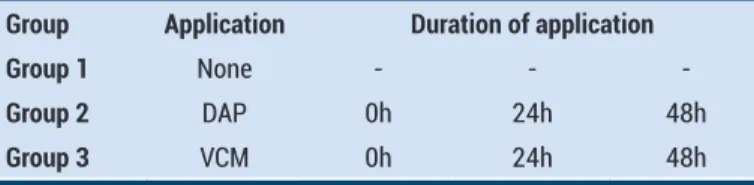
Table 1. Experimental design. Applied antibiotics and duration of application
Group Application Duration of application
Group 1 None - -
-Group 2 DAP 0h 24h 48h
Group 3 VCM 0h 24h 48h
DAP: Daptomycin and VCM: Vancomycin symbolized
Case Selection Criteria
The osteochondral tissues to be used for primary chondrocyte culturing were not taken from patients known to be hypersensitive to vancomycin, linezolid, or teicoplanin. Apart from this, no tissues were taken from the parkinson patients treated with monoamine oxidase inhibitors such as rasajilin, selejiline and moclobemide within the last 14 days. In addition, the tissues of patients using antidepressant drugs or the tissues of patients with malignancy were also not used for the preparation of primary cell cultures (1,7,8).
Cases [The average age of the patients was 65.18±4.71 (mean±standard deviation), n=4] who did not respond to medical and conservative treatments and had large osteophytes, were graded using the Kellgren–Lawrence Radiological Grading Scale (stage-IV) (14-16).
Preparation of primary human chondrocyte cultures:
Osteochondral tissues were resected from femur distal and proximal ends of tibia, which were resected in total knee arthroplasty. Afterwards, standard human primary chondrocyte cultures were carried out (15-17).
During prosthesis application, chondrocyte cultures were prepared from chondral tissues whose articular surfaces were undamaged. This undamaged, intact tissue contains not only cartilage cells but also the environment such as the extracellular matrix.
The counted cells, with the Thoma slide in the presence of the Trypan blue, were seeded at 1.4 x 10⁴ cells per well in 96 well plates for performing proliferation analysis with the MTT ELISA assay. The cells were transferred to 24 well plates for analysis of fluorescence microscopy used for inverted light microscopy and AO / PI staining. Here, cells were seeded at 3.4x10⁴ cells per well. The cells were also seeded at 4.4 x10⁶ cells per dish for RNA isolation and quantitative real-time polymerase chain reaction (qRT-PCR) analyses. After seeding the counted cells in the culture containers specific to each analysis, all samples were incubated for a period of 24 h. At the end of this period, the drug was applied to the cell samples that became confluent and adhered to the floor of the container.
Preparation of Drugs
DAP (Cubicin®, Novartis) and VCM (Vankomisin®, ABBOTT), main stock solutions, were freshly prepared in Dulbecco’s Modified Eagle’s medium in flow cabin as 500 mg and 1000 mg, respectively. These main stock solutions were then transferred to dark glass bottles and letter coding was performed. In this way, researchers did not know which medicines were applied to cell cultures, or which medicines were involved in which samples, while analyzes were carried out. In other words, the researchers were blind to both the drug and analyzes.
Final concentrations in which these drugs were applied in human primary chondrocyte cultures were prepared in regard to minimal bactericidal concentrations with the purpose imitating clinical practices. Final concentration was applied as 200 μg/ml for DAP and 200 μg/ml for VCM (1,18).
Administration of DAP and VCM to cultures
No drug administered-chondrocyte cultures used as control groups for all tests and was named as Group I. Experimental design of the study were given in Table 1.
Analyses
Cell adhesion and morphology were evaluated by an inverted microscope and micro- photographed.
Vitality tests were performed via a commercial MTT kit (3-[4,5-dimethyltiazol-2-yl]-2,5-diphenyltetrazolium bromide (Vybrant MTT Cell Proliferation Assay, Cat#V13154, Thermo Fisher Scientific) using the manufacturer’s instruction. The kit’s functioning principle is explained as follows: tetrazolium ring is cleaved by mitochondrial dehydrogenase enzymes yielding blue formazan crystals, which do not occur in dead cells. MTT analyses were performed before (control group=Group I) and after the drugs were added (Group II and III) using an enzyme linked immunosorbent assay (ELISA/540 nm) microplate reader (Mindray MR 96 A, PRC) (1,15-17.19,20). Then, letter labels designated to each sample were transcribed by the project coordinator. Vitality of the control group was assumed to be 100% before the transfer of antibiotics (0h) to the culture medium. Viability, toxicity, and proliferation tests were performed at the 24th hour after application of antibiotics to culture samples. The results were presented as nanometer (nm) in terms of absorbance. Then, proliferation analyses for hours (24h-48h) were recorded.
To determine cell viability and confirm MTT results, nucleic acid binding dyes AO and PI were used. AO stains all nucleated cells both live and dead and generates green fluorescence. PI can only penetrate to dead cells with poor membrane integrity, and stains nucleated cells to generate red fluorescence. Cells stained with both AO/PI all live nucleated cells fluoresce green and all dead nucleated cells fluoresce red.
To prepare AO/PI stain 4 mg AO (dissolved in 2ml 99% ETOH), 10g sodium-ethylenediaminetetraacetic acid, 4
mg PI and 50 ml Fetal calf serum are mixed well and to reach a 200 ml final volume sterile distillated water are added.
Total ribonucleic acid (RNA) was extracted from cultured primary human chondrocytes using PureLink RNA Mini kit (Ambion, Cat#12183018A) and 2-Mercaptoethanol (Thermo Fisher Scientific, Cat#31350010). To obtain complementary DNA (cDNA), 50 ng RNA were reverse transcribed with High–Capacity cDNA Reverse Transcription Kit (Thermo Fisher Scientific, Cat#4368814) using thermal cycler (ProFlex, Thermo Fisher Scientific). PCR reaction was performed using 10× reverse transcription buffer (10μl) 25× deoxyribonucleotide triphosphate (4μl), 10× random primers (10μl), 50U/μl reverse transcriptase (5μl), nuclease free water (5μl) and RNA (20μl) reaction mix and PCR program as at 25 0C for 10 min hold and 370C for 120 min hold. All genes were amplified using TaqMan® Gene Expression Assays for CHAD (Cat#4448892), HIF-1α (Cat#4453320), COL2A1 (Cat#4453320) and internal control gene [housekeeping genes- actin beta (ACTB) (Cat#4453320)]. RT-PCR reaction mix prepared with 1μl TaqMan Gene Expression Assay, 10 μl TaqMan Gene Expression Master Mix (Cat#4369016), 4 μl cDNA template and Ultrapure DNase/RNase-Free Distilled Water (Cat#10977035) for each gene in MicroAmp Fast Optical 96-Well Reaction Plates (Cat#4346906). Reaction protocol was as follows; 2 min hold at 500C, 10 min hold at 950C, 15 secs at 950C, and 1 min at 600C for 40 cycles and performed with Applied Biosystems 7300/7500 Real-Time PCR System (15,16,21,22).
Statistical Analysis
The analyses were performed by Minitab 16 program and the data were evaluated with 95% confidence interval. Descriptive statistics were given as mean ± standard deviation (SD). Variance Analysis was used while comparing the groups. Tukey HSD was used in the evaluation of significance. Alpha significance level was considered as <0.05.
RESULTS
Morphological analysis of chondrocyte cultures: Inverted microscopy and AO/PI staining:
Cell death and changes in cell morphology in cultures were observed depending on the antibiotic application. However, it was also determined that surviving cells continued to proliferate after 48 hour (Figure 1).
Cell vitality was evaluated by MTT analysis as well as by AO/PI staining. In experimental groups where applications were performed by an inverted microscopy, it was observed that the number of death cells increased after 24 h, yet cell regeneration started after 48th h. AO/PI staining (Figure 2).
Statistical evaluation of proliferation of MTT data
MTT analysis was carried out at the 0th, 24th and 48th hours and data were statistically evaluated. The results of the statistical evaluation supported the morphologic
evaluation. It was found that vitality rate was related to vitality and similar for two antibiotics types at 0th and 48th hours. Table 2 and Figure 3.
Figure 1. Inverted light microscope images.
Figure 2. AO / PI staining.
Table 2. Statistical evaluation
Source DF Adj SS Adj MS F-Value P-Value Hour 2 0.030676 0.015338 14.05 0.000*
Application 2 0.009632 0.004816 4.41 0.015*
Hour*Hour 4 0.006601 0.001650 1.51 0.204*
Error 99 0.108044 0.001091
Total 107 0.154952
Hours N Mean ± SD Grouping 0 36 0.356000 ±0.00 B**
24 36 0.391583 ± 0.01 A**
48 36 0.355667 ± 0.03 B**
Application N Mean ± SD Grouping DAP 36 0.377917 ± 0.01 A***
Control 36 0.370167 ± 0.03 A/B***
VCM 36 0.355167 ± 0.04 B***
*:Analysis of Variance; ** Tukey HSD Pairwise Comparisons of hours;***:
Tukey HSD Pairwise Comparisons of application. SD: Standard deviation
Figure 3. MTT-cell viability, toxicity, and proliferation results.
qRT-PCR evaluation
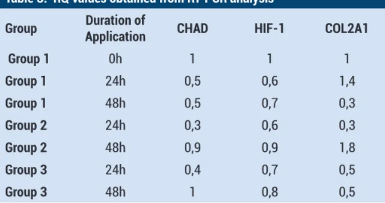
CHAD, HIF-1α and COl2A1 gene expression levels of control group were considered as 100% [Relative Quantity (RQ)=1)]. In experimental groups, gene expression was given in the table as RQ. Group I was not applied any kinds of drugs where it was observed that CHAD and HIF-1α expressions decreased at the 24th and 48th hours. In this group, COL2A1 expression increased 1.4 times at the 24th hour whereas it decreased at the 48th hour (RQ=0.3). Three genes decreased at the 24th hour in the experimental group where DAP was used. CHAD and HIF-1α expression values were similar to that of the control group at the 48th hour (RQ=0.9). COL2A1 increased 1.8 times at the 48th hour.
It was observed a decrease in 3 gene expression after 24 h in the cultures where VCM was used. Decrease in question reverted in the genes called CHAD and HIF-1α. A 50% decrease in the COL2A1 expression continued after 48 h. RQ values obtained from RT-PCR analysis were given in Table 3.
Table 3. RQ values obtained from RT-PCR analysis
Group ApplicationDuration of CHAD HIF-1 COL2A1
Group 1 0h 1 1 1 Group 1 24h 0,5 0,6 1,4 Group 1 48h 0,5 0,7 0,3 Group 2 24h 0,3 0,6 0,3 Group 2 48h 0,9 0,9 1,8 Group 3 24h 0,4 0,7 0,5 Group 3 48h 1 0,8 0,5
DISCUSSION
With the developments in pharmaceutics technology and regenerative medicine, toxicity of drugs and repair of damaged tissues have been areas of research (1). Regenerative studies have been carried out in surgical sites as well as branches in medicine. These studies make use of tissue engineering with the purpose of preserving facet articular cartilage, intervertebral disc structure and vertebral bone tissue or repairing damaged tissues (7,8). As such studies are being carried out, toxic effects of drugs on healthy cartilage and bone tissues are being studied.
In researches in which toxicity has been studied, experimental setups are frequently comprised of tissues obtained from animal tissues. However, due to the fact that the characteristics of sensitiveness in tissues differ from human tissues, the results of such studies may be different and misleading. Besides, as cell line systems contain mono type cells, have complex coordination mechanisms between cells and their micro environments and inhibit interaction between cells and extracellular matrix structures, in vitro test results have been controversy in terms of compliance. The main disadvantage of these cell lines is that they do not contain all phenotypic and genotypic characteristics since they are genetically modified cells (15-24).
In the current research, commercial cell line was not used but primary human cartilage cell cultures. It was aimed to answer whether DAP and/or VCM was cytotoxic to chondrocytes and extra cellular matrix contents. The second stage of our study was to determine whether DAP and VCM application had effects on COL2A1, HIF-1α and CHAD gene expressions.
COL2A1 gene encodes an important cartilage-specific extracellular matrix protein that is synthesized from proliferative chondrocytes, which are essential for chondrogenesis (25-27).
Furthermore, the HIF-1α transcription factor, which is a regulatory protein with a key role in the development of hypoxia adaptive response, and which is involved in the passage of DAP and VMC from cell respiration to anaerobic glycolysis during cellular energy production, was examined first time in the literature.
In addition to all these, CHAD interacts with collagen type II and a2β1 integrin and plays a role in the binding of connective tissue cells to matrix components. This effect is carried out through a small leucine-rich repeat protein (SLRP) which is a soluble low-density lipoprotein receptor-related protein. Thus, CHAD can protect the cellular phenotype and regulate tissue hemostasis (25). Although it seems that preparing in vitro experimental setup is a limitation, we are of the opinion that the results are reliable as we did not use cell lines or animal cell cultures. This study may have two limitations, first limitation is that no comparison was made between patient and healthy cartilage tissues, because it is not ethically possible to obtain healthy cartilage tissue from a healthy person. The second is that the cell cultures prepared in this study were formed as a result of processing of tissues obtained from four patients. The fact that both the number of samples was small (n = 4) and the people from whom these samples were obtained had the same race is the real limitation of this study.
Infections seen in orthopedics and/or neurosurgery branches generally require long term antibiotics use. Following a serious operation, the presence of infections may deteriorate surgical benefits unless an efficient antibiotherapy is used.
In cytotoxic researches, only glycopeptide antibacterial agents and some antibiotics in quinolone group have been evaluated (1,27-29).
Infections resulted from gram-positive factors have been on the increase in the world. Treatment alternatives for resistant gram-positive factors are limited. DAP is the first cyclical lipopeptide agent used in clinics.
Staphylococcus aureus, resistant to methicilline effect
spectrum, Staphylococcus aureus, medium resistant to VCM, Staphylococcus aureus, resistant to VCM and enterococcis, resistant to VCM, are Gram-positive bacterium. DAP has been approved for complicated skin and soft tissue infections, bacteremia’s and right heart infective endocarditis.
Infection after spinal surgery is a major complication, and every effort should be made by physician to prevent and/or minimize it (30). In articular infections resulted from prosthesis, DAP and VCM have been reported to be used in antimicrobial treatment guides (31,32). There are studies indicating that DAP is efficient for Staphylococcus aureus factor in osteoarticular tissues (33).
However, in the treatment of infections in bone and articulation tissues, there is less experience concerning the use of DAP. However, in the treatment of bone and joint tissue infections, there is less experience concerning the use of DAP. Although there is no clinical study, findings obtained from in vitro studies suggest that it may well infuse into synovial tissue and bone tissue (9,34).
It is known that inferior unit of HIF-1α creates dimer with HIF-1β by replacing from cytoplasm to nuclear under hypoxic conditions. Thus, HIF-1connects to the specific row which is identified as “hypoxia response agent” on DNA with the purpose of connecting to other cofactors in cell nucleus. Then, expression of target genes is triggered. Inferior unit of HIF-1α is degraded in proteasome by means of ubiquitination under normoxia conditions. Low glucose level increases rapidly in oxidative stress and hypoxic environments (35,36).
Group I was the control group where no kinds of drugs were applied. It was observed that CHAD and HIF-1α expressions decreased at the 24th and 48th hours. In the group where DAP was applied, there was a decrease in HIF-1α at the 24th hour and it was found to be (RQ=0.9) at the 48th hour, which was similar to the control group. There was a decrease in gene expression in the group in which VCM was applied at the 24th hour, yet it inverted at the 48th hour.
Human CHAD gene identifies progenese which is pertain to biophysical and structural features of the extracellular matrix. Besides, it is known that human CHAD gene identifies phenotype existing in the cell development process. It takes these effects on signal pathways in the arrangement of collagen fibril sled by SLRPs (37).
In literature, it is reported that CHAD affects collagen fibrillogenesis and modulates chondrocyte differentiation.
It is known that negative regulatory role of CHAD affects stabile extracellular matrix formation. In addition, members of leucine-rich repeat protein family in articular cartilage interact with many collagen type thanks to CHAD and collagen fibrillogenesis are affected (46,47).
It was found that CHAD expression decreased at the 24th and 48th hours in Group 1. In the group where DAP was applied, there was a decrease in HIF-1α at the 24th hour and it was found to be (RQ=0.9) at the 48th h, which was similar to the control group. There was a decrease in gene expression in the group in which VCM was applied at the 24th hour, yet it inverted at the 48th hour.
COL2A1 is essential in the development of extracellular matrix development (40). It was observed that COL2A1 expression increased 1.4 times at the 24th hour whereas it decreased at the 48th h (RQ=0.3). In the group where VCM was applied, COL2A1 decreased at the 24th hour, yet it increased 1.8 times at the 48th hour. In VCM applied cultures, it was found that COl2A1 expression 50% decreased at the 24th hour continued at the 48th hour. With the help of RT-PCR analysis which was applied for these 3 genes it might be concluded that these genes decreased at the 24th hour after drug use. Likewise, cell vitality and proliferation diminished at the 24th hour. However; it was observed that culturalized cells were recoveried and continued proliferation at the 48th hour. the current study was performed using primary chondrocyte cultures.
These cultures contain all the cell types of the original tissue as well as extracellular matrix elements. The fact that the research was carried out in primary cultures is a powerful aspect of our study. Because the results we obtained are very close to those to be obtained from the original tissue. We can explain the regaining of the regression of proliferation, increased cell death and decreased gene expression at the 24th hour at a very close level of the control group at the 48th hour with a fact that the primary cultures contained a heterogeneous cell population.
Administered drug doses were calculated considering the amount to be delivered to tissues through a systemic application. Since our study was not a dose-response study, the applications were performed in a single dose. It can be said that the clinical use of the indicated doses is safe in terms of chondrocyte viability. However, in the light of application data at the 24th hour, it can be maintained that the increase in doses or tandem applications may diminish live cell population and have even negative effect on the gene expression level.
CONCLUSIONS
Our in vitro study revealed the effects of active ingredients of DAP and VCM, which are particularly used in treatments of methicillin-resistant Staphylococcus aureus-active infections on human chondrocyte tissue’s morphology and gene expressions of CHAD, Col2A1 and HIF-1α. As
a conclusion, in clinics, time and doses of these drugs should be evaluated considering their effects on cartilage tissues.
Competing interests: The authors declare that they have no competing interest.
Financial Disclosure: There are no financial supports
Ethical approval: Namik Kemal University Medical Faculty Ethics Committee (Date: 25/12/2014; Number: 127)
REFERENCE
1. Dogan M, Isyar M, Yilmaz I, et al. Are the leading drugs against Staphylococcus aureus really toxic to cartilage? J Infect Public Health 2016; 9:251-8.
2. Tascini C, Tagliaferri E, Di Paolo A, et al. Three-times weekly teicoplanin as outpatient treatment of chronic osteoarticular infections. J Chemother 2009;21:421-5.
3. Baaj AA, Dakwar E, Le TV, Smith DA, Ramos E, Smith WD, et al. Complications of the mini-open anterolateral approach to the thoracolumbar spine. J Clin Neurosci 2012; 19(9): 1265-7.
4. Kullar R, Chin JN, Edwards DJ, et al. Pharmacokinetics of single-dose daptomycin in patients with suspected or confirmed neurological infections. Antimicrob Agents Chemother 2011;55:3505-9.
5. Sterner B, Harms M, Wöll S, et al. The effect of polymer size and charge of molecules on permeation through synovial membrane and accumulation in hyaline articular cartilage. Eur J Pharm Biopharm 2016;101:126-36.
6. Levick JR. Microvascular architecture and exchange in synovial joints. Microcirculation 1995;2:217-33.
7. Gumustas SA, Yilmaz İ, Isyar M, et al. Assessing the negative impact of phenyl alkanoic acid derivative, a frequently prescribed drug for the suppression of pain and inflammation, on the differentiation and proliferation of chondrocytes. J Orthop Surg Res 2016;11:70.
8. Gumustas F, Yilmaz I, Sirin DY, et al. Chondrocyte proliferation, viability and differentiation is declined following administration of methylphenidate utilized for the treatment of attention-deficit/hyperactivity disorder. Hum Exp Toxicol 2017;36:981-92.
9. Montange D, Berthier F, Leclerc G, et al. Penetration of daptomycin into bone and synovial fluid in joint replacement. Antimicrob Agents Chemother 2014;58:3991-6.
10. Ehlicke F, Freimark D, Heil B,et al. Intervertebral disc regeneration: influence of growth factors on differentiation of human mesenchymal stem cells (hMSC). Int J Artif Organs 2010;33:244-52.
11. Rosenzweig DH, Tremblay Gravel J, Bisson D, et al. Comparative analysis in continuous expansion of bovine and human primary nucleus pulposus cells for tissue repair applications. Eur Cell Mater 2017;33:240-51.
12. Feng G, Jin X, Hu J, et al. Effects of hypoxias and scaffold architecture on rabbit mesenchymal stem cell differentiation towards a nucleus pulposus-like phenotype. Biomaterials 2011;32:8182-9.
13. Zhou X, Tao Y, Liang C, et al. BMP3 alone and together with TGF-β promote the differentiation of human mesenchymal stem cells into a nucleus pulposus-like phenotype. Int J Mol Sci 2015;16:20344-59.
14. Kellgren JH, Lawrence JS. Radiological assessment of rheumatoid arthritis. Ann Rheum Dis 1957;16:485-93. 15. Sirin DY, Kaplan N, Yilmaz I, et al. The association between
different molecular weights of hyaluronic acid and CHAD, HIF-1α, COL2A1 expression in chondrocyte cultures. Exp Ther Med 2018;15:4205-12
16. Karaarslan N, Yilmaz I, Sirin DY, Do we damage nucleus pulposus tissue while treating cerebrovascular ischemic neurological deficits. Annals of Medical Research 2018;25:266-73.
17. Isyar M, Yilmaz I, Yasar Sirin D, et al. A practical way to prepare primer human chondrocyte culture. J Orthop 2016;13:162-7.
18. Barcia-Makay M, Seral C, Mingeot-Leclercq MP, et al. Pharmacodynamic evaluation of theintracellular activities of antibiotics against Staphylococcus aureus in a model of THP-1 macrophages. Antimicrob Agents Chemother 2006;50:841-51.
19. Oznam K, Sirin DY, Yilmaz I, et al. Iopromide- and gadopentetic acid-derived preparates usedin MR arthrography may be harmful to chondrocytes. J Orthop Surg Res 2017;12:98. 20. Komur B, Akyuva Y, Karaslan N, et al. Can a biodegradable
ımplanted bilayered drug delivery system loaded with BMP-2/BMP-12 take an effective role in the biological repair process of bone-tendon ınjuries? a preliminary report. J Pharm (Cairo) 2017;2017:7457865.
21. Sirin DY, Karaarslan N, Evaluation of the effects of pregabalin on chondrocyte proliferation and CHAD, HIF-1α, and COL2A1 gene expression. Arc Med Sci 2018.
22. Karaarslan N, Yilmaz I, Ozbek H, et al. Are specific gene expressions of extracellular matrix and nucleus pulposus affected by primary cell cultures prepared from intact or degenerative intervertebral disc tissues? Turk Neurosurg 2018;22.
23. Guzelant AY, Isyar M, Yilmaz İ, et al. Are chondrocytes damaged when rheumatologic inflammation is suppressed? Drug Chem Toxicol 2017;40:13-23.
24. Gökçe A, Yılmaz I, Gökay NS, et al. Does insulin, transferrin and selenous acid preparation effect chondrocyte proliferation? Acta Orthop Traumatol Turc 2014; 48:313-9. 25. Batista MA, Nia HT, Önnerfjord P, et al. Nanomechanical
phenotype of chondroadherin-null murine articular cartilage. Matrix Biol 2014;38:84-90.
26. Ushijima T, Okazaki K, Tsushima H, et al. CCAAT/ enhancer-binding protein beta regulates the repression of type II collagen expression during the differentiation from proliferative to hypertrophic chondrocytes. J Biol Chem 2014;289:2852-63.
27. Stueber T, Karsten J, Stoetzer C, et al. Differential cytotoxic properties of drugs used for intra-articular injection on human chondrocytes: an experimental in-vitro study. Eur J Anaesthesiol 2014;31:640-5.
28. Goto K, Imaoka M, Goto M, et al. Effect of body-weight loading onto the articular cartilage on the occurrence of quinolone-induced chondrotoxicity in juvenile rats. Toxicol Lett 2013;216:124-9.
29. Goto K, Yabe K, Suzuki T, et al. Gene expression profiles in the articular cartilage of juvenile rats receiving the quinolone antibacterial agent ofloxacin. Toxicology 2008; 249:204-13. 30. Marimuthu C, Abraham VT, Ravichandran M, et al.
Antimicrobial prophylaxis in ınstrumented spinal fusion surgery: a comparative analysis of 24-hour and 72-hour dosages. Asian Spine J 2016;10:1018-22.
31. Zimmerli W, Trampuz A, Ochsner PE. Prosthetic joint infection. N Engl J Med 2004;351:1645-54.
32. Gilbert DN, Moellering RC, Eliopoulos GM, Chambers HF, Saag MS, editors. The sanford guide to antimicrobial therapy. In: Antimicrobial Prophylaxis. 47th edition, Sperryville: Antimicrobial Therapy Inc 2009. pp. 201.
33. Barreau S, Benaboud S, Kerneis S, et al. Staphylococcus aureus osteo-articular infection: usefulness of the determination of daptomycin serum concentration to explain a treatment failure. Int Clin Pharmacol Ther 2016;54:923-7. 34. Suleyman G, Kenney R, Zervos MJ, et al. Safety and efficacy
of outpatient parenteral antibiotic therapy in an academic infectious disease clinic. J Clin Pharm Ther 2017;42:39-43. 35. Wang P, Zhang F, He Q, et al. Flavonoid compound ıcariin
activates hypoxia ınducible factor-1α in chondrocytes and promotes articular cartilage repair. PLoS One 2016;11:e0148372.
36. Bai R, Zhao AQ, Zhao ZQ, et al. MicroRNA-195 induced apoptosis in hypoxic chondrocytes by targeting hypoxia-inducible factor 1 alpha. Eur Rev Med Pharmacol Sci 2015;19:545-51.
37. Heppner JM, Zaucke F, Clarke LA. Extracellular matrix disruption is an early event in the pathogenesis of skeletal disease in mucopolysaccharidosis I. Mol Genet Metab 2015;114:146-55.
38. Tillgren V, Ho JC, Önnerfjord P, et al. The novel small leucine-rich protein chondroadherin-like (CHADL) is expressed in cartilage and modulates chondrocyte differentiation. J Biol Chem 2015;290:918-25.
39. Mansson B, Wenglén C, Mörgelin M, et al. Association of chondroadherin with collagen type II. J Biol Chem 2001;276:32883-8.
40. Unguryte A, Bernotiene E, Bagdonas E, Garberyte S, Porvaneckas N, Jorgensen C. Human articular chondrocytes with higher aldehyde dehydrogenase activity have stronger expression of COL2A1 and SOX9. Osteoarthritis Cartilage 2016;24:873-82.