RESEARCH ARTICLE
Artificial oocyte activation with calcium ionophore for frozen sperm cycles
Seda Karabuluta,b, Özlem Aksüngerc, Can Atac, Yusuf Sağıroglud, andİlknur Keskina,baSchool of Medicine,İstanbul Medipol University, İstanbul;bREMER (Regenerative and Restorative Medicine Research Center),İstanbul Medipol University,İstanbul, Turkey;cMemorial Antalya Hastanesi/Tüp Bebek Merkezi, Antalya, Turkey;dIVF Unit, Florence Nightingale Hospital Kadıkoy, İstanbul, Turkey
ABSTRACT
Fertilization problems are the major problems that may be faced in 30–55% of the patients during an intracytoplasmic sperm injection (ICSI) cycle. A successful oocyte activation depends on factors related to both sperm and oocyte, and one of the important factors that mediates the process is Ca2+ concentration within the oocyte. Artificial oocyte activation (AOA) is a method used for fertilization problems that commonly involve the usage of Ca2+ionophores and is usually used in problems such as total fertilization failure (TFF) and globozoospermia. The aim of the present study was to investigate possible effects of AOA for different groups of patients with fertilization failure. Four groups of patients (previous TFF, low oocyte number, severe sperm quality, and frozen sperm (FS) group) that underwent ICSI with AOA were included in the study. All groups had similar control groups with same indications except TFF, where AOA was not performed. Fertilization rates were significantly higher in the TFF group than those observed in other AOA groups. Fertilization rates and quality of embryos observed in the remaining AOA groups were higher than those of the controls, which were statistically insignificant. Prgenancy rates were higher in all AOA groups compared to the controls, although the differences were significant in FS group only. Quality of embryos and pregnancy rates were lower in the TFF group compared to the remaining AOA groups indicating possible concomitant problems. Fertilization rates, quality of embryos and pregnancy rates seemed to be increased in all indication groups suggesting that not only TFF patients but also a wide variety of patients with different indications may benefit from AOA.
Abbreviations: ICSI: Intracytoplasmic sperm injection; ARTs: Assisted reproductive techni-ques; Ca: Calcium; AOA: Artificial oocyte activation; TFF: Total fertilization failures; OAT: Oligoasthenoteratozoospemia; IVF: In vitro fertilization; SOAT: Severe OAT; LON: Low ooctye number; FS: Frozen sperm; hCG: human chorionic gonadotrophin; PVP: polyvinylpyrrolidone; HSA: human serum albumin
ARTICLE HISTORY
Received 26 November 2017 Revised 16 February 2018 Accepted 17 February 2018
KEYWORDS
Artificial oocyte activation; ICSI; fertilization; sperm
Introduction
Since the introduction of intracytoplasmic sperm injection (ICSI) into assisted reproductive techni-ques (ARTs), male factor infertility is treated more effectively. There are numerous studies examining the effects of the sperm cell on activation of the oocyte for fertilization (Berkovitz et al., 2006; Moaz et al. 2006; Nasr Esfahani et al. 2007). The mean fertilization rate after microinjection is known to be 70–80%, which depends on the quality of the gametes and culture conditions (Palermo et al.2009; Neri et al. 2014). Fertilization failure after ICSI is suggested to be the result of an oocyte activation deficiency (Neri et al. 2014; Swain and Pool 2008; Yanagida 2004; Rawe et al. 2000). A successful oocyte activation depends on the factors related
both to the sperm and oocyte, and is mediated by oscillations in Ca2+ concentration in the cytoplasm of the oocyte. It is triggered by the sperm factor phospholipase C zeta (Saunders et al. 2002; Tesarik et al. 2002; Kashir et al. 2010, 2015; Amdani et al. 2013). Ca2+ signaling and Ca2+ release from intra-cellular stores are crucial for oocyte activation and further embryonic development (Kline and Kline 1992; Ozil and Huneau 2001; Marangos and Carroll 2004; Ducibella et al. 2006). Oocyte activat-ing ability of the spermatozoon is a critical factor for fertilization. During natural fertilization, oocyte activation is achieved by the entrance of the sperm leaving its membrane outside the oocyte. Ooplasmic factors can thereby interact with the sperm nucleus. However, during ICSI, the sperm cell is introduced into the ooplasm as a whole. Fertilization problems CONTACTSeda Karabulut [email protected] Department of Histology and Embryology, Kavacık mah, İstanbul Medipol University, International School of Medicine, Ekinciler cd. No: 19 Beykoz,İstanbul, Turkey.
2018, VOL. 64, NO. 5, 381–388
https://doi.org/10.1080/19396368.2018.1452311
are suggested to appear as a result of the impair-ment in the release of oocyte activating factors from the sperm following ICSI (Nasr Esfahani et al. 2007).
Artificial oocyte activation (AOA) is the most com-mon treatment strategy used for fertilization problems (Ebner et al. 2012) to achieve successful fertilization, especially in problems of total fertilization failure (TFF) and in decreased fertilization rates. TFF cases are observed in routine practice of ARTs with an incidence of ~1–3% of ICSI cycles and may be reobserved in subsequent cycles (Flaherty et al. 1998; Esfandiari et al.2005; Kashir et al.2010). Sperm-related problems that may result in TFF are non-viability, altered chro-matin status, inability to activate the oocyte, and decon-densation failure (Nasr Esfahani et al.2010). The main oocyte-related factor that leads to TFF is failed activa-tion. Oocyte activation process is a result of complex interactions triggered by the entrance of the sperm cell into the oocyte. Intracellular Ca2+rise observed shortly after spermatozoon–oocyte fusion is the initiator mechanism of oocyte activation (Miyazaki and Ito 2006; Ramadan et al.2012).
Cases of failed fertilization following ICSI are rare, and are mainly caused by the lack of oocyte activation (Tosti and Ménézo 2016). Impairment of activating factors would result in delayed or abnormal oocyte activation, which may lead either to fertilization failure or to various abnormalities in embryonic development (Tesarik, 1998a, 1998b) The aim of AOA is to mimic physiological mechanisms that take place within the oocyte, based mainly on calcium changes (Tosti and Ménézo2016). Several chemical, mechanical, or physi-cal stimuli may be used to promote oocyte activation during an ICSI cycle to overcome failed fertilization (van den Meerschaut et al. 2014b). Several studies have reported an increase in fertilization rates and cleavage-stage embryos with AOA techniques (Sfontouris et al. 2015). Most common AOA techni-ques involve the usage of calcium ionophore (CaI) (Heindryckx et al. 2008; van den Meerschaut et al. 2014b; Ebner et al. 2012; Montag et al. 2012; Nasr Esfahani et al.2010; Van den Meerschaut et al. 2012), strontium (Yanagida et al.2006; Kyono et al. 2008), a modified ICSI technique (Ebner et al.2004), or electric pulses (Zhang et al.1999).
Currently, CaI (A23187) is the most commonly used technique to overcome failed oocyte activation follow-ing ICSI (Eldar Geva et al. 2003). CaI (A23187) is a carboxylic acid ionophore that transfers Ca2+and mag-nesium from the medium through the cell membrane into the oocyte (Reed and Lardy1972). Influx of Ca2+ results in an increase in the concentration of calcium in
the oocyte cytoplasm that acts as a signal to promote DNA synthesis and cell division (Luckasen et al.1974). Several studies have demonstrated that AOA by CaI may elevate free intracellular calcium and trigger oocyte activation by this way (Heindryckx et al. 2005). The technique has been demonstrated to be nondestructive in the study of Van den Meerschaut in which neonatal and neurodevelopmental outcome of children aged between 3 and 10 years whose conception was provided with the help of assisted reproduction techniques and assisted oocyte activation was analyzed, and mean out-comes were observed to be within the expected ranges (van den Meerschaut et al.2014a).
AOA is used mainly for cases with TFF and globo-zoospermia. Data analyzing the effects of AOA on the outcomes of ICSI procedure for different indications are limited, and these studies focus mostly on testicular sperm and low fertilization cases. We therefore aimed to investigate the possible effects of AOA on a wide variety of patients including frozen sperm (FS), severe oligoasthenoteratozoospemia (SOAT), and low oocyte number (LON) in which fertilization rates are expected to benefit more. We also aimed to analyze the distribu-tion of cases with overall TFF among couples who underwent an ICSI cycle.
Results
Overall TFF cases distribution
A total of 1837 couples who underwent an ICSI cycle were analyzed. TFF is observed in 122 (6.6%) cycles. Mean age was 37 years and mean mature oocyte count were 6.4. Seventy (57.3%) of the patients had no history of a previous in vitro fertilization (IVF) attempt, whereas the remaining 57 (46.72%) had at least one previous ICSI cycle. Among those with a previous cycle, 19 (33.3%) had experienced a previous fertilization fail-ure, whereas 38 (66.6%) had not.
AOA results for different indications
We analyzed and compared ICSI outcomes of four groups (group 1: cases with previousTFF, group 2: cases with low oocyte count, group 3: cases with SOAT, and group 4: cases where FS were used) to their controls. Outcomes of the group with TFF were also compared to the remaining three groups of AOA, in order to observe the possible difference between this group and others. Patient characteristics including women age, mature oocyte count, and number of embryos transferred were similar in all groups and
controls, except the LON group in which mean age was higher and oocyte count were lower (Table 1).
Demographic data of the patients and ICSI outcome parameters (fertilization rate, top quality embryo rate, preganancy rate) are presented inTable 1and shown in Figure 1. Fertilization rates of SOAT, LON, and FS groups were found to be similar compared to their controls (69–65.5%, 80.2–66.7%, 61.2–59.2%, respectively) (p > 0.05); however, TFF group had significantly higher fertilization rates (74.1%) compared to the remaining AOA groups and controls (p = 0.03; p = 0.000, respectively).
Rates of top quality embryos were analyzed in order to investigate the efffect of AOA on the quality of embryos in different indication groups. We observed that SOAT and FS groups had higher rates of top
quality embryos compared to their controls (50–20%, 77.7–50%, respectively), although the differences were not statistically significant (p > 0.05). Unlike fertiliza-tion rates, rate of top quality embryos in TFF group was significantly lower (22.8%) compared to the remaining AOA groups (50%, 32%, 77.7%, respectively) (p < 0.05). Pregnancy rates were the third ICSI parameter ana-lyzed. We have observed that FS group had significantly higher pregnancy rates compared to its control (100%, 40% respectively) (p = 0.000). Other AOA groups had also distinctly higher pregnancy rates than their con-trols although the differences were not statistically sig-nificant (p > 0.05). The pregnancy rates were found lower in the TFF group (28.6%) than SOAT and FS AOA groups (66.6%, 100%, respectively) as well but without statistical significance.
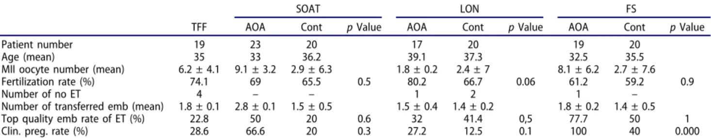
Table 1.Patient characteristics and ICSI outcome parameters dispersion according to the subgroups that AOA is performed.
SOAT LON FS
TFF AOA Cont p Value AOA Cont p Value AOA Cont p Value
Patient number 19 23 20 17 20 19 20
Age (mean) 35 33 36.2 39.1 37.3 32.5 35.5
MII oocyte number (mean) 6.2 ± 4.1 9.1 ± 3.2 2.9 ± 6.3 1.8 ± 0.2 2.4 ± 7 8.1 ± 6.2 2.7 ± 7.6
Fertilization rate (%) 74.1 69 65.5 0.5 80.2 66.7 0.06 61.2 59.2 0.9
Number of no ET 4 – – 1 2 1 –
Number of transferred emb (mean) 1.8 ± 0.1 2.8 ± 0.1 1.5 ± 0.5 1.5 ± 0.4 1.4 ± 0.2 1.8 ± 0.2 1.4 ± 0.5
Top quality emb rate of ET (%) 22.8 50 20 0.6 32 41.4 0,5 77.7 50 1
Clin. preg. rate (%) 28.6 66.6 20 0.3 27.2 12.5 0.1 100 40 0.000
Fertilization rate, top quality embryo rates of ET and pregnancy rates were given as mean (%) and MII oocyte number, number of transferred embryos were given as mean±SD.
TFF: total fertilization failure; SOAT: severe oligoasthenoteratozoospermia; LON: low ooctye number; FS: frozen sperm; ET: embryo transfer; Clin. Preg. rate: clinical pregnancy rate.
0 10 20 30 40 50 60 70 80 90 100
Fertilization Top Quality Clin. Pregnancy
SOAT LON
*
FS SOAT SOAT LON FS FS ** LONFigure 1.ICSI outcome parameters dispersion according to the subgroups that AOA is performed.
Fertilization rate, top quality embryo rates of ET, and pregnancy rates were given as mean (%).
Abbreviations: TFF, total fertilization failure; SOAT, severe oligoasthenoteratozoospermia; LON, low ooctye number; FS, frozen sperm; ET, embryo transfer; Clin. Preg. rate, clinical pregnancy rate; ICSI, intracytoplasmic sperm injection.
* Representp < 0.05. ** Representsp < 0.005.
Discussion
Fertilization takes place following a successful oocyte activation and it depends on both sperm- and oocyte-related factors (Saunders et al.2002; Tesarik et al.2002; Kashir et al.2010). In case of a problem in one of these factors, TFF or decreased rate of fertilization may be observed. As TFF and all other fertilization problems are frustrating both for the couples and clinicians, a treatment strategy is of importance for counseling the patients and for future management. Although fertili-zation may be possible in the subsequent attempts (Kinzer et al.2008; Pabuccu et al.2016), IVF specialists usually do not have the chance to wait and see the results of a new cycle because of the high cost, the time, and the reduced hope of the couples. The main treatment strategy to solve these problems may be ‘AOA’ by CaI, in order to avoid recurrent fertilization problems.
Several studies have reported the usage of AOA with CaI as a safe and a reliable method (Ebner et al.2012). According to the results of the studies evaluating the efficacy of AOA (Moaz et al.2006; Nasr Esfahani et al. 2008; Borges et al.2009), it has been considered to be useful in a selected patient population who have experi-enced TFF or low fertilization outcome in the previous ICSI attempts (Taylor et al. 2010; Sermondade et al. 2011). Furthermore, it has been reported to be benefi-cial in patients with a compromised fertilization due to globozoospermia (Ebner et al.2012; Montag et al.2012; Nasr-Esfahani et al., 2009). However, there is limited data on the efficacy of AOA for different indications including patients having severely decreased sperm parameters (concentration: <1 mil/ml, total motility: <50%, and normal morphology: <1%), FS, and low oocyte count. In the present study, we investigated possible effects of AOA on these groups of patients to achieve and/or enhance fertilization and pregnancy rates while analyzing the incidence and distribution of TFF cases among infertile patients.
Cases of TFF were observed to be 6.6% among total cycles, and among those 2% had at least one previous cycle with no fertilization problem. This data shows that TFF may be observed in different cycles of the same patient couple, and therefore couples with a pre-vious cycle and successful fertilizations may be affected as well. We observed that 68% of these patients had low oocyte count (≤3), 23% had severe sperm parameters, 7% had the cycle with FS, and 3% had no detectable reason. This led us to the idea that AOA may be beneficial fort them.
We observed significantly higher fertilization rates in the TFF group compared to the remaining groups,
suggesting that TFF group benefits most from AOA with regard to the success in fertilization. Several stu-dies have also reported a significant increase in fertili-zation rates in patients with TFF which is in accordance with our data (Montag et al.2012; van den Meerschaut et al.2014b). Furthermore, rates of top quality embryos in the TFF group were found to be lower compared to those observed in the remaining AOA groups, suggest-ing that although fertilization is achieved by AOA in this group, the quality of the embryos was not well enough which may point to other possible problems accompanying Ca2+insufficiency. The pregnancy rates observed in the group with TFF were found to be 28.6%, which was lower compared to the remaining AOA groups (SOAT: 66.6%, FS: 100% respectively); this confirms the suggestion that other possible pro-blems may accompany Ca2+insufficiency in this group. The effects of AOA were studied for several more indications including poor semen quality, low fertiliza-tion rates, and poor ovarian reserve. Investigating the effects of AOA on patients with sperm problems, Ebner et al. (2012) showed that cryptozoospermic men benefit tremendously from AOA with CaI with respect to the fertilization rates. We found a 3.5% increase in fertili-zation rates of SOAT group which appears to be in accordance with this study. Rate of top quality embryo within SOAT group was 30% higher compared to their controls, which was insignificant. Ebner et al. (2012) have reported no significant difference in the embryo quality of AOA performed in cryptozoospermic cases. Different data obtained in the present study may be due to the patient population where only cryptozoospermic men were selected for AOA. Several studies have reported a significant increase in fertilization rates of patients with severe teratozoospermia (Moaz et al. 2006; Nasr Esfahani et al. 2007) in which ionomycin is used for AOA; however, sperm morphology was the only parameter considered. The present study reports the first results of AOA use in cases of SOAT.
Montag et al. (2012) found a significant increase in fertilization rates in the patients having a previous cycle with low fertilization rates while Van den Meerschaut et al. (2012) observed no difference.
There is only one study that has investigated the effects of AOA in diminished ovarian reserve in which no benefit of AOA is observed (Caglar Aytac et al. 2015). We found a 13.5% increase in fertilization rates in the AOA-performed LON group (p = 0.06) in which the difference was close to significance level. Different results obtained may be due to the patient inclusion criteria in which the study population was mainly poor responders in the study of Caglar Aytac rather than oocyte count. There were no significant
differences in the pregnancy rates of AOA in LON group. This low pregnancy rates of LON group may be partly due to the bad cohort of patients with higher ages (mean 39.1 years) and LON (mean 1.8).
There is no previous data about the effects of AOA in cycles that used FS cycles, so this is the first data evaluating the effect of AOA on this group. We found no significant difference in fertilization rates but top quality embryo rates were increased by 27.7% although the differences were not significant predicting another potential benefit of AOA for a different patient popula-tion of FS.
When pregnancy rates were considered, we have analyzed that all the AOA groups have distinctly higher pregnancy rates than their controls although the differ-ences were only strongly significant in the FS group (p = 0.000).
According to our results, benefit of TFF group from AOA is one more time confirmed. The results obtained are not unexpected for TFF group although the rates are lower than normal ICSI cycles. Among all, TFF and FS groups seem to benefit from AOA by means of fertilization, embryo quality, and pregnancy rates.
We introduce for the first time a new group of patient with FS that benefits from AOA significantly concluding that sperm’s fertilization capacity decreases with the cryopreservation process but a good develep-mental potential is triggered once the fertilization occurs by the help of AOA. The overall results point out a new potential for AOA use in a wide variety of patients that will have no or little chance to achieve a pregnancy otherwise.
ICSI outcome measures were positively affected from AOA in all groups with a special regard to FS although some of the results were below the signifi-cance value indicating that not only TFF patients but also a wide variety of patients with different indications may benefit from AOA and therefore should be con-sidered as an alternative technique for FS cycles.
Consequently, due to low sample size of our study, further studies including ongoing pregnancy and live-birth outcomes with larger sample sizes are required. Future development in the field of AOA has enormous potential for clinical benefit, especially to enlarge the group that may benefit from this technique.
Materials and methods
Patients
This multicentered study was conducted on infertile couples undergoing ICSI treatment at Florence Nightingale Hospital, IVF center, İstanbul, Turkey,
and Memorial Antalya Hospital IVF Center, Antalya, Turkey, between March 2015 and March 2017. A total of 138 cycles were included in the study, 78 of them were AOA performed and 60 of them were control patients. The patients were divided into four groups according to the reason of AOA procedure: (1) pre-vious TFF, (2) LON, (3) SOAT, and (4) FS.
Cycles in which testicular sperm is used, preimplan-tation genetic screening and preimplanpreimplan-tation genetic diagnosis were performed. Exclusion criteria included the following: a female age of≥40 years; BMI< 25 kg/m2; a history of ovarian surgery; grade 3–4 endometriosis and endometrioma, adenomyomas, myomas, congenital uterine anomaly, and tubal pathology; and couples that carry any kind of genetic abnormality.
TFF group included the patients that have a pre-vious ICSI cycle with TFF, SOAT group included the patients that have severe decreased sperm parameters (sperm concentration <1 mil/ml, total motility <30%, and normal sperm morphology <4%), LON group included the patients that have ≤3 oocyte number, and FS group included the patients in which cryo-preserved sperm samples were used. All of the groups have a control group in which AOA is not performed although the patients have the same indications. Twenty patients were randomly chosen for each con-trol group. Patient randomization was performed by a single researcher with the support of a computer-generated program.
All patients signed a written consent form and the research project was approved by the ethics committee of Medipol University, under approval number of 10840098-604.01.01-E.15394 on 23 June 2017.
Ovarian stimulation and oocyte pickup
A GnRH agonist or an antagonist treatment was applied for controlled ovarian stimulation according to the patient characteristics. Recombinant follicle-sti-mulating hormone (GONAL-f, Merck Serono, Geneva, Switzerland) was used in all cases. Human chorionic gonadotropin (hCG) (Ovitrelle, Merck Serono) was administered subcutaneously when the dominant folli-cle had reached a mean diameter of ~18 mm.
Oocyte pickup was performed 36 h after hCG administration, under general anesthesia, with a 17-G dual lumen needle (Swemed) through transvaginal ultrasound guidance.
Retrieved oocytes were collected in a MOPS (3-(N-morpholino)propanesulfonic acid) buffered medium (G-MOPS, Vitrolife, Gothenburg, Sweden) under oil (Ovoil, Vitrolife), at 37°C.
After denudation of the cumulus–oocyte complexes by using two different diameters of denudation pipettes (170–140 µm) and by enzymatic denudation with 40 IU hyaluronidase (Vitrolife) 2 h after oocyte pickup, oocytes were placed in a CO2–O2-controlled incubator (ESCO, Singapore) for incubation. All consequent incu-bation steps were handled under oil (Ovoil, Vitrolife).
Sperm evaluation and preparation
Semen samples were freshly collected on the day of oocyte retrieval. After allowing liquefaction at 37°C, an initial evaluation was performed. Sperm concentration, motility, progression, and morphology were evaluated according to the World Health Organization 2010 guidelines. Samples were prepared by density centrifugation method. One milliliter of each sample was put on two different density layers (90% and 45%) of PureSperm solution (Nidacon, Sweden) and centrifuged. The pellet is placed in another tube and washed twice with G-IVF medium to get rid of washing solution. A second evaluation after preparation was performed and recorded. The sperm solution was placed in a microdrop containing 5 µl PVP (polyvinylpyrrolidone) solution in an ICSI dish (Falcon 1006, BD, Borough, New York, USA). PVP was used for sperm immobilization (ICSI, Vitrolife).
ICSI procedure
All laboratory procedures were carried out on Falcon Product Line (BD) plasticware. ICSI was performed in HEPES-buffered medium (G-Gamete, Vitrolife). For all oocytes, a motile spermatozoon exhibiting normal mor-phology was selected under an inverted microscope under ×40 magnification (Olympus, Japan), equipped with an Narishige micromanipulation set. Microinjection was
performed after aggressive sperm immobilization as pre-viously described (Palermo et al.1996).
Oocyte activation by CaI
The injected oocytes from each patient were incubated for 15 min in the 30 µl of pre-equilibrated drops of medium containing a ready-to-use bicarbonate-buf-fered reagent including CaI (GM508 Cult-Active, Gynemed, Lensahn, Germany), immediately after ICSI according to the instruction manual. After at least two rinsing steps, oocytes were transferred to the microdro-plets of fresh fertilization medium (G-IVF), supplemen-ted with 5% (w/v) human serum albumin (HSA).
Embryo culture and evaluation
All embryos were cultured for 2–3 days in a 37°C incubator (ESCO) with 6% CO2, 5% O2, 89% N2, and humidified atmosphere. Fertilization was evaluated 16–18 h after ICSI. Two clearly visible pronuclei and two polar bodies were considered evidence of normal fertilization. Fertilization rates were calculated as (the number of normally fertilized oocytes/the number of metaphase II oocytes injected) ×100. Fertilized oocytes were transferred to the pre-equilibrated microdroplets of fresh cleavage medium (G1) supplemented with 5% (w/v) HSA under oil and placed in a CO2–O2 -con-trolled incubator for embryo culture.
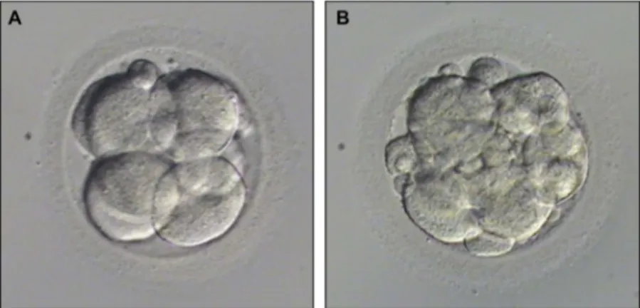
On day 2 and 3, another evaluation was performed to assess embryo quality. Embryos with less than 10% fragmentation and consisting of seven to eight even-sized blastomeres on day 3 without any vacuolization, granulation, and multinucleation were considered as top quality embryo and others as bad quality embryos (Figure 2). The percentage of top quality embryos was
Figure 2.Embryo quality examples were given: (A) Top quality embryo (Less than 10% fragmentation and consisting of 7–8 even-sized blastomeres on day 3 without any vacuolization, granulation and multinucleation; (B) Bad quality embryo (More than 10% fragmentation and/or uneven-sized blastomeres and/or with vacuolization, granulation and multinucleation.
calculated as (the number of top quality embryos on day 3/the number of transferred embryos) ×100.
Statistical analysis
All data were extracted from the patients’ electronic medical records. The statistical analysis was done with SPSS 22.0 software for windows (IBM Software, New York, NY, USA). The results are expressed as mean for fertilization rates, good quality embryo rates, and preg-nancy assessment. Mann–Whitney U test was used for abnormally distributed variables and Student’s t-test for normally distributed variables. Chi-square test was used to compare the pregnancy rates between the groups. The results were evaluated in 95% confidence interval and the statistical significance was defined asp < 0.05.
Disclosure statement
No potential conflict of interest was reported by the authors.
Notes on contributors
Contributed to the design, acquisition of data, analysis and interpretation of data, drafting of manuscript, and critical revision of the manuscript: SK; contributed to the acquisition of data: ÖA, CA; contributed both to statistics and acquisition of data: YS; contributed to the design, analysis and interpreta-tion of data, and critical revision of the manuscript:İK.
References
Amdani SN, Jones C, Coward K.2013. Phospholipase C zeta (PLCζ): oocyte activation and clinical links to male factor infertility. Adv Biol Regul. 53(3):292–308.
Berkovitz A, Eltes F, Lederman H, Peer S, Ellenbogen A, Feldberg B, Bartoov B. 2006. How to improve IVF-ICSI outcome by sperm selection. Reprod Biomed Online. 12:634–638.
Borges E Jr, de Almeida Ferreira Braga DP, de Sousa Bonetti TC, Iaconelli A Jr, Franco JG Jr. 2009. Artificial oocyte activation with calcium ionophore A23187 in intracyto-plasmic sperm injection cycles using surgically retrieved spermatozoa. Fertil Steril. 92:131–136.
Caglar Aytac P, Kilicdag EB, Haydardedeoglu B, Simsek E, Cok T, Parlakgumus HA. 2015. Can calcium ionophore “use” in patients with diminished ovarian reserve increase fertilization and pregnancy rates? A randomized, con-trolled study. Fertil Steril. 104(5):1168–1174.
Ducibella T, Schultz RM, Ozil JP. 2006. Role of calcium signals in early development. Semin Cell Dev Biol. 17:324–332.
Ebner T, Köster M, Shebl O, Moser M, Van Der Ven H, Tews G, Montag M.2012. Application of a ready-touse calcium
ionophore increases rates of fertilization and pregnancy in severe male factor infertility. Fertil Steril. 98:1432–1437. Ebner T, Moser M, Sommergruber M, Jesacher K, Tews G.
2004. Complete oocyte activation failure after ICSI can be overcome by a modified injection technique. Hum Reprod. 19:1837–1841.
Eldar Geva T, Brooks B, Margalioth EJ, Zylber Haran E, Gal M, Silber SJ.2003. Successful pregnancy and delivery after calcium ionophore oocyte-activation in a normozoosper-mic patient with previous repeated failed fertilization after intracytoplasmic sperm injection. Fertil Steril. 79(Suppl 3):1656–1658.
Esfandiari N, Javed MH, Gotlieb L, Casper RF. 2005. Complete failed fertilization after intracytoplasmic sperm injection—analysis of 10 years’ data. Int J Fertil Womens Med. 50:187–192.
Flaherty SP, Payne D, Matthews CD.1998. Fertilization fail-ures and abnormal fertilization after intracytoplasmic sperm injection. Hum Reprod. 13(Suppl 1):155–164. Heindryckx B, de Gheselle S, Gerris J, Dhont M, de Sutter P.
2008. Efficiency of assisted oocyte activation as a solution for failed intracytoplasmic sperm injection. Reprod Biomed Online. 17:662–668.
Heindryckx B, Van der Elst J, de Sutter P, Dhont M. 2005. Treatment option for sperm-or oocyte-related fertilization failure: assisted oocyte activation following diagnostic het-erologous ICSI. Hum Reprod. 20:2237–2241.
Kashir J, Heindryckx B, Jones C, de Sutter P, Parrington J, Coward K. 2010. Oocyte activation, phospholipase C zeta and human infertility. Hum Reprod Update. 16:690–703.
Kashir J, Nomikos M, Swann K, Lai FA. 2015. PLCζ or PAWP: revisiting the putative mammalian sperm factor that triggers egg activation and embryogenesis. Mol Hum Reprod. 21(5):383–388.
Kinzer DR, Barrett CB, Powers RD. 2008. Prognosis for clinical pregnancy and delivery after total fertilization fail-ure during conventional in vitro fertilization or intracyto-plasmic sperm injection. Fertil Steril. 90(2):284–288. Kline D, Kline JT.1992. Repetitive calcium transients and the
role of calcium in exocytosis and cell cycle activation in the mouse egg. Dev Biol. 149:80–89.
Kyono K, Kumagai S, Nishinaka C, Nakajo Y, Uto H, Toya M, Sugawara J, Araki Y. 2008. Birth and follow-up of babies born following ICSI using SrCl2 oocyte activation. Reprod Biomed Online. 17:53–58.
Luckasen JR, White JG, Kersey JH.1974. Mitogenic proper-ties of a calcium ionophore, A23187. Proc Natl Acad Sci U S A. 71:5088–5090.
Marangos P, Carroll J.2004. Fertilization and InsP3-induced Ca2+ release stimulate a persistent increase in the rate of degradation of cyclin B1 specifically in mature mouse oocytes. Dev Biol. 272:26–38.
Miyazaki S, Ito M.2006. Calcium signals for egg activation in mammals. J Pharmacol Sci. 100:545–552.
Moaz MN, Khattab S, Foutouh IA, Mohsen EA. 2006. Chemical activation of oocytes in different types of sperm abnormalities in cases of low or failed fertilization after ICSI: a prospective pilot study. Reprod Biomed Online. 13:791–794.
Montag M, Köster M, van der Ven K, Bohlen U, van der Ven H. 2012. The benefit of artificial oocyte activation is dependent on the fertilization rate in a previous treatment cycle. Reprod Biomed Online. 24:521–526.
Nasr Esfahani MH, Deemeh MR, Tavalaee M.2010. Artificial oocyte activation and intracytoplasmic sperm injection. Fertil Steril. 94:520–526.
Nasr Esfahani MH, Razavi S, Javdan Z, Tavalaee M.2008. Artificial oocyte activation in severe teratozoospermia undergoing intracytoplasmic sperm injection. Fertil Steril. 90:2231–2237.
Nasr Esfahani MH, Razavi S, Mardani M, Shirazi R, Javanmardi S. 2007. Effects of failed oocyte activation and sperm protamine deficiency on fertilization post-ICSI. Reprod Biomed Online. 14:422–429.
Neri QV, Lee B, Rosenwaks Z, Machaca K, Palermo GD.
2014. Understanding fertilization through intracytoplasmic sperm injection (ICSI). Cell Calcium. 55:24–37.
Ozil JP, Huneau D. 2001. Activation of rabbit oocytes: the impact of the Ca2+ signal regime on development. Development. 128:917–928.
Pabuccu GE, Sinem Caglar G, Dogus Demirkiran O, Pabuccu R. 2016. Uncommon but devastating event: total fertilisa-tion failure following intracytoplasmic sperm injecfertilisa-tion. Andrologia. 48(2):164–170.
Palermo GD, Neri QV, Takeuchi T, Rosenwaks Z.2009. ICSI: where we have been and where we are going. Semin Reprod Med. 27:191–201.
Palermo GD, Schlegel PN, Colombero LT, Zaninovic N, Moy F, Rosenwaks Z. 1996. Aggressive sperm immobilization prior to intracytoplasmic sperm injection with immature spermatozoa improves fertilization and pregnancy rates. Hum Reprod. 11(5):1023–1029.
Ramadan WM, Kashir J, Jones C, Coward K. 2012. Oocyte activation and phospholipase C zeta (PLCf): diagnostic and therapeutic implications for assisted reproductive technol-ogy. Cell Commun Signal. 10:12.
Rawe VY, Olmedo SB, Nodar FN, Doncel GD, Acosta AA, Vitullo AD. 2000. Cytoskeletal organization defects and abortive activation in human oocytes after IVF and ICSI failure. Mol Hum Reprod. 6:510–516.
Reed PW, Lardy HA.1972. A23187: a divalent cation iono-phore. J Biol Chem. 247:6970–6977.
Saunders CM, Larman MG, Parrington J, Cox LJ, Royse J, Blayney LM, Swann K, Lai FA. 2002. PLC zeta: a sperm-specific trigger of Ca2+ oscillations in eggs and embryo development. Development. 129:3533–3544.
Sermondade N, Hafhouf E, Dupont C, Bechoua S, Palacios C, Eustache F, Sifer C.2011. Successful childbirth after intra-cytoplasmic morphologically selected sperm injection
without assisted oocyte activation in a patient with globo-zoospermia. Hum Reprod. 26:2944–2949.
Sfontouris IA, Nastri CO, Lima ML, Tahmasbpourmarzouni E, Raine Fenning N, Martins WP. 2015. Artificial oocyte activation to improve reproductive outcomes in women with previous fertilization failure: a systematic review and meta-analysis of RCTs. Hum Reprod. 30:1831–1841. Swain JE, Pool TB.2008. ART failure: oocyte contributions to
unsuccessful fertilization. Hum Reprod Update. 14:431– 446.
Taylor SL, Yoon SY, Morshedi MS, Lacey DR, Jellerette T, Fissore RA, Oehninger S.2010. Complete globozoospermia associated with PLCf deficiency treated with calcium iono-phore and ICSI results in pregnancy. Reprod Biomed Online. 20:559–564.
Tesarik J. 1998a. Oocyte activation after intracytoplasmic injection of mature and immature sperm cells. Hum Reprod. 13:117–127.
Tesarik J.1998b. Oscillin-reopening the hunting season. Mol Hum Reprod. 4:1007–1009.
Tesarik J, Rienzi L, Ubaldi F, Mendoza C, Greco E.2002. Use of a modified intracytoplasmic sperm injection technique to overcome sperm-borne and oocyteborne oocyte activa-tion failures. Fertil Steril. 78:619–624.
Tosti E, Ménézo Y.2016. Gamete activation: basic knowledge and clinical applications. Hum Reprod Update. 22(4):420–439. Van den Meerschaut F, D’haeseleer E, Gysels H, Thienpont
Y, Dewitte G, Heindryckx B, de Sutter P.2014a. Neonatal and neurodevelopmental outcome of children aged 3–10 years born following assisted oocyte activation. Reprod Biomed Online. 28:54–63.
Van den Meerschaut F, Nikiforaki D, de Gheselle S, Dullaerts V, van den Abbeel E, Gerris J, Heindryckx B, de Sutter P.
2012. Assisted oocyte activation is not beneficial for all patients with a suspected oocyte-related activation defi-ciency. Hum Reprod. 27:1977–1984.
Van den Meerschaut F, Nikiforaki D, Heindryckx B, de Sutter P.2014b. Assisted oocyte activation following ICSI fertilization failure. Reprod Biomed Online. 28:560–571. Yanagida K. 2004. Complete fertilization failure in ICSI.
Hum Cell. 17:187–193.
Yanagida K, Morozumi K, Katayose H, Hayashi S, Sato A.
2006. Successful pregnancy after ICSI with strontium oocyte activation in low rates of fertilization. Reprod Biomed Online. 13:801–806.
Zhang J, Wang CW, Blaszcyzk A, Grifo JA, Ozil J, Haberman E, Adler A, Krey LC. 1999. Electrical activation and in vitro development of human oocytes that fail to fertilize after intracytoplasmic sperm injection. Fertil Steril. 72:509–512.