68
Ankara Üniversitesi Tıp Fakültesi Mecmuası 2010, 63(2) DAHİLİ BİLİMLER / MEDICAL SCIENCES
Olgu Sunumu / Case Report
Ultrasound and MRI findings of a large combined laryngeal mucocele were described with special emphasis on the diagnostic features and differential diagnosis.
Key Words : Ultrasound, Magnetic resonance, Laryngocele, Larynx
Büyük kombine laringeal bir mukoselin ultrason ve manyetik rezonans bulguları tanısal özellikleri ve ayırıcı tanı üzerinde durularak sunulmuştur.
Anahtar Sözcükler: Ultrason, Manyetik Rezonans, Laringosel, Laringeal
Ankara Üniversitesi Tıp Fakültesi Radyodiagnostik Anabilim Dalı
Ultrasound and MRI Findings in a Large Combined Laryngeal
Mucocele: Case Report
Büyük Kombine Bir Laringeal Mukoselde Ultrason ve Manyetik Rezonans Görüntüleme Bulguları: Olgu Sunumu
Evren Üstüner, Ebru Düşünceli Atman, Hasan Özcan, İlhan Erden
A laryngocele is a rare lesion with an esti-mated incidence of 1 in 2.5 million persons per year which represents an abnormal dilatation of the saccule in contact with the laryngeal space(1, 2). The name laryngeal mucocele or saccu-lar cyst is coined when a saccu-laryngocele is filled with mucous and fluid instead of air (2, 3). An internal laryngocele is lim-ited to the interior of larynx; an external laryngocele extends beyond the confines of the thyroid cartilage through the thy-rohyoid membrane and penetrates at the site of the entry of superior laryngeal nerve and artery. If both external and internal components exist a combined laryngocele is present (4).
Herein we discuss the ultrasound and MR findings of a large combined type la-ryngeal mucocele in a 28-year-old male with special emphasis on the di-agnostic and clinical features, and dif-ferential diagnosis of laryngoceles.
Case Report
A 28-year-old male presented with hoarse-ness, frequent cough, dyspnea and for-eign body sensation of two months du-ration worsening at nights while lying down and claimed that the symptoms had started after a severe upper respira-tory tract illness. A non-pulsatile mass
was externally visible on the right an-terolateral portion of the neck which enlarged with Valsalva’s maneuver. He had been smoking twenty cigarettes a day for the last 10 years. The rest of the physical exam and laboratory findings were unremarkable. He did not recall any incidence of overusing his voice. An ultrasound study was requested to
de-termine the nature of the neck mass, which revealed an infrahyoid anecho-ic, fluid filled, cystic lesion involving the right aryepiglottic fold, filling the right vestibule, extending externally through the thyrohyoid membrane. Hyperechoic mucous was also identi-fied within the cystic lesion (Fig-1). No evidence of a lymphadenopathy or an associated solid mass was detected. A diagnosis of combined laryngeal mucocele was made.
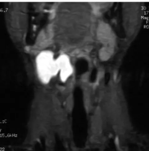
Later an MRI study was done to exclude an associated malignancy, in which on T2-weighted FSEIR images, the laryngeal mucocele was clearly visible due to homogeneous signal hyper-intensity of the cystic mass reflecting the fluid content in a background of fat suppression. No air was present within the cystic lesion. The laryngeal mucocele was extending from the val-lecula to the true vocal cords,
displac-Received: 28.07.2010 • Accepted: 25.10.2010 Corresponding author
Uz.Dr.Evren Üstüner
Ankara University Faculty of Medicine, Department of Radiology, Ankara, 06100, Turkey
Phone : +90 312 508 27 36 Fax : +90 312 310 71 17 E-mail Address : [email protected]
69
Evren Üstüner, Ebru Düşünceli Atman, Hasan Özcan, İlhan Erden Journal Of Ankara University Faculty of Medicine 2010, 63(2)
ing the false vocal cords, and filling the pyriform sinus and the ventricle on the right. Extralaryngeal component pen-etrated through the thyrohyoid mem-brane (Fig -2). There was no evidence of lymph node disease or obstructing soft tissue component suggestive of malignancy. The patient underwent an uncomplicated resection.
Figure 1: An anechoic, fluid filled, combined
type laryngocele is depicted in this US image in the transverse plane extending beyond the thyrohyoid membrane laterally and limited by the thyroid cartilage anteriorly. Medially the laryngeal folds are in apposition.
Figure 2: On T2 FSEIR sequences in coronal
plane the laryngocele is clearly visible as a cystic lesion of high signal intensity with regu-lar, well-defined borders in the background of fat suppression, minimally displacing the laryngeal air space medially, extending from the vallecula to the vocal cords filling the ventricle and extending laterally beyond the larynx through the thyrohyoid membrane.
Discussion
Laryngoceles are frequently observed in men, with a male to female ration of 5:1 and most come to clinical at-tention in the 5th or 6th decade (1). Combined laryngoceles account for 44% of all laryngoceles and 10% may
become infected (2). Laryngoceles are either congenital or acquired. Increase in endolaryngeal pressure like in glass blowers, players of wind instruments, singers and street hawkers or presence of a lesion that causes stenosis of the saccule neck may predispose to laryn-gocele development (4, 5). Although most laryngoceles are small in size and asymptomatic, they may present with hoarseness, dysphagia, pain and for-eign body sensation. External laryngo-celes depending on the size may pres-ent as a visible neck mass that change size in Valsalva’s maneuver (1). Com-pression of the mass may cause a gur-gling sound known as Bryce’s sign (2). Size, presence of infection and possible
as-sociation with malignancy should be evaluated with scrutiny during imag-ing and other cystic laryngeal lesions should be ruled out. Size partially influ-ences the severity of symptoms (1). Se-rious, life-threatening upper airway ob-struction due to a giant laryngocele and even death secondary to mucous aspira-tion in a critically narrowed airway has been reported (1, 6). Therefore airway patency needs to be carefully assessed. Superimposed infection may cause acute
flare or exacerbation of the existing symptoms therefore imagers should be aware of the signs of infection (2). CT imaging is especially advantageous in this regard because radiological signs of inflammation such as wall thicken-ing or rim enhancement of the laryn-gocele could be easily demonstrated (2,7). Neck ultrasound proves to be useful because it allows quick and real time diagnosis. The most useful input of the US exam is differentiation of a cystic mass from a solid lesion and US diagnosis is most accurate if the space is filled with fluid instead of air or mucous. The dimensions of the le-sion could be easily assessed whether it is external or internal, the content is better visualized either purulent, sim-ple fluid, mucous or air and also ultra-sound may act as a guide for aspiration of cysts contents (7).
In our case nor history or occupation in-dicated an acquired laryngocele sec-ondary to increased intralaryngeal
pressure. Therefore, on imaging, we specially focused on ruling out an as-sociated malignancy or an obstructing lesion involving the saccule and our patient had direct laryngoscopic ex-amination to rule out laryngeal cancer prior to imaging. MRI or CT imaging with contrast are especially helpful in ruling out other possible associated lesions with laryngocele such as laryn-geal carcinoma, squamous cell carci-noma, amyloidosis, papilloma, cord leukoplakia, tuberculosis, scleroma, adenolymphoma, granular cell tumor, chordoma (2,4,8). These imaging modalities offer better resolution and the relation of the lesion with the sur-rounding tissues is better displayed es-pecially when the lesion is totally filled with air which is disadvantageous for ultrasound imaging. Lymph node sta-tions should also be carefully evaluated and US, CT and MRI are all successful in this regard. Fortunately preopera-tive imaging studies and postoperapreopera-tive pathologic studies did not reveal any focus of malignancy in this patient. Laryngoceles may rupture; rupture may
cause emphysema of the subcutaneous tissues (5) or lateral pharyngeal space infection (1). Imagers should also be aware of other cystic lesions that are included in the differential diagnosis of laryngoceles such as thyroglossal duct cysts, branchial cleft cyst, cystic higroma, tracheocele and parapharyn-geal abscess (2, 5). If the cystic nature of the lesion could not be inferred from the imaging studies solid lesions such as paraganglioma, schwannoma, lipoma and lymphadenopathy should also be considered in the differential diagno-sis (2, 5). The typical localization and radiological pattern of these lesions are most helpful during decision making. In conclusion, laryngoceles are saccular
cysts which are either limited to the lar-ynx or extend beyond the confines of the larynx. US is a practical and often diagnostic imaging modality. Imagers should be aware that although laryngo-cele is a benign entity, possible associa-tion with a malignancy or life threaten-ing upper airway obstruction warrants further examination and imaging.
70 Ultrasound and MRI Findings in a Large Combined Laryngeal Mucocele: Case Report
Ankara Üniversitesi Tıp Fakültesi Mecmuası 2010, 63(2)
REFERENCES
1- Pennings RJE, Van den Hoogen FJA. Marres HAM. Giant laryngoceles: a cause of upper airway obstruction. Eur Arch Otorhinolar-yngol 2001; 258: 137-140.
2- Jahendran J, Sani A, Rajan P, Mann GS, Ap-poo B. Intravenous neck injections in a drug abuser resulting in infection of a laryngocele. Asian J Surgery 2005; 28 (1): 41-44. 3- Swartz JD, D’Angelo AJ Jr, Harnsberger
HR, Zwillenberg S, Marlowe FT. The la-ryngeal mucocele. Imaging analysis of a rare lesion. Clin Imaging 1990; 14(2): 110-115.
4- Cankaya H, Egeli E, Unal O, Kiris M. La-ryngeal amyloidosis a rare cause of laryngo-cele. J Clin Imaging 2002; 26: 86-88. 5- Nazaroglu H, Ozates M, Uyar A, Deger E,
Simsek M. Laryngopyocele : signs on com-puted tomography. Eur J Radiol 2000; 33: 63-65.
6- Satou T, Naito T, Hayashi Y, Hashimoto S, Sugiyama S. A sudden death case of laryn-gocele accompanied by infarction of the dor-so-lateral portion of the medulla oblangata. Legal Med 1999; 1: 257-261.
7- Van Vierzen PB, Joosten FB, Manni JJ. So-nographic, MR and CT findings in a large laryngocele: a case report. Eur J Radiol 1994; 18: 45-47.
8- Papila I, Acioglu E, Karaman E, Akman C. Laryngeal chondroma presenting as a la-ryngopyocele. Eur Arch Otorhinolaryngol 2005; 262: 473-476.