129
Ankara Üniversitesi Tıp Fakültesi Mecmuası 2009, 62(3) DAHİLİ BİLİMLER / MEDICAL SCIENCES
Olgu Sunumu / Case Report
Received: 09.02.2010 • Accepted: 30.04.2010 Corresponding author
Uz.Dr.Bengü Nisa AKAY
Ankara Üniversitesi Tıp Fakültesi İbni Sina Hastanesi Deri ve Züh-revi Hastalıkları Anabilim Dalı Samanpazarı 06100 Ankara Türkiye Phone : +90 312 508 25 58
E-mail Address : [email protected]
Pitted keratolysis is a superficial bacterial infection of the skin. Corynebacteria infection of the skin is generally the cause although other bacteria have been isolated. Clinically there are many skin coloured, punched out depressions measuring 2 to 30 mm in diameter. The most common localization is the plantar skin where pressure bearing areas, are most involved. Palmar lesions are very rare. Here we report a patient with pitted keratolysis associated with palmar hyperhidrosis developing after using battery heated hand warmer. Dermatoscopic examination revealed nu-merous black circles in a parallel pattern on the ridges of the skin markings. To our knowledge, the dermatoscopic features of pitted keratolysis have not been described previously.
Key Words : Dermatoscopy, hyperhidrosis, pitted keratolysis
Pitted keratoliz derinin yüzeyel bakteriyel enfeksiyonudur. Derinin korinebakteriyel enfeksiyonu genellikle etken patojen olup, başka bakteriler de izole edilmiştir. Klinik olarak çok sayıda, 2-30 mm çapında çukurcuklar mevcuttur. En sık, daha fazla basınca maruz kalan ayak tabanı tutulur. Palmar lezyonlar çok nadirdir. Burada, pilli el ısıtıcısı kullanımı sonrası palmar pitted keratoliz gelişen bir olgu sunulmaktadır. Dermatoskopik incelemede deri çizgilerinin sırt kısımlarında, çok sayıda, paralel dağılım paterninde, siyah daireler gözlenmiştir. Bilgilerimiz dahilinde, bugüne ka-dar pitted keratolizisin dermatoskopik bulguları bildirilmemiştir.
Anahtar Sözcükler: Dermatoskopi, hiperhidrozis, pitted keratoliz
1Ankara Üniversitesi Tıp Fakültesi Deri ve Zührevi Hastalıkları
Anabilim Dalı
Dermatoscopic Findings of Palmar Pitted Keratolysis Due to
Battery Heated Hand Warmer
Pilli El Isıtıcısına Bağlı Palmar Pitted Keratolizde Dermatoskopik Bulgular
Bengü Nisa Akay, Hatice Şanlı
Pitted keratolysis (PK) is generally a dis-order of the soles caused by a series of gram positive bacteria, in particular several Corynebacterium spp(1-3). All share a common feature, which enables them to open small tunnels in the stratum corneum. Prolonged occlusion with shoes associated with hyperhidrosis or not, is a triggering factor. Involvement of the palms has rarely been reported(3).
The diagnosis of PK is made on clinical grounds. Bacterial culture is not clini-cally pertinent due to the presentation of multipl bacterial species(4). Derma-toscopy is a noninvasive technique that has been used to enhance the diagnos-tic accuracy of pigmented skin lesions. However, newer use of fields, are being continually explored. In this report, a case of PK associated with palmar hy-perhidrosis is discussed together with its dermatoscopic findings.
Case Report
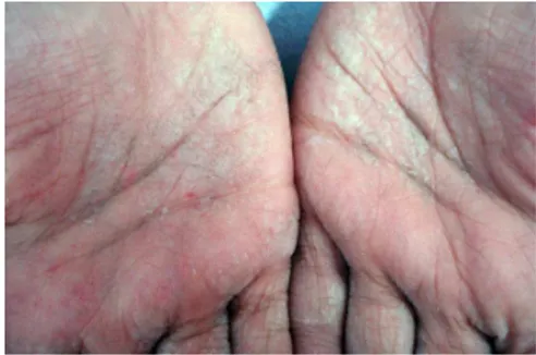
A 10-year-old boy presented with a one-month history of increased sweat pro-duction on his palms. Hyperhidrosis was induced after using battery heat-ed hand warmers for a period of two weeks. Afterwards superficial depres-sions were accompanied on the palmar surface of the hands (Figure 1). Physi-cal examination revealed foPhysi-cal macer-ated areas of the palmar region with
Açıklama:Figure 1. Focal macerated areas of the palmar region with numerous depressed microspots.
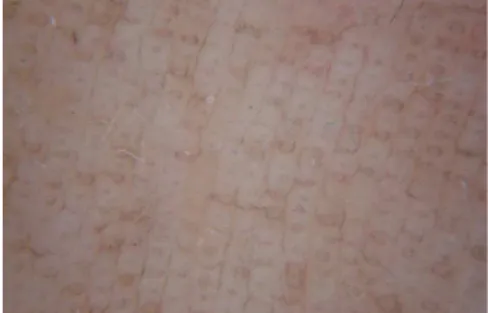
Açıklama:Figure 2. Numerous black circles in a parallel pattern on the ridges of the skin markings (documente
Figure 1: Focal macerated areas of the palmar region with numerous depressed microspots.
130 Dermatoscopic Findings of Palmar Pitted Keratolysis Due to Battery Heated Hand Warmer Ankara Üniversitesi Tıp Fakültesi Mecmuası 2009, 62(3)
numerous depressed microspots. The affected skin shows coral red fluores-cence upon Wood’s light examination. The starch iodine test performed on the palmar surfaces of the hands and was immediately and strongly positive. PK with palmar hyperhidrosis was di-agnosed. Dermatoscopic examination revealed numerous black circles in a parallel pattern on the ridges of the skin markings (Figure 2). Treatment with topical aluminium hydroxide and topical erythromycin lead to resolving of the lesions.
Discussion
PK is a non-inflammatory, superficial bac-terial infection of the skin, confined to the stratum corneum of the soles, characterized clinically by multifocal, discrete, superficial crateriform pits and superficial erosions. The cause is usually attributed to a member of Co-rynebacterium species, Micrococcus sedentarius (now renamed as Kytococ-cus sedentarius), and Dermatophilus
congolensis(3,5-7). All of these share a common feature, which enables them to open small tunnels in the stratum corneum.
PK is reported to be more common among barefooted laborers/farmers, marine workers, soldiers and indus-trial workers wearing occluded shoes for prolonged periods. The disease is mostly confined to the stratum corne-um of the soles. It can rarely occur on the palms(3). Our patient developed palmar hyperhidrosis after using bat-tery heated hand warmer. Here, heat and humidity produce a microenvi-ronment that predisposes to PK. Hy-perhidrosis is reported the commonest symptom of PK reported by 70% of the cases(3).
The diagnosis of PK is made on clini-cal grounds. Bacterial culture is not clinically relevant as multiple bacterial species are usually present(4). Skin bi-opsies are not performed routinely, as the diagnosis can be made easily by the unique clinical presentation. Histolog-ical evaluation reveals a crater limited to the stratum corneum. Filaments and coccoid organisms may be seen in the base and margin of the same with H/E stain, however, the organisms can be detected more easily with special stains like Gram stain, Periodic acid-Schiff (PAS), or methenamine silver stains(8). Wood’s ultraviolet light ex-amination is not consistently helpful, but the affected area displays a
char-acteristic coral red fluorescence due to water soluble coproporphyrin III pro-duced by the organisms. In the present case the affected skin shows coral red fluorescence under Wood’s light. To our knowledge, dermatoscopic
find-ings of PK have not been reported. Dermatoscopy revealed numerous black circles in a parallel pattern on the ridges of the skin markings. Cra-ters limited to the stratum corneum explain the circles seen by dermatos-copy. Some bacteria produce pigments which can be seen after they grow into colonies. Here, the pigment seen as black circles by dermatoscopy may correspond to the pigment produced by coccoid organisms. Although in the present case, a proof of infectious agents which justify the diagnosis of a pitted keratolysis was notconducted and also numerous black circles seen by dermatoscopy could be similar in other cornification disorders like seb-orrheic keratosis and linear epidermal nevus, typical clinical appearance and rapid healing of the lesions with topi-cal aluminium hydroxide and topitopi-cal erythromycin led us to consider pitted keratolysis in the diagnosis.
In conclusion, dermatoscopy may add ad-ditional information to the clinical ex-amination in PK. However, we would recommend further investigations with histopathological correlation in patients with PK to confirm this der-matoscopic sign.
REFERENCES
1. Zaias N, Taplin D, Rebell G. Pitted kera-tolysis. Arch Dermatol 1965;92:151-154. 2. Zaias N. Pitted and ringed keratolysis; a
review and update. J Am Acad Dermatol 1982;7:787-791.
3. Takama H, Tamada Y, Yano K, et al. Pit-ted keratolysis clinical manifestations in 53 cases. Br J Dermatol 1997;137:282-285.
4. Heid E, Cribier B, Kessler A. Les coryne-bacterioses cutanees. Ann Dermatol Vene-reol 1994;121:855-858.
5. Nordstrom KM, Mc Ginley KJ, Cappello L, et al. Pitted keratolysis. The role of Mi-crococcus sedentarius. Arch Dermatol 1987;123:1320-1325.
6. Rubel LR. Pitted keratolysis and Der-matophilus congolensis. Arch Dermatol 1972;105:584-586.
7. Wohlrab J, Rohrbach D, Marsch WC. Kera-tolysis sulcata (pitted keraKera-tolysis): clinical symptoms with different histological corre-lates. Br J Dermatol 2000; 143:1348-1349. 8. Weedon D, Strutton G. Pitted keratolysis.
In: Michael J, Houston, editors. Weedon Skin Pathology. 2nd ed. London: Churchill Livingstone.; 2002. p. 623-624.
Açıklama:Figure 1. Focal macerated areas of the palmar region with numerous depressed microspots.
Açıklama:Figure 2. Numerous black circles in a parallel pattern on the ridges of the skin markings (documente
Figure 2: Numerous black circles in a paral-lel pattern on the ridges of the skin markings (documente by Dermlite foto)