*Corresponding author: Bedia Cakmakoglu. Department of Molecular Medicine, Aziz Sancar Institute for Experimental Medicine Research, Istanbul University, Istanbul, Turkey. Tel: +90-2124142000; Fax: +90-2125324171; Email: [email protected]
Iranian Journal of Basic Medical Sciences
ijbms.mums.ac.ir
An answer to colon cancer treatment by mesenchymal stem cell
originated from adipose tissue
Elif Sinem Iplik
1, Baris Ertugrul
2, Ilknur Kozanoglu
3, Yusuf Baran
4, 5, Bedia Cakmakoglu
2*
1 Department of Pharmaceutical Microbiology, Faculty of Pharmacy, Istanbul Yeni Yuzyil University, Istanbul, Turkey2 Department of Molecular Medicine, Aziz Sancar Institute for Experimental Medicine, Istanbul University, Istanbul, Turkey 3 Department of Hematology, Baskent University, Adana, Turkey
4 Faculty of Life and Natural Sciences, Abdullah Gul University, Kayseri, Turkey
5 Department of Molecular Biology and Genetics, Izmir Institute of Technology, Urla, Izmir, Turkey A R T I C L E I N F O A B S T R A C T
Article type:
Original article Objective(s): Colon cancer is risen up with its complex mechanism that directly impacts on its treatment as well as its common prevalence. Mesenchymal stem cells (MSCs) have been considered as a therapeutic candidate for conventional disease including cancer. In this research, we have focused on apoptotic effects of adipose tissue-derived MSCs in colon cancer.
Materials and Methods: MSCs were obtained from adipose tissue and characterized by Flowcytometer
using suitable antibodies. MSCs, HT-29, HCT-116, RKO and healthy cell line MRC5 were cultured by different seeding procedure. After cell viability assay, changes in caspase 3 enzyme activity and the level of phosphatidylserine were measured.
Results: For cell viability assay, a 48 hr incubation period was chosen to seed all cells together. There
was a 1.36-fold decrease in caspase 3 enzyme activity by co-treatment of RKO and MSCs in addition to 2.02-fold decrease in HT-29 and MSCs co-treatment, and 1.103-fold increase in HCT-116 and MSCs. The results demonstrated that HCT-116 led to the highest rate of apoptotic cell death (7.5%) compared with other cells.
Conclusion: We suggest that MSCs might remain a new treatment option for cancer by its differentiation
and repair capacity.
Article history: Received: Sep 6, 2017 Accepted: Jan 10, 2018 Keywords: Apoptosis Colon cancer Stem cells Cell death
►Please cite this article as:
Iplik ES, Ertugrul B, Kozanoglu I, Baran Y, Cakmakoglu B. An answer to colon cancer treatment by mesenchymal stem cell originated from adipose tissue. Iran J Basic Med Sci 2018; 21:465-468. doi: 10.22038/IJBMS.2018.26152.6420
Introduction
Colon cancer is the third most common cancer among the leading cause of cancer related death in the world (1-3). Some of the risk factors for colon cancer include age, personal history, family history, and racial and ethnic background (4, 5). Moreover, life style and diet-related factors contribute to the development of colon cancer.
Generally, the most typical treatment options for colon cancer are surgery, chemotherapy, and radiotherapy (6). The main aim while treating the cancer is not to harm other body parts or healthy cell environment. The therapy is chosen by tumor type, tumor location, and stage as well as patients conditions (7). However, there are several treatment options for the colon cancer, e.g. a personalized therapy depending on several individual attributes, which may alter positive outcome and the treatment success. The other dilemma for colon cancer treatment is the different characteristic of tumor. One tumor might respond positively while the other could have negative or aggressive feedback for the chosen therapy. For these reasons, not only for colon cancer treatment but also for all types of cancer treatments the attempt was to find new therapeutic approaches such as mesenchymal stem cells therapy.
Mesenchymal stem cells (MSCs) are adult stem cells that have multipotent differentiation potential. Even though, they could be isolated primarily from
the bone marrow, it is well possible that to have MSCs from umbilical cord blood, adipose tissue, adult muscle, and the dental pulp of deciduous baby teeth. Recently, MSCs has turned out popular for cancer treatment as well as tissue damage. Nevertheless, some studies have shown that MSCs may induce the apoptosis directly or indirectly by related pathways. In addition, there are some evidences indicating that MSCs could help the tumor growth (8).
In this research, we aimed to find out this ongoing dilemma about antiproliferative and apoptotic effects of MSCs on colon cancer. Our research might be the first study combining adipose tissue-isolated MSCs and colon cancer cell lines including HT-29, HCT-116, and RKO and also MRC5 as a control cell line.
Materials and Methods MSCs isolation and cultivation
The MSCs were obtained from adipose tissue of the mouse. The tissue taken from the mouse was separated into two consecutive petri dishes and 5 ml phosphate buffered saline (PBS) including 2% penicillin-streptomycin was added on the tissues. Type-I Collagenase (0.075%) was used to isolate MSCs from the tissue (9).
Characterization of MSCs by flow cytometer
Iran J Basic Med Sci, Vol. 21, No. 5, May 2018 Iplik et al. Colon cancer & mesenchymal stem cell
466
completed using anti-CD34 PE-Cy7 (BD Biosciences, San Diego, CA, USA), CD45 APC-Cy7 (BD Biosciences Pharmingen, San Diego, CA, USA), anti-CD73 PE (BD Biosciences Pharmingen), and anti-NG2 PE (Beckman Coulter, Marsellia, France) antibodies by flowcytometry (BD FACSCanto-II, USA) and DiVA software (BD Biosciences Pharmingen) (10,11).
Cell culture for HT-29, HCT-116, RKO and healthy cell line MRC5
The colon cancer cell lines HT-29, HCT-116, and RKO and MRC5 as healthy cell line were purchased from American Type Culture Collection (ATCC, Manassas, VA). McCoy’s 5a modified medium was used for culturing for HT-29 and HCT-116 cells; Eagle’s Modified medium was used for culturing RKO and Dulbecco’s modified medium was used to culture MRC5 cells. All medium were prepared by adding supplemented materials including fetal bovine serum (FBS), and penicillin/streptomycin. Culture conditions were 37 °C and 5% CO
2.
MSCs and HT-29, HCT-116, RKO and healthy cell line MRC5 seeding procedure and cell viability assay
Three different groups were organized for seeding at a density of 1×10⁶ cells/well in 24-well plates for all
cell lines including healthy cell line. In the first group, all cells (HT-29, HCT-116, RKO and MRC5) were seeded on different plates and incubated for 24 hr period. After attachment of the cells to the plate, MSCs were seeded as a treatment agent in concentration of 104– 106 cells/
well.
In the second group, MSCs were seeded first at a density of 104– 106 cells/well and incubated for 24 hr
period. After incubating, the cells (HT-29, HCT-116, RKO and MRC5) were seeded at a density of 1×10⁶ cells/well.
All cells including MSCs were seeded all together in the third group at a density of 1×10⁶ cells/well (HT-29,
HCT-116, RKO and MRC5) and in concentration of 104–
106 MSCs cells/well.
After seeding the all groups, WST-1 colorimetric cell proliferation assay was conducted after each incubation period; 24 hr, 48 hr and 72 hr. For this purpose, 25 ul of WST-1 solution was added to each well. According to the assay procedure, wells were incubated 4 hr at 37 °C. To measure color development, microplate
reader (Thermo Fisher Scientific, Germany) was used at 450 nm. Percentage of proliferation was calculated compared to the untreated cells.
Changes in Caspase 3 enzyme activity
Change in Caspase 3 enzyme activity as an important sign for apoptosis process was examined by Caspase 3 colorimetric assay kit (BioVision Research Products,USA) and BCA protein assay kit. The absorbance values were normalized to protein concentrations and determined using Bradford assay. The main principles of the kit to measured of labeled substrate DEVD-pNA. The final reaction was measured spectrophotometrically after necessary incubation period at 400 nm wavelengths for Caspase 3 and 562 nm wavelengths for BCA on an ELISA reader (Thermo Electron Corporation Multiskan Spectrum, Finland).
Level of phosphatidylserine to detect apoptosis
Cell death may lead to several changes in components of the inner and outer side of the members such as phosphatidylserine (PS). Normally, PS is located in the cell. While apoptosis starts, PS changes its location through the cell surface. By changes in the PS level, apoptosis can be detected by staining with the green fluorescent dye Annexin V-FITC (BD Pharmingen, Germany). After cells and dyes were suspended and incubated for almost one hour, the mixtures were immediately analyzed using flowcytometry.
Statistical analysis
Results are expressed as the mean standard error of the mean (SEM). The data were analyzed using one-way ANOVA. P-value<0.05 was considered statistically significant.
Results
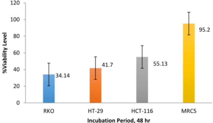
Viability level of colon cancer and healthy cell lines after treatment with MSCs in a time- and dose-dependent manner
Cytotoxic effects of MSCs were determined by WST-1 colorimetric assay. The three different seeding group that mentioned earlier were incubated with increasing concentration of MSCs (104– 106)for 24 hr, 48 hr and 72
hr. The results showed that the third group (the group in which all cells including MSCs seeded all together) was the statistically important effects in a time-dependent manner (*P-value<0.05). The concentration of MSCs was 105 cells/well and the concentrations of HT-29,
HCT-116, RKO and MRC5 were 106 cells/well during 48
hr incubation period (*P-value<0.05) (Figure 1).
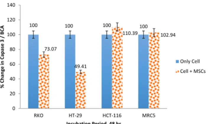
Changes in caspase 3 enzyme activity in a time- and dose-dependent manner
Caspase-3 enzyme activity was measured after 48 hr incubation period. The values are normalized to protein concentrations and determined using a Bradford assay. There were a 1.36-fold decrease in caspase-3 activity in RKO and MSCs and 2.02-fold decrease after 48 hr in HT-29 and MSCs. On the other hand, we have also found 1.103-fold increase in caspase-3 activity 48 hr after incubation in HCT-116 and MSCs (Figure 2). MRC5 cell line was used to show the relationship between MSCs and healthy cell line. There was almost no important change after 48 hr treatment (*P-value<0.05).
34.14 41.7 55.13 95.2 0 20 40 60 80 100 120 RKO HT-29 HCT-116 MRC5 % Vi ab ili ty Level Incubation Period, 48 hr
Figure 1. Viability level of colon cancer cell lines HT-29, HCT-116, RKO and healthy cell line MRC5 after treatment with MSCs. (Chosen seed-ing group and the time of incubation is shown)
467
Iran J Basic Med Sci, Vol. 21, No. 5, May 2018
Colon cancer & mesenchymal stem cell Iplik et al.
Level of phosphatidylserine for apoptosis
PS level was measured by flowcytometry to detect apoptosis after double staining with the green fluorescent dye Annexin V-FITC (BD Pharmingen, Germany). The results demonstrated that highest levels of apoptotic cell death was observed in HCT-116 (7.5%) compared to other cells. Also, 5.15% apoptosis and 0.15% necrosis was reported in HT-29 cells. Differently, RKO had low level of cell death including necrosis (1.65% for apoptotic cells; 0.10% for necrotic cells) (*P-value<0.05) (Figure 3).
Discussion
MSCs have several abilities due to their differentiation capacity. Not only tissue damage role of MSCs has still being researched, but also growth factor and cytokines production have become a number of area that need to be lighten up by its relation in the pathological conditions such as neoplastic damage, inflammation process, and tissue repair (12). Although, there are some questions about MSCs for the treatments, some preclinical and early clinical trials were come up promising results for cardiovascular area, nervous system, skin and also lung (13,14). Complicatedly, MSCs-related treatments have lots of questions and dilemma including its impact on tumor growth and apoptosis. In this research, for the first time we aimed to find out the apoptosis level of adipose-derived MSCs when given as an anticancer agent in different colon cancer cell lines and also in a healthy cell line.
Khakoo et al. studied antitumorigenic effects of human MSCs at in vivo condition on nude mice in a model of Kaposi’s sarcoma. They injected MSCs into mice
with Kaposi’s sarcoma, and isolated and co-cultured MSCs and Kaposi’s sarcoma cells in vitro. They found out decreasing level of proliferation of Kaposi’s sarcoma cells by affecting Akt-signaling (15) Oppositely, Xu et al. worked on osteosarcoma and MSCs by injection of MSCs into nude mice and showed increasing stimulation of tumor growth (16). These different results might be related to different characteristic of derivation area of MSCs such as adipose tissue, bone marrow, and umbilical cord. Furthermore, it also might be due to both tumor type and MSCs characteristics. A limited number of studies have tried to explain underlying mechanism of this interaction (8).
Spaeth et al. focused on MSCs contribution to fibrovascular network expansion and tumor progression on human ovarian carcinoma in tumor-bearing nude mice and observed the larger tumor capacity after treatments (17). Zhu et al. worked on bone marrow MSCs and human colon carcinoma at in vivo condition and showed the stimulation of growth (18). In contrast, our study showed apoptotic cell death in HCT-116 and HT-29 that both are characteristic condition of colon, while no change was observed in MRC5 as healthy cell line. RKO is a colon cancer cell line too although characteristic condition is only carcinoma, resulted instead of apoptotic cell death it ended up low level of necrotic death. In our opinion, it is theoretically thought that MSCs treatments might have a strong relation with the cancer type specifically.
Lanza et al. worked on neuroprotective role and antioxidant capacity of MSCs and successfully showed that MSCs activate the antioxidant system; hence, protection is possible from oxidative stress (19). The study might have pointed out an important key for cancers because of the relation between antioxidant systems.
Hogan et al. showed the role of plasminogen activator inhibitor type 1 (PAI-1) in MSCs on colon cancer using HT-29 and HCT-116 cell lines (20). They focused on secretion of PAI-1 in different concentrations, which resulted in different outcomes on both cell lines. They reported that MSCs-secreted PAI-1 in HCT-116 cells resulted in decreasing proliferation in all concentration, while MSCs-secreted PAI-1 in HT29 cells led to increasing proliferation activity by higher PAI-1 levels (20). Lu et
al. studied the growth inhibitory impact of MSCs at in vitro and in vivo conditions on murine hepatoma and
lymphoma cells and rat insulinoma cells. They showed upregulation of p21 and caspase 3 in all cell types (21). Similarly, we found out that co-treatment of HCT-116 and MSCs increased caspase 3 activity, while the enzyme activity decreased in other colon cancer cell lines. Again, we ended up with same dilemma that MSCs have effects on cancer cells due to its differentiation characteristic. However, further studies are needed to confirm the possible effects.
Similar to other researches, we need to examine its potential effects on colon cancer and also underlying mechanistic pathways and the possible role of cytokines and chemokines until their role is clearly lightened up.
Conclusion
There are several doubtful questions on many
100 100 100 100 73.07 49.41 110.39 102.94 0 20 40 60 80 100 120 140 RKO HT-29 HCT-116 MRC5 % C han ge in C ap ase 3 / B CA Incubation Period, 48 hr Only Cell Cell + MSCs
Figure 2. Change in Capase 3 (%) in colon cancer cell lines HT-29, HCT-116, RKO and healthy cell line MRC5
1.65 5.15 7.5 0.1 0.15 0.6 0 10 RKO HT-29 HCT-116 % Le ve l o f c el l d eath Incubation Period, 48 hr Apoptotic Cells Necrotic Cells
Iran J Basic Med Sci, Vol. 21, No. 5, May 2018 Iplik et al. Colon cancer & mesenchymal stem cell
468
researches about MSCs. On the other hand, differentiation potential and repair capacity of MSCs may have beneficial therapeutic effect in some diseases including cancer. We suggest that, after working on detailed pathways of HCT-116, HT-29, and RKO cell lines, it might be considered as a new treatment option. Prospectively, the results would lead to new treatment options by a view on lack of immunosuppression using MSCs of patients isolated from their own adipose tissue.
Acknowledgements
This work was funded by Istanbul University Scientific Research Project number 39247. We would like to thank Mr David F. Chapman for editing the manuscript.
Conflict of interest
The authors declare that they have no conflict of interest.
References
1. Nakaji S, Umeda T, Shimoyama T, Sugawara K, Tamura K, Fukuda S, et al. Environmental factors affect colon carcinoma and rectal carcinoma in men and women differently. Int J Colorectal Dis 2003; 18: 481–486.
2. Saif MW, Chu E. Biology of colorectal cancer. Cancer J 2010;16:196–201.
3. Koliaraki V, Pallangyo CK, Greten FR, Kollias G. Mesenchymal cells in colon cancer. Gastroenterology 2017; 152:964-979. 4. Niv Y, Goel A, Boland CR. JC virus and colorectal cancer: a possible trigger in the chromosomal instability pathways. Curr Opin Gastroenterol 2005; 21: 85–89.
5. Giovannucci E. Insulin, insulin-like growth factors and colon cancer: a review of the evidence. J Nutr. 2001;131:3 109–120. 6. Slattery ML. Diet, lifestyle, and colon cancer. Semin Gastrointest Dis 2000;11:142–146.
7. Mishra J, Drummond J, Quazi SH, Karanki SS, Shaw JJ, Chen B, et al. Prospective of colon cancer treatments and scope for combinatorial approach to enhanced cancer cell apoptosis. Crit Rev Oncol Hematol 2013; 86: 232–250.
8. Bergfeld SA, DeClerck YA. Bone marrow-derived mesenchymal stem cells and the tumor microenvironment. Cancer Metastasis Rev 2010; 29: 249–261.
9. Sunay O, Can G, Cakir Z, Denek Z, Kozanoglu I, Erbil G, et
al. Autologous rabbit adipose tissue-derived mesenchymal
stromal cells for the treatment of bone injuries with distraction osteogenesis. Cytotherapy 2013; 15: 690-702.
10. Bieback K, Kem S, Kocaomer A, Ferlik K, Bugert P. Comparing mesenchymal stromal cells from different human tissues: bone marrow, adipose tissue and umbilical cord blood. Biomed Mater Eng 2008; 18: 71-76.
11. Kozanoglu I, Boga C, Ozdogu H, Sozer O, Maytalman E, Yazici AC, et al. Human bone marrow mesenchymal cells express NG2: possible increase in discriminative ability of flow cytometry during mesenchymal sstromal cell identification. Cytotherapy 2009;11:527-533.
12. Kalinina NI, Sysoeva VY, Rubina KA, Parfenova YV, Tkachuk VA. Mesenchymal stem cells in tissue growth and repair. Acta Naturae 2011; 3: 30-37.
13. Williams AR, Hare JM. Mesenchymal stem cells: Biology, pathophysiology, translational findings, and therapeutic implications for cardiac disease. Circ Res 2011;109: 923-940. 14. Hong HS, Kim YH, Son Y. Perspectives on mesenchymal stem cells: Tissue repair, immune modulation, and tumor homing. Arch Pharm Res 2012;35:201.
15. Khakoo AY, Pati S, Anderson SA, Reid W, Elshal MF, Rovira II, et al. Human mesenchymal stem cells exert potent antitumorigenic effects in a model of Kaposi’s sarcoma. J Exp Med 2006; 203: 1235–1247.
16. Xu WT, Bian ZY, Fan QM, Li G, Tang TT. Human mesenchymal stem cells (hMSCs) target osteosarcoma and promote its growth and pulmonary metastasis Cancer Lett 2009; 281: 32–41.
17. Spaeth EL, Dembinski JL, Sasser AK, Watson K, Klopp A, Hall B, et al. Mesenchymal stem cell transition to tumor-associated fibroblasts contributes to fibrovascular network expansion and tumor progression. PLoS One 2009; 4:e4992. 18. Zhu W, Xu W, Jiang R, Qian H, Chen M, Hu J, et al. Mesenchymal stem cells derived from bone marrow favor tumor cell growth
in vivo. Exp Mol Pathol 2006; 80: 267–274.
19. Lanza C, Morando S, Voci A, Canesi L, Principato MC, Serpero LD, et al. Neuroprotective mesenchymal stem cells are endowed with a potent antioxidant effect in vivo. J Neurochem 2009; 110:1674–1684.
20. Hogan NM, Joyce MR, Murphy JM, Barry FP, O’Brien T, Kerin MJ, et al. Impact of Mesenchymal Stem Cell secreted PAI-1 on colon cancer cell migration and proliferation. Biochem Biophys Res Commun 2013; 435: 574–579.
21. Lu YR, Yuan Y, Wang XJ, Wei LL, Chen YN, Cong C, et al. The growth inhibitory effect of mesenchymal stem cells on tumor cells in vitro and in vivo. Cancer Biol Ther 2008; 7: 245–251.