INTRODUCTION
Synovial sarcoma (SS) is an aggressive soft-tissue tumor with a tendency to recur locally and high potential for distant metastasis. Morphologically, these tumors consist of spin-dle-like cells positive by immunohistochemical staining for epithelial markers. SS constitutes 5-10% of all soft-tissue sarco-mas and typically affects adolescents and young adults [1-4]. This sarcoma is most often found in the lower extremities (especially the knee), upper extremities, and the head and neck (mostly in the parapharyngeal region). SS can be classi-fied into three histological types: monophasic (pure epithelioid or fibroblastic cells), biphasic, and poorly differentiated [1-5], and is diagnosed upon histopathological findings of epithelial
differentiation or immunostaining for cytokeratin or epithe-lial membrane antigen (EMA). While some investigators have reported transducer-like enhancer of split 1 (TLE1) to be an extremely sensitive biomarker for SS [1,2,6], others showed a low specificity of TLE1 in diagnosing SS [7]. Recently, the t(X;18) (SYT-SSX) translocation has been recommended as the most reliable diagnostic tool [1,2,8].
The prognosis for SS is usually poor, with 5- and 10-year survival rates between 36-76% and 20-63%, respectively [3,9-11]. Factors associated with a better prognosis include age <20 years, tumor size smaller than 5 cm, more distal location along the extremities, lower tumor stage, and appropriate excision. Predictors of poor prognosis include less-differenti-ated tumor areas, presence of necrosis, and high mitotic activ-ity, i.e., >10 mitoses/10 high power fields (HPFs) [3,5,9,12].
Enhancer of zeste homologue 2 (EZH2) belongs to the polycomb group (PcG) proteins of cell cycle regulators that suppress transcription. An excessive expression of EZH2 is found in various carcinomas, lymphomas, and soft tissue *Corresponding author: Ulviye Yalçınkaya, Department of Surgical
Pathology, Faculty of Medicine, Uludağ University, Gorukle 16059 Bursa, Turkey. Phone: +905322736031. Fax: +90224295 0019.
E-mail: [email protected]
Submitted: 16 January 2017/Accepted: 18 February 2017
Enhancer of zeste homologue 2 (EZH2) expression in
synovial sarcomas as a promising indicator of prognosis
Ulviye Yalçınkaya1*, Nesrin Uğraş1, Gonca Özgün2, Gökhan Ocakoğlu3, Adem Deligönül4, Sibel Kahraman Çetintaş5,
Muhammed Sadık Bilgen6
1Department of Surgical Pathology, Faculty of Medicine, Uludağ University, Bursa, Turkey, 2Department of Surgical Pathology, Faculty
of Medicine, Başkent University, Ankara, Turkey, 3Department of Biostatistics, Faculty of Medicine, Uludağ University, Bursa, Turkey, 4Department of Medical Oncology, Faculty of Medicine, Uludağ University, Bursa, Turkey, 5Department of Radiation Oncology, Faculty of
Medicine, Uludağ University, Bursa, Turkey, 6Department of Orthopedics, Faculty of Medicine, Uludağ University, Bursa, Turkey
ABSTRACT
Synovial sarcoma (SS) is a type of soft-tissue sarcoma, often linked to poor survival. Although overexpression of enhancer of zeste homologue 2 (EZH2) has been associated with poor prognosis in different tumors, a few studies investigated this link in SS. Here, we analyzed the rela-tionship between EZH2 expression and prognostic factors in SS. We included 29 patients with SS. Immunostaining of EZH2 was performed with (D2C9) XPTM Rabbit mAb antibody, and the results were classified as low EZH2 expression (negative or weak expression) and high EZH2
expression category (moderate or strong expression). Analysis of survival in relation to prognostic factors was performed with Kaplan-Meier survival curves and Cox proportional hazard regression analysis. Our sample included 19/29 female and 10/29 male patients, with age range 16-63 years. The tumor diameter ranged from 2 to 15 cm. Necrosis was observed in 15/29 cases. Sixteen cases had >10 mitoses per 50 high-power fields (HPFs). Out of 29 cases, 14 showed low and 15 had high EZH2 expression. Statistically significant results were obtained for the association between the presence of metastasis and necrosis (p = 0.042), high EZH2 expression and distant metastasis (p = 0.018), high EZH2 expression and necrosis (p = 0.016), and high EZH2 expression and the tumor size >5 cm versus tumor size ≤5 cm (p = 0.014). Patients with all of the follow-ing: the tumor size ≤5 cm, low EZH2 expression, and without necrosis and distant metastasis had significantly longer survival time. Our results are consistent with previous studies, suggesting that EZH2 overexpression is an indicator of poor prognosis in SS.
KEY WORDS: Enhancer of zeste homologue 2 (EZH2); immunohistochemistry; prognostic factors; synovial sarcoma; survival analysis
sarcomas, and growing evidence suggests that this overex-pression correlates with the aggressiveness and poor clinical outcome of such tumors [13-22]. However, few studies have investigated the expression of EZH2 in SS [23-25]. The aim of this study was to investigate the relationship between the EZH2 expression as evaluated by immunohistochemistry and known prognostic indicators in SS.
MATERIALS AND METHODS
We retrospectively analyzed the data for 29 patients diag-nosed with SS between 2002 and 2014 at our pathology depart-ment. All slides were reviewed and re-evaluated. The following information was collected from the medical records: age, gen-der, tumor site, tumor size, tumor grade, follow-up duration, recurrence, metastasis, and survival time. We used the World Health Organization (WHO) guidelines for the histological type and Fédération Nationale des Centers de Lutte Contre le Cancer (FNCLCC) criteria to determine the tumor grade (dif-ferentiation, mitotic index, and necrosis). Tumors were clas-sified histologically into three groups: biphasic, monophasic fibrous, or poorly differentiated. The study was conducted in accordance with the Institutional Ethical Guidelines.
Immunohistochemical study
Four-micron-thick serial sections were obtained from formalin-fixed, paraffin-embedded (FFPE) blocks. Immunohistochemical staining was performed on a Leica BOND-MAX Autostainer (Leica Microsystems, Berlin, Germany), and peroxidase/DAB Bond Polymer Refine Detection System (Leica Microsystems) was used for visu-alization. EZH2 immunohistostaining was done with D2C9XPTM Rabbit mAb (1:100 dilution) primary antibodies,
while other sections were reserved for hematoxylin and eosin (H&E) staining.
Scoring system
To assess the immunohistochemical labeling of EZH2, the immunostained slides were evaluated using a 10× magnifica-tion. The nuclear staining was scored as follows: a) negative, no visible staining, b) weak, 1-25% of nuclei were positive, c) moderate, 25-75%, and d) strong, over 75% [19]. The EZH2 staining was classified into two categories according to the nuclear staining: a “low EZH2 expression category” with neg-ative or weak expression, and a “high EZH2 expression cate-gory” with moderate or strong expression.
Statistical analysis
The association between EZH2 expression and the pres-ence of distant metastasis was tested by the Fisher’s exact
test and Fisher-Freeman-Halton test where appropriate. The log-rank test was used to determine the difference between the Kaplan-Meier curves for survival time. Median survival time was reported. To determine the prognostic factors that affect the overall survival time, we performed the Cox pro-portional hazard regression analysis with backward selec-tion procedure following the Kaplan-Meier analysis. The significant predictors obtained in the Kaplan-Meier analysis were entered into the Cox proportional hazard regression, and the results of the final step were reported. The results were reported as hazard ratios with 95% confidence intervals (CIs) and the related p-values. All analyses were performed using IBM SPSS Statistics for Windows, Version 21.0. (IBM Corp., Armonk, NY). A value of p < 0.05 was considered as statistically significant.
RESULTS
Clinical data
The study included 19/29 female (65.5%), and 10/29 male (34.5%) patients with age range 16-63 (mean age: 35). The mean diameter of the tumors was 8 cm, ranging from 2 to 15 cm. Eight tumors measured ≤5 cm, 21 tumors were >5 cm. The tumors occurred in the periphery in 22/29 cases (76%) and centrally in 7/29 cases (24%).
The specimens consisted of 15 marginal excisions and 14 wide resections. In 4 cases, recurrence was treated by tion, including two hemipelvectomies and two finger amputa-tions. Twenty-two patients were treated with both chemo and radiation therapy following the surgery.
The follow-up information for a minimum of 13 months was available for all 29 cases (13-147 months range). Out of the 29 cases, 15 (52%) patients died of the tumor, 9 (31%) had local recurrence, and 5 (17.2%) had distant metastasis.
Pathological features
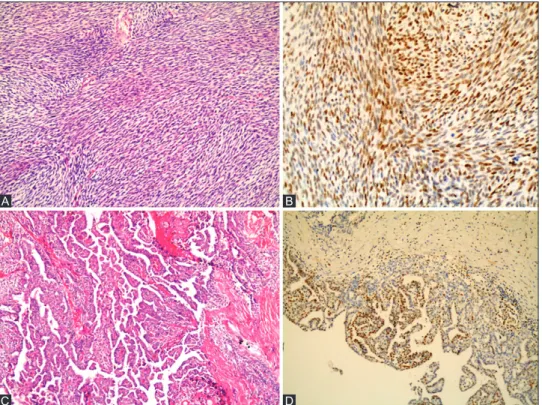
Histopathologically, 14/29 cases were monophasic (48.3%), 11/29 biphasic (37.9%), and 4/29 poorly differentiated (13.8%) (Figure 1 and 2). In 1 case, there were large areas of epithelial differentiation, and 3 cases localized on the periphery had large areas of calcification.
Out of the 29 cases, 14 (48.3%) were evaluated as Grade II and 15 (51.7%) as Grade III. We observed necrosis in 15 (51.7%) cases, the mitotic rate per 50 HPFs ranged from 4 to 39. In addition, 16 cases had >10 mitoses per 50 HPFs.
We examined the effects of clinicopathological parameters on distant metastasis, and found that there was a statistically significant association between metastases and the presence of necrosis (p = 0.042). Other clinicopathological parameters of the tumor (i.e., age, gender, tumor location, tumor size,
histological type, grade, and mitoses) did not show significant association with distant metastasis (Table 1).
Immunohistochemical findings and the
relationship with clinicopathological features
Out of the 29 patients, 14 (48.3%) cases were classified as low EZH2 expression and 15 (51.7%) were classified as high EZH2 expression. The relationship between the EZH2 expres-sion and clinicopathological features in SS patients is summa-rized in Table 2 and Figure 1 and 2.
There were statistically significant correlations between high EZH2 expression and distant metastasis (p = 0.018) and the presence of necrosis (p = 0.016).
In addition, high EZH2 expression was significantly more frequently detected in patients with the tumor size >5 cm compared with the tumor size ≤5 cm (p = 0.014). Patients with low EZH2 expression were predominantly female (p = 0.050).
On the other hand, no statistically significant relationship was observed between EZH2 expression and other clinico-pathological factors, including age, tumor location, size, local recurrence, histological type, grade, and mitoses.
Survival analysis
Using the Kaplan-Meier analysis, the overall median fol-low-up for the entire study population was 68 months (95%, CI: 15.1-34.9 months). The survival was not affected by the age,
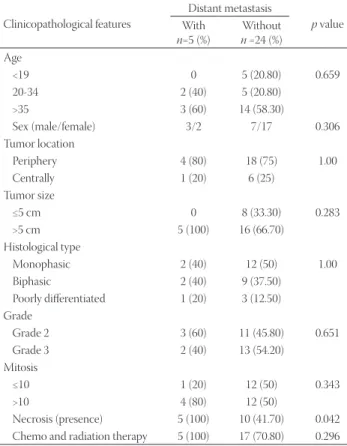
TABLE 1. Clinicopathological features of patients with distant metastasis compared with patients without distant metastasis
Clinicopathological features Distant metastasis p value With n=5 (%) n =24 (%)Without Age <19 0 5 (20.80) 0.659 20-34 2 (40) 5 (20.80) >35 3 (60) 14 (58.30) Sex (male/female) 3/2 7/17 0.306 Tumor location Periphery 4 (80) 18 (75) 1.00 Centrally 1 (20) 6 (25) Tumor size ≤5 cm 0 8 (33.30) 0.283 >5 cm 5 (100) 16 (66.70) Histological type Monophasic 2 (40) 12 (50) 1.00 Biphasic 2 (40) 9 (37.50) Poorly differentiated 1 (20) 3 (12.50) Grade Grade 2 3 (60) 11 (45.80) 0.651 Grade 3 2 (40) 13 (54.20) Mitosis ≤10 1 (20) 12 (50) 0.343 >10 4 (80) 12 (50) Necrosis (presence) 5 (100) 10 (41.70) 0.042 Chemo and radiation therapy 5 (100) 17 (70.80) 0.296
TABLE 2. Demographic and clinicopathological characteristics of patients with respect to EZH2 expression
Demographic and clinicopathological characteristics EZH2 expression p value Low n=14 (%) High n=15 (%) Age <19 2 (14.30) 3 (20) 0.462 20-34 2 (14.30) 5 (33.30) >35 10 (71.40) 7 (46.70) Sex (male/female) 2/12 8/7 0.050 Tumor location Periphery 9 (64.30) 13 (86.70) 0.215 Centrally 5 (35.70) 2 (13.30) Tumor size ≤5 cm 7 (50) 1 (6.70) 0.014 >5 cm 7 (50) 14 (93.30) Histological type Monophasic 8 (57.10) 6 (40) 0.612 Biphasic 5 (35.70) 6 (40) Poorly differentiated 1 (7.10) 3 (20) Grade Grade 2 9 (64.30) 5 (33.30) 0.096 Grade 3 5 (35.70) 10 (66.70) Mitosis ≤10 9 (64.30) 4 (26.70) 0.066 >10 5 (35.70) 11 (73.30) Necrosis (presence) 4 (28.60) 11 (73.30) 0.016 Local recurrence (presence) 3 (21.40) 6 (40) 0.427 Distant metastasis (presence) 0 5 (33.30) 0.018
EZH2: Enhancer of zeste homologue 2
gender, tumor location, local recurrence, histological type, grade, mitoses, chemo and radiation therapy, and type of sur-gery (Table 3). However, patients with the tumor size ≤5 cm, without necrosis, without distant metastasis and low EZH2 expression had a significantly longer survival time (p = 0.022,
p = 0.006, p = 0.001, and p = 0.017, respectively; Table 3).
Moreover, the analysis of independent risk factors affecting survival showed that patients with distant metastasis had a 3.59 risk factor for the total survival time.
DISCUSSION
EZH2 is a PcG protein. This family consists of transcrip-tional suppressor regulators responsible for the repair of DNA damage, cellular differentiation, cellular aging, and apoptosis. PcG proteins are involved in the maintenance of stem cell character and in the tumor development. Specifically, EZH2 acts as a histone methyltransferase targeting the N-terminal tail of histone H3 to produce a characteristic H3-Lys27 trimethylation (H3K27me3) pattern. EZH2 is expressed at high levels in cells exhibiting an embryonic gene expression pattern, and its expression declines with tissue maturation and differentiation. Recent studies have suggested that EZH2 over-expression might be related to aggressive behavior and poor
prognosis in various carcinomas, lymphomas, and soft tissue sarcomas [13-25].
Different clinical and pathological factors have been sug-gested as indicators of SS prognosis. Recent studies including
a large number of cases with long follow-up periods have shown that the advanced stage, high grade, male gender, age >40 years, tumor >5 cm in diameter, poor differentiation, as well as occurrence at the proximal sites along the extremities
FIGURE 1. Enhancer of zeste homologue 2 (EZH2) expression in two histological types of synovial sarcoma (SS). (A) Monophasic fibrous SS consisting of relatively homogeneous spindle cells arranged in a fascicular pattern (H&E, ×100), (B) EZH2 immunostaining is strong in the same tumor (×200), (C) biphasic SS, (H&E, ×100), and (D) EZH2 immunostaining shows a moderate expression of EZH2 in the glan-dular epithelium (×100).
B
D A
C
FIGURE 2. (A) Poorly differentiated synovial sarcoma (SS) with a prominent hemangiopericytoma-like vascular pattern (H&E, ×200), (B) in the same tumor, enhancer of zeste homologue 2 (EZH2) immunostaining is weak (×200), (C) monophasic fibrous SS with extensive tumor calcification (H&E, ×100), and (D) in the same tumor, EZH2 immunostaining is negative (×100).
B
D A
are associated with poor prognosis [4,5,9,26-28]. On the other hand, younger age (adolescence), small size, distal location, calcification, and thorough resection are indicators of good prognosis [3,9,29].
SS is aggressive, with a 50-70% tendency to recur locally or to metastasize to the lungs, bones, and sometimes to the lymph nodes [3,4,9]. In this study, we found that patients with the tumor size ≤5 cm (p = 0.022) and without distant metasta-sis (p = 0.001) had better survival. We also demonstrated that the patients with distant metastasis had a 3.59 risk factor for the total survival time. Although the t(X;18) translocation involv-ing the SSX2 gene has been correlated with better outcomes, no studies have confirmed that it is an independent prognostic factor for SS [3,27,28]. Moreover, necrosis, vascular invasion, high mitotic activity, and high Ki-67 proliferative index are
accepted histopathological predictors of the shortened sur-vival [9,13,27]. Our results showed that the patients without necrosis had a significantly higher survival time (p = 0.006).
In the last decade, several new immunohistochemical and molecular markers were investigated as indicators of SS prognosis and potential therapeutic targets [9]. To this end, we aimed to evaluate whether immunohistochemical EZH2 expression correlates with known prognostic indi-cators of SS.
A histological type may serve as a prognostic indicator for SS. Tumors with >20% of poorly differentiated histology show more aggressive behavior [5,8,9]. Similarly, Paulino [30] reported that biphasic histology is associated with a better clinical outcome more frequently than the monophasic his-tology; in addition, Krieg et al. [3] found that a histological
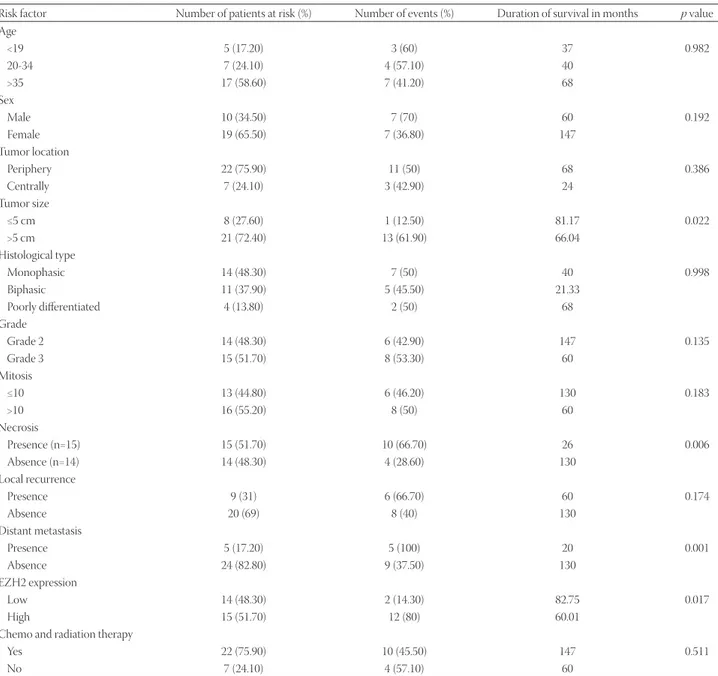
TABLE 3. Median survival time in months according to the Kaplan-Meier analysis in relation to demographic and clinicopathological characteristics
Risk factor Number of patients at risk (%) Number of events (%) Duration of survival in months p value Age <19 5 (17.20) 3 (60) 37 0.982 20-34 7 (24.10) 4 (57.10) 40 >35 17 (58.60) 7 (41.20) 68 Sex Male 10 (34.50) 7 (70) 60 0.192 Female 19 (65.50) 7 (36.80) 147 Tumor location Periphery 22 (75.90) 11 (50) 68 0.386 Centrally 7 (24.10) 3 (42.90) 24 Tumor size ≤5 cm 8 (27.60) 1 (12.50) 81.17 0.022 >5 cm 21 (72.40) 13 (61.90) 66.04 Histological type Monophasic 14 (48.30) 7 (50) 40 0.998 Biphasic 11 (37.90) 5 (45.50) 21.33 Poorly differentiated 4 (13.80) 2 (50) 68 Grade Grade 2 14 (48.30) 6 (42.90) 147 0.135 Grade 3 15 (51.70) 8 (53.30) 60 Mitosis ≤10 13 (44.80) 6 (46.20) 130 0.183 >10 16 (55.20) 8 (50) 60 Necrosis Presence (n=15) 15 (51.70) 10 (66.70) 26 0.006 Absence (n=14) 14 (48.30) 4 (28.60) 130 Local recurrence Presence 9 (31) 6 (66.70) 60 0.174 Absence 20 (69) 8 (40) 130 Distant metastasis Presence 5 (17.20) 5 (100) 20 0.001 Absence 24 (82.80) 9 (37.50) 130 EZH2 expression Low 14 (48.30) 2 (14.30) 82.75 0.017 High 15 (51.70) 12 (80) 60.01
Chemo and radiation therapy
Yes 22 (75.90) 10 (45.50) 147 0.511
No 7 (24.10) 4 (57.10) 60
subtype can also serve as an independent prognostic factor for SS [3,30]. Changchien et al. [23] observed higher EZH2 expression in poorly differentiated SS and suggested that EZH2 expression might correlate with aggressive clinical behavior [23]. We found high EZH2 expression in 3 out of 4 cases of poorly differentiated SS, but there was no statisti-cally significant relationship between EZH2 expression and the histological subtype.
Although some studies suggest that gender does not cor-relate with prognosis in SS, other authors report that male gender is associated with a worse prognosis [3,31,32]. In this study, patients with low EZH2 expression were predomi-nantly female (p = 0.050), which is consistent with the previ-ous studies.
Similar to other soft tissue sarcomas, SS tends to have a better prognosis in younger patients [12,26,30,31]. A study from a single institution on 271 cases of SS found a significant correlation between advanced age and poor prognosis [26]. Similarly, another study on 121 SS cases reported that the age over 25 years was correlated with the lower disease-free sur-vival [9]. Furthermore, SS located in the extremities is reported to have a better prognosis than the tumor in the head-neck region [3,30,33,34]. For example, in children and adolescents SS in the extremities had a better prognosis than the tumors in other locations, and it was suggested that the treatment should be planned accordingly [34,35]. In our study, no signif-icant relationship was observed between the age, tumor loca-tion, and EZH2 expression.
The standard treatment for SS is wide resection of the tumor and surrounding tissue. SS recurs in 70-83% of cases with inadequate marginal resection. Recurrence may increase the risk of metastasis, and thus negatively influence the prog-nosis. Aggressive surgery accompanied by chemo or radia-tion therapy increases disease-free survival [3,4,26,33-35]. We found no statistically significant relationship between EZH2 expression and treatment modality and local recurrence, and we demonstrated a significant correlation between high EZH2 expression and distant metastasis (p = 0.018).
In most studies of carcinoma, lymphoma, and soft-tissue sarcomas, EZH2 overexpression was an indicator of poor prognosis [13-15,19-25]. Similarly, we observed that low EZH2 expression was associated with prolonged survival (p = 0.017).
Generally accepted indicators of prognosis in SS include tumor stage at presentation, tumor size, and FNCLCC tumor grade [5,27,28]. The tumor size >5 cm is strongly associated with poor prognosis [3,5,9,28,33], as well as the presence of distant metastasis at diagnosis and FNCLCC grade 3 [3,27,28]. Changchien et al. [23] reported that tumors overexpressing EZH2 were also >5 cm in diameter and had distant metasta-ses, suggesting that EZH2 overexpression is associated with greater tumor size, presence of distant metastasis, and poor
prognosis [23]. We also found a significant correlation between high EZH2 expression and tumor size >5 cm (p = 0.014), and the presence of distant metastasis (p = 0.018).
Tumor necrosis and high mitotic counts (>10 mitosis/10 HPFs) have been reported as indicators of a poor prognosis in SS [5,9,27]. We found no significant relationship between EZH2 expression and mitosis, but there was a significant cor-relation between high EZH2 expression and the presence of necrosis (p = 0.016).
CONCLUSION
Our findings suggest that patients with the tumor size ≤5 cm, without necrosis, without distant metastasis, and with low EZH2 expression had a significantly longer survival time. In addition, patients with distant metastasis had a 3.59 risk fac-tor for the total survival time. Furthermore, we demonstrated a significant correlation between high EZH2 expression and tumor size >5 cm, necrosis, and distant metastasis. This is con-sistent with the previous studies suggesting that EZH2 overex-pression is an indicator of poor prognosis in SS. Our findings warrant the confirmation by future studies with larger cohorts to determine whether EZH2 expression is an indicator of prognosis in SS.
ACKNOWLEDGMENTS
This study was supported by the Uludag University, Commission of Scientific Research Projects (Project HDP (T)-2013/2).
DECLARATION OF INTERESTS
The authors declare no conflict of interests.
REFERENCES
[1] Suurmeijer AJ, de Bruijn D, Geurts van Kessel A, Miettinen MM. Synovial sarcoma. In: Fletcher CD, Bridge JA, Hogendoor P, Mertens F, editors. WHO Classification of Tumours of Soft Tissue and Bone. 4th ed. Lyon: IARC; 2013. p. 213-5.
[2] Goldblum JR, Folpe AL, Weiss SW, editors. Malignant soft tissue tumors of uncertain type. Enzinger and Weiss’s Soft Tissue Tumors. 6th ed. Philadelphia, PA: Saunders; 2013. p. 1052-70.
[3] Krieg AH, Hefti F, Speth BM, Jundt G, Guillou L, Exner UG, et al. Synovial sarcomas usually metastasize after >5 years: a multicenter retrospective analysis with minimum fol-low-up of 10 years for survivors. Ann Oncol 2011;22(2):458-67. https://doi.org/10.1093/annonc/mdq394.
[4] Lamm W, Schur S, Köstler WJ, Amann G, Pokrajac B, Funovics P, et al. Initially localized synovial sarcoma in adults: a retrospective single-center analysis of 26 patients registered at the Department of Oncology, University of Vienna between 2004 and 2013. Oncology 2014;87(1):48-57. https://doi.org/10.1159/000363185.
[5] Machen SK, Easley KA, Goldblum JR. Synovial sarcoma of the extremities: A clinicopathologic study of 34 cases,
including semi-quantitative analysis of spindled, epithelial, and poorly differentiated areas. Am J Surg Pathol 1999;23(3):268-75. https://doi.org/10.1097/00000478-199903000-00004.
[6] Terry J, Saito T, Subramanian S, Ruttan C, Antonescu CR, Goldblum JR, et al. TLE1 as a diagnostic immunohistochem-ical marker for synovial sarcoma emerging from gene expres-sion profiling studies. Am J Surg Pathol 2007;31(2):240-6. https://doi.org/10.1097/01.pas.0000213330.71745.39.
[7] Kosemehmetoglu K, Vrana JA, Folpe AL. TLE1 expression is not specific for synovial sarcoma: A whole section study of 163 soft tissue and bone neoplasms. Mod Pathol 2009;22(7):872-8. https://doi.org/10.1038/modpathol.2009.47.
[8] van de Rijn M, Barr FG, Xiong QB, Hedges M, Shipley J, Fisher C. Poorly differentiated synovial sarcoma: an analysis of clin-ical, pathologic, and molecular genetic features. Am J Surg Pathol 1999;23(1):106-12.
https://doi.org/10.1097/00000478-199901000-00012.
[9] Bergh P, Meis-Kindblom JM, Gherlinzoni F, Berlin O, Bacchini P, Bertoni F, et al. Synovial sarcoma: identification of low and high risk groups. Cancer 1999;85(12):2596-607.
https://doi.org/10.1002/ (SICI)1097-0142 (19990615) 85:12 <2596: AID-CNCR16>3.0.CO;2-K.
[10] Trassard M, Le Doussal V, Hacène K, Terrier P, Ranchère D, Guillou L, et al. Prognostic factors in localized primary synovial sarcoma: a multicenter study of 128 adult patients. J Clin Oncol 2001;19(2):525-34.
https://doi.org/10.1200/JCO.2001.19.2.525.
[11] Lewis JJ, Antonescu CR, Leung DH, Blumberg D, Healey JH, Woodruff JM, et al. Synovial sarcoma: a multivariate analysis of prognostic factors in 112 patients with primary localized tumors of the extremity. J Clin Oncol 2000;18(10):2087-94.
https://doi.org/10.1200/JCO.2000.18.10.2087.
[12] Sultan I, Rodriguez-Galindo C, Saab R, Yasir S, Casanova M, Ferrari A. Comparing children and adults with synovial sarcoma in the Surveillance, Epidemiology, and End Results program, 1983 to 2005: an analysis of 1268 patients. Cancer 2009;115(15):3537-47. https://doi.org/10.1002/cncr.24424.
[13] Yamaga K, Osaki M, Kidani K, Shomori K, Yoshida H, Ito H. High expression of enhancer of zeste homologue 2 indicates poor prognosis in patients with soft tissue sarcomas. Mol Med Rep 2008;1(5):633-9.
https://doi.org/10.3892/mmr_00000004.
[14] Chase A, Cross NC. Aberrations of EZH2 in cancer. Clin Cancer Res 2011;17(9):2613-8.
https://doi.org/10.1158/1078-0432.CCR-10-2156.
[15] Chang CJ, Hung MC. The role of EZH2 in tumour progression. Br J Cancer 2012;106(2):243-7.
https://doi.org/10.1038/bjc.2011.551.
[16] Sasaki H, Setoguchi T, Matsunoshita Y, Gao H, Hirotsu M, Komiya S. The knock-down of overexpressed EZH2 and BMI-1 does not pre-vent osteosarcoma growth. Oncol Rep 2010;23(3):677-84. [17] Bracken AP, Pasini D, Capra M, Prosperini E, Colli E, Helin K. EZH2
is downstream of the pRB-E2F pathway, essential for proliferation and amplified in cancer. EMBO J 2003;22(20):5323-35.
https://doi.org/10.1093/emboj/cdg542.
[18] Kanno R, Janakiraman H, Kanno M. Epigenetic regulator polycomb group protein complexes control cell fate and cancer. Cancer Sci 2008;99(6):1077-84.
https://doi.org/10.1111/j.1349-7006.2008.00797.x.
[19] Alford SH, Toy K, Merajver SD, Kleer CG. Increased risk for distant metastasis in patients with familial early-stage breast cancer and high EZH2 expression. Breast Cancer Res Treat 2012;132(2):429-37. https://doi.org/10.1007/s10549-011-1591-2.
[20] Sudo T, Utsunomiya T, Mimori K, Nagahara H, Ogawa K, Inoue H, et al. Clinicopathological significance of EZH2 mRNA expression in patients with hepatocellular carcinoma. Br J Cancer
2005;92(9):1754-8.
https://doi.org/10.1038/sj.bjc.6602531.
[21] Lessard J, Sauvageau G. Bmi-1 determines the proliferative capacity of normal and leukaemic stem cells. Nature 2003;423(6937):255-60. https://doi.org/10.1038/nature01572.
[22] Varambally S, Dhanasekaran SM, Zhou M, Barrette TR, Kumar-Sinha C, Sanda MG, et al. The polycomb group pro-tein EZH2 is involved in progression of prostate cancer. Nature 2002;419(6907):624-9.
https://doi.org/10.1038/nature01075.
[23] Changchien YC, Tátrai P, Papp G, Sápi J, Fónyad L, Szendroi M, et al. Poorly differentiated synovial sarcoma is associated with high expression of enhancer of zeste homologue 2 (EZH2). J Transl Med 2012;10:216.
https://doi.org/10.1186/1479-5876-10-216.
[24] Kawano S, Grassian AR, Tsuda M, Knutson SK, Warholic NM, Kuznetsov G, et al. Preclinical evidence of anti-tumor activity induced by EZH2 inhibition in human models of synovial sarcoma. PLoS One 2016;11(7):1-22.
https://doi.org/10.1371/journal.pone.0158888.
[25] Shen JK, Cote GM, Gao Y, Choy E, Mankin HJ, Hornicek FJ, et al. Targeting EZH2-mediated methylation of H3K27 inhibits proliferation and migration of synovial sarcoma in vitro. Sci Rep 2016;6:25239. https://doi.org/10.1038/srep25239.
[26] Ferrari A, Gronchi A, Casanova M, Meazza C, Gandola L, Collini P, et al. Synovial sarcoma: a retrospective analysis of 271 patients of all ages treated at a single institution. Cancer 2004;101(3):627-34. https://doi.org/10.1002/cncr.20386.
[27] Guillou L, Benhattar J, Bonichon F, Gallagher G, Terrier P, Stauffer E, et al. Histologic grade, but not SYT-SSX fusion type, is an important prognostic factor in patients with synovial sarcoma: a multicenter, retrospective analysis. J Clin Oncol 2004;22(20):4040-50. https://doi.org/10.1200/JCO.2004.11.093.
[28] ten Heuvel SE, Hoekstra HJ, Bastiaannet E, Suurmeijer AJ. The clas-sic prognostic factors tumor stage, tumor size, and tumor grade are the strongest predictors of outcome in synovial sarcoma: no role for SSX fusion type or ezrin expression. Appl Immunohistochem Mol Morphol 2009;17(3):189-95.
https://doi.org/10.1097/PAI.0b013e31818a6f5c.
[29] Varela-Duran J, Enzinger FM. Calcifying synovial sarcoma. Cancer 1982;50(2):345-52.
https://doi.org/10.1002/1097-0142(19820715)50:2<345:AID- CNCR2820500231>3.0.CO;2-Y.
[30] Paulino AC. Synovial sarcoma prognostic factors and patterns of failure. Am J Clin Oncol 2004;27(2):122-7.
https://doi.org/10.1097/01.coc.0000047130.91699.DC.
[31] Campbell C, Gallagher J, Dickinson I. Synovial sarcoma - Towards a simplified approach to prognosis. ANZ J Surg 2004;74(9):727-31. https://doi.org/10.1111/j.1445-1433.2004.03144.x.
[32] Wright PH, Sim FH, Soule EH, Taylor WF. Synovial sarcoma. J Bone Joint Surg Am 1982;64(2):112-22.
https://doi.org/10.2106/00004623-198264010-00016.
[33] Canter RJ, Qin LX, Maki RG, Brennan MF, Ladanyi M, Singer S. A synovial sarcoma-specific preoperative nomogram supports a survival benefit to ifosfamide-based chemotherapy and improves risk stratification for patients. Clin Cancer Res 2008;14(24):8191-7. DOI: 10.1158/1078-0432.CCR-08-0843.
[34] Ferrari A, Bisogno G, Alaggio R, Cecchetto G, Collini P, Rosolen A, et al. Synovial sarcoma of children and adolescents: the prognostic role of axial sites. Eur J Cancer 2008;44(9):1202-9.
https://doi.org/10.1016/j.ejca.2008.03.016.
[35] Soole F, Maupain C, Defachelles AS, Taque S, Minard-Colin V, Bergeron C, et al. Synovial sarcoma relapses in children and ado-lescents: prognostic factors, treatment, and outcome. Pediatr Blood Cancer 2014;61(8):1387-93.