Percutaneous Repair in A Patient with Iatrogenic Subclavian
Artery Pseudoanerysm
İyatrojenik Subklavyen Arter Psödoanevrizmasında Perkütanöz Tamir
Veysel Özgür Barış
1, Ozgür Ulaş Özcan
1, Hüseyin Göksülük
1, Deniz Kumbasar
11 Ankara University School of Medicine , Department of Cardiology
Central vein catheterization is a common procedure but it has potential fatal complications such as pneumo-thorax, hemothorax or arterial injury. We present a 74 year old female who was suffered a subclavien artery pseudoaneurysm because of accidental arterial puncture during the subclavien venous catheterizations. The patient was treated by stent graft by percutaneously due to high surgical risk.
Key Words: Subclavien Artery Pseudoaneurysm, Percutaneous intervention
Santral ven kateterizasyonu sıklıkla kullanılan fakat pnömotoraks, hemotoraks veya arteryel hasar gibi ciddi komplikasyonları olabilecek bir işlemdir. Bu yazıda, subklavyen venöz kateterizasyon sırasında gelişen subk-lavyen arter psödoanevrizması olan 74 yaşında kadın hastayı takdim ettik. Hastya yüksek cerrahi risk nedeniyle perkütan yol ile greft stent ile tedavi edildi.
Anahtar Sözcükler: Subklavyen Arter Psödoanevrizması, Perkütan Girişim
A 74-year-old female was admitted to emergency service with dyspnea,
orthopnea and fatigue of nearly two weeks history. She had a history of atrial fibrillation and dilated cardiomyipathy. Her medications included ramipril, metoprolol and warfarin. Physical examination revealed right jugular venous distention, 3+ pitting pretibial edema, 4/6 pansystolic murmur at apex and inspiratory crackles at the base and middle zone of the lung. Electrocardiography showed atrial fibrillation with rapid ventricular rate. Transthoracic echocardiography revealed dilated left ventricle and EF was 15%. After diagnosis of acute heart failure, the patient was referred coronary intensive care unit for intravenous diuretic and inotrop therapy. Right subclavien venous catheterizations were attempted to measure central venous pressure. However, accidental arterial puncture during the procedure was noted. After intravenous inotrop therapy was stopped, subclavien vein introducer sheath was removed. Two days later, the patients developed a continuous right upper chest pain with radiation to the right forearm. Repeated physical
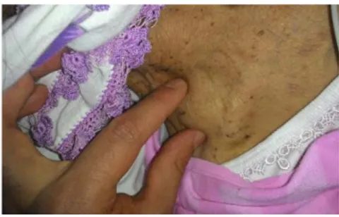
examination revealed a pulsatile mass measuring nearly 3x3 cm in the right supraclavicular region with to and fro murmur and absent right radial artery pulsation (Figure 1) .Color Doppler ultrasound revealed a 35x24 mm sized pseudoaneurysm communicating between the right subclavien artery (SCA). After multidisciplinary consultation, the patient was transferred to the coronary angiography unite for endovascular treatment.
Figure 1: Physical examination revealed a pulsatile mass measuring nearly 3x3 cm in the right supraclavicular region
Under local anesthesia, the right common femoral artery as accessed percutane-ously and an 8-Fr introducer was placed. With a 0.038-inch 150-cm guide wire the
Received : June 28,2016 • Accepted: Oct 03,2016 Corresponding Author:
Barış, Veysel Özgür MD
E-mail: [email protected] Phone: +90312-508 68 89
Ankara University School of Medicine Ankara İbn-i Sina Hospital 12th Floor Sıhhiye, Ankara
Ankara Üniversitesi Tıp Fakültesi Mecmuası 2016, 69 (3)
DOI: 10.1501/Tıpfak_000000945
DAHİLİ BİLİMLERİ/ MEDICAL SCIENCES
Ankara Üniversitesi Tıp Fakültesi Mecmuası 2016, 69 (3)
Percutaneous Repair in a Patient with Iatrogenic Subclavian Artery Pseudoanerysm
208
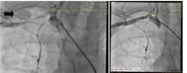
tip of a 6-F Right Judkins catheter was positioned at the origin of the right SCA. Selective angiography of the right SCA confirmed the pseudoaneurysm with a narrow neck between the anterior wall of the right SCA (Figure 2). A 5 mm x 25 mm balloon-expandable Viabahn graft-stent (W. L. Gore and Associates, Flagstaff, Arizona) was implanted to the right SCA to exclude the pseudoaneu-rysm from the arterial lumen. The final right SCA angiogram demonstrated suc-cessful total occlusion of the pseudoan-eurysm and patency of vertebral, inter-nal mammarian artery and distal SCA (Figure 3). After the procedure, right forearm pain was improved and right ra-dial pulsation was recovered. Two days later, the patient was discharged with as-pirin, clopidogrel, warfarin and standard medications for heart failure.
Central vein catheterization is a common procedure performed for measurement of central venous pressure or ultra filtra-tion therapy for acute decompansated heart failure (1). Commonly blind tech-nique guided by surface landmarks was used by cardiology specialists (2). This technique can lead to multiple punctures that may result in potential complica-tions such as pneumothorax, hemotho-rax and arterial injury (2). Especially, in a non-compressible artery such as SCA, arterial puncture may cause formation of a pseudoaneurysm, arteriovenous fis-tula, and uncontrolled hemorrhage into soft tissues and adjacent pleural space (3). Small pseudoaneurysms of SCA may be asymptomatic while pulsatile mass, hematoma, neurological and air-way compression, mediastinal syn-drome, upper limb ischemia, and heart
failure may ensue from large pseudoan-eurysms (3). Such complications may warrant urgent treatment.
Percutaneous treatments (stent graft, embo-lization, balloon temporary occlusion, thrombin injection, and closure device) have become attractive alternatives to conventional surgery for the treatment of pseudoaneurysms (2). In our case, surgical repair was considered to be high risk because of patient clinical condition and large size of the pseudoaneurysm. Therefore, percutaneous treatment with covered stent was considered the first line treatment.
Figure 2: Selective angiography of the right SCA confirmed the
pseudoaneurysm with a narrow neck between the anterior wall of the right SCA Figure 3:The final right SCA angiogram demonstrated successful total occlusion of the pseudoaneurysm and patency of vertebral, internal mammarian artery and distal SCA
REFERENCES
1. Lin, C.H., Wu HS, Chan DC, et al., The mechanisms of failure of totally implantable central venous access system: analysis of 73 cases with fracture of catheter. Eur J Surg Oncol, 2010. 36: p. 100-103.
2. Petrocheilou, G., Kokkinis, C., Statphopoulou, S.,et al., Iatrogenic pseudoaneurysm of the brachiocephalic artery: a rare complication of Hickman line insertion. Int Urol Nephrol, 2008. 40: p. 1107-1110.
3. Kusminsky, R.E., Complications of central venous catheterization. J Am Coll Surg, 2007. 204: p. 681-696.