http://journals.tubitak.gov.tr/medical/ © TÜBİTAK
doi:10.3906/sag-1308-20
Effects of ozone therapy and taurine on ischemia/reperfusion-induced
testicular injury in a rat testicular torsion model
Tolga Reşat AYDOS1,*, Mehmet Murad BAŞAR2, Oğuz KUL3, Hasan Tarık ATMACA3, Tuba UZUNALİOĞLU3, Üçler KISA4, Oğuzhan Ekin EFE1
1Department of Pharmacology, Faculty of Medicine, Başkent University, Ankara, Turkey 2Department of Urology/Andrology, Memorial Şişli Hospital, İstanbul, Turkey 3Department of Pathology, Veterinary Faculty, Kırıkkale University, Kırıkkale, Turkey 4Department of Clinical Biochemistry, Faculty of Medicine, Kırıkkale University, Kırıkkale, Turkey
1. Introduction
Testicular torsion is one of the common urological emergencies that must be diagnosed and treated as early as possible to preserve fertility. Testicular damage is directly correlated with the degree and duration of testicular torsion. However, correction of torsion and restoration of normal blood flow may sometimes fail to avoid the development of permanent spermatogenic failure and infertility (1). Ischemia/reperfusion (I/R) injury due to testicular torsion and detorsion causes some biochemical and morphological changes in the testicular tissue (2). For example, the increase in intratesticular reactive oxygen species leads to gonadal necrosis and germ cell apoptosis (3,4). Previous studies have shown that testicular injury develops especially during the reperfusion period (5).
Ozone therapy, applied as a gas mixture of oxygen and ozone, has been widely used in a number of diseases such
as chronic cutaneous ulcers, peritonitis, infected wounds, ischemic diseases, and joint problems (6). Ozone therapy acts as an effective oxidative stress regulator, mainly by stimulating the antioxidant system of the cell (7–10).
Taurine is an antioxidant agent obtained from the metabolism of sulfur-containing amino acids such as methionine and cysteine. It protects the tissues against oxidative damage by stabilization of biomembranes and neutralization of free oxygen radicals (11–13). Instead of using pharmacological antioxidant agents, such as vitamin E or melatonin, taurine, the major intracellular free β-amino acid, was preferred in the present study as an endogenous agent.
The aim of this study was to investigate the effects of ozone therapy and/or taurine treatment comparatively on testicular damage due to I/R injury in an experimental torsion model in rats.
Background/aim: To investigate the effect of ozone and/or taurine treatment comparatively on testicular damage due to ischemia/
reperfusion (I/R) injury in an experimental torsion model in rats.
Materials and methods: Adult Wistar rats with and without torsion/detorsion were used. In order to monitor the effect of ozone and/
or taurine treatment on testicular damage due to I/R injury, following histopathological investigation apoptotic indexes were scored by TUNEL method. Moreover, tumor necrosis factor receptor 1 (TNFR1), caspase 3, caspase 8, endothelial nitric oxide synthase (eNOS), tumor necrosis factor alpha (TNFα), and cytochrome C immunostainings were performed and the levels of malondialdehyde, glutathione peroxidase, superoxide dismutase, catalase, total sulfhydryl, and nitric oxide were determined in the testicular tissue.
Results: Intraperitoneal ozone and/or taurine treatment prevented both histopathological damage and increase in the apoptotic index.
Torsion did not exert an effect on the levels of TNFα and cytochrome C. Ozone and/or taurine treatment prevented increases in TNFR1, caspase 3, and caspase 8. The level of oxidative stress markers was unchanged. The increases in NO level and eNOS expression were prevented by ozone and/or taurine treatment in I/R groups.
Conclusion: Using ozone therapy and/or taurine before reperfusion may be a solution for germ cell degeneration resulting from
testicular torsion and related infertility.
Key words: Ozone therapy, taurine, ischemia/reperfusion, rat, testis, oxidative stress
Received: 05.08.2013 Accepted: 01.10.2013 Published Online: 15.08.2014 Printed: 12.09.2014
2. Materials and methods
Animal studies were conducted in the Research Unit of the Başkent University Experimental Animals Breeding and Research Center after being approved by the Local Ethics Committee of Kırıkkale University for Experimental Animals (09.02.2010, No. 10/07).
Fifty-four male adult Wistar rats (316 ± 4.30 g) were assigned to 9 groups (n = 6 for each). Surgical interventions were performed under ketamine (50–60 mg/kg, intraperitoneal [i.p.]) and xylazine (7–10 mg/kg, i.p.) anesthesia. Taurine (Sigma, USA; 7.5 mL/kg in 10% water solution) was administered intraperitoneally. Ozone gas used for ozone therapy (1 mg/kg, i.p.) was obtained from an ozone generator device (EVOZONE Basic Plus, Germany). Ozone-resistant silicon-treated polypropylene syringes were used throughout the procedure. Both taurine and ozone therapies were administered at the end of the ischemia period (15 min before reperfusion).
Experimental groups were as follows: T1: Control.
T2: Sham.
T3: I/R. The scrotum was opened surgically; the left testis was exposed and whirled 720° clockwise. The scrotum was then closed after the testis was fixed in the scrotum. The scrotum was reopened after 2 h of ischemia, twisted in the opposite direction, and reperfused for 4 h.
T4: Taurine. T5: Ozone therapy. T6: I/R + taurine. T7: I/R + ozone therapy. T8: Taurine + ozone therapy. T9: I/R + taurine + ozone therapy.
Apoptosis (with the TUNEL method), endothelial nitric oxide synthase (eNOS), caspase 8, caspase 3, tumor necrosis factor alpha (TNFα), tumor necrosis factor receptor 1 (TNFR1), and cytochrome C expressions in testicular tissue sections were investigated histopathologically. Malondialdehyde (MDA), superoxide dismutase (SOD), glutathione peroxidase (GPx), catalase, nitric oxide (NO), and total sulfhydryl (t-SH) levels were determined biochemically in isolated testicular tissues.
2.1. Pathological examinations
Testicular tissues were isolated after 4 h of reperfusion and fixed in 4% buffered paraformaldehyde solution for 24 h for pathological examinations. Later, they were embedded in paraffin.
2.2. Tissue microarray method
Samples 2 mm in thickness were obtained from 3 different parts of each paraffin-embedded testicular tissue and placed into recipient paraffin blocks prepared formerly (Galileo TMA CK 3500). Fifty-four tissues taken from the donor blocks were then placed into recipient blocks and they were sectioned at 4–5 µm in thickness.
2.3. Immunohistochemical analyses
A commercial indirect immunoperoxidase streptavidin/ biotin kit (catalogue number RE/110-K, Novocastra, HRP, USA) was used for the detection of each marker in the tissues and all the stages were performed according to the suggestions of the manufacturer. Briefly, tissue sections were incubated in 3% H2O2 for 5 min and boiled in antigen retrieval solution (citrate buffer, pH 6.0) for 40 min. Tissues were incubated in normal goat serum for 7 min to block nonspecific bindings. Afterwards, primary antibody incubation was performed using the following commercial primary antibodies prepared against the targeted antigenic structure: caspase 3 (mouse, antirat), caspase 8 (mouse, antirat), TNFα (mouse, antirat), TNFR1 (mouse, antirat), cytochrome C (mouse, antirat), and eNOS (mouse, antirat). They were then incubated with biotin-marked polyvalent secondary antibody for 10 min and with streptavidin-peroxidase for 10 min. Aminoethyl carbazole chromogen and substrate were applied for 10–20 min. Mayer’s hematoxylin was administered to the sections for 2 min for counterstaining and adhered with ImmunoMount.
Positive tissue sections were also included in the test at each time for all the tested section groups. Nonimmunized rat serum was used instead of primary antibody for the isotype serum negative control.
2.4. TUNEL method
For the evaluation of apoptotic cells in the examined testis tissues, the TUNEL technique was used according to the TUNEL kit procedure (In Situ Cell Death Detection Kit, POD, 11 684 817 910, Roche, Germany).
2.5. Histomorphometric analysis
For histomorphometric analysis, the immunopositive cells were counted and semiquantitatively scored according to the following criteria under 20× objective fields: 0) no immunoreactive cells; 1) 1–10 immunoreactive cells; 2) 11–20 immunoreactive cells; 3) ≥21 immunoreactive cells. The apoptotic index was calculated using the mean number of TUNEL reaction-positive germ cells in the whole 1 mm2 area for each testis tissue.
2.6. Biochemical analysis
All samples used for biochemical investigation were frozen in liquid nitrogen and stored at –80 °C until the assays. The tissue was homogenized (Labortechnik, Germany) in 1 mL of 0.9% NaCl solution in ice. The homogenized tissue was centrifuged at 1500 × g for 10 min at 4 °C. The supernatants were used for protein, MDA, NO, t-SH, SOD, GPx, and catalase determinations. The protein level was measured by the method of Lowry et al. (14). MDA was measured by the method described by Armstrong and Al-Awadi (15), which was modified from the Yagi method. The calibration curve was prepared with 1,1,3,3-tetraethoxypropane (Sigma) standards of 1–25 nmol/L dilutions. The results
were measured as nM/g protein. The nitrite/nitrate levels were measured as described by Miranda et al. (16). Nitrate was reduced to nitrite with vanadium(III) and then the nitrite level was measured using Griess reagents. This reflects the total amount of nitrate and nitrite in the sample. Serial dilutions of 0.5–250 μM Na nitrate (Merck, Germany) were used as standards and the results were expressed as μmol/g protein. t-SH in tissue with Ellman’s reagent was measured as described by Sedlak and Lindsay (17). SOD, GPx, and catalase were determined using a commercially available quantitative enzyme-linked immunosorbent assay kit (Cayman Chemical Company, USA).
2.7. Statistical analysis
Data were statistically analyzed by using the GraphPad Prism 5.0 package program. The groups were compared by Kruskal–Wallis analysis of variance test followed by post-hoc tests or the Mann–Whitney U test. The results were shown as mean ± standard deviation. P < 0.05 was considered as statistically significant.
3. Results
3.1. Histopathological findings
On histopathological evaluation, the testicular tissues of the animals in the taurine and/or ozone therapy groups (T4, T5, and T8) along with the control and sham groups were found to have normal testicular structure and to show uniform seminiferous tubule morphology. Furthermore, the structures of the Sertoli cells, primary and secondary spermatocytes, spermatids, and spermatozoa were normal. In the I/R (T3) group, the seminiferous tubule diameters were decreased, the germinal epithelial cells were detached from the basal membrane and cell losses were determined in the germinal epithelium due to desquamation, and the lumens were filled with desquamated cells. Peritubular polymorphonuclear leukocyte infiltrations were observed in some regions. Moreover, irregular formations including degenerated germinal cells accompanied by necrosis and calcification were detected in seminiferous tubules.
Although we observed a few epithelial cells detached from the basal lamina and spilled into tubule lumen in the I/R + taurine (T6) and I/R + ozone therapy (T7) groups, the histopathological damage in the testicular tissue that resulted from I/R injury was prevented by taurine and/or ozone therapy application (T6, T7, and T9 groups).
3.2. Immunohistochemical findings
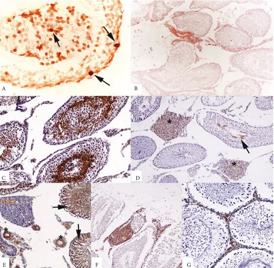
TUNEL assay and immunohistochemical analysis revealed positive findings at varying degrees in all animals in the examined groups (Figures 1A–1G).
3.3. Apoptotic changes examined with the TUNEL method in rat testicular tissue
We observed a significant increase in the apoptotic index value in I/R group (T3) compared to the control (T1) in
the left testicular tissue of rats in which torsion/detorsion was applied. Neither taurine nor ozone treatment altered the apoptotic index values in the testicular tissues significantly. The increase in apoptosis due to I/R injury was significantly prevented by taurine (T6) and ozone (T7) administrations. Coadministration of taurine and ozone (T9) also decreased the elevated apoptosis due to I/R injury; however, this finding was not statistically significant (Table 1).
3.4. Analyses of TNFR1, caspase 3, caspase 8, eNOS, TNFα, and cytochrome C alterations in rat testicular tissue
Ozone therapy and/or taurine administration significantly prevented the increase in TNFR1 and caspase 8, the extrinsic inducers of apoptosis, and caspase 3, a common inducer of the intrinsic and extrinsic pathways, due to I/R injury. On the contrary, I/R did not change the levels of TNFα, which plays a role as an extrinsic inducer, or cytochrome C, which is produced in the mitochondria-mediated intrinsic pathway of apoptosis. I/R also increased the eNOS amount significantly, which was reversed by taurine and ozone therapy. However, taurine or ozone therapy neither alone nor together caused any changes in these markers in the control animals. The apoptotic markers and eNOS values in the rat testicular tissue are shown in Table 2.
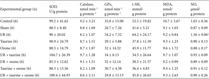
3.5. Changes in the oxidative stress markers in the rat testicular tissue
In order to assess the effect of the oxidative stress damage in the testis tissue, the MDA, GPx, SOD, catalase, t-SH, and NO levels were determined. However, no change was observed in not only the antioxidant enzyme activities such as catalase, SOD, and GPx, which convert reactive oxygen radicals to less toxic products, but also in the MDA level, which is formed as a result of membrane lipid peroxidation, as well. Moreover, I/R damage, ozone therapy, and taurine did not cause any changes in levels of antioxidant molecular substances such as t-SH in testicular tissues. On the contrary, I/R injury increased the NO level, which was reversed by taurine and/or ozone therapy. The biochemical findings in rat testicular tissue are presented in Table 3.
4. Discussion
In the present study, torsion and detorsion-induced I/R injury was found to cause morphological changes and increase the polymorphonuclear cells in the seminiferous tubules, consisting with the literature findings (18). We also observed germinal epithelial cell loss and degenerative changes in spermatogonia and spermatids. Our immunohistochemical studies revealed a significant increase in the immunopositivity of inflammatory cells in testicular tissue. Degeneration in the germinal epithelium,
E G C
A B
D
F
Figure 1. A) TUNEL-positive cells in I/R-induced testicular injury (arrows). TUNEL assay, POD converter, DAB chromogen. B)
TNFR1 immunoreactivity in inflammatory cells between tubules in I/R-induced testicular injury. ABC method (anti-TNFR1), counterstaining with Mayer’s hematoxylin. C) Caspase 3 antigen-positive spermatogonia and spermatocytes. ABC method (anticaspase 3), counterstaining with Mayer’s hematoxylin. D) Caspase 8 immunopositivity in the interstitial tissue (stars), spermatogonia, and spermatocytes (arrow). ABC method (anticaspase 8), counterstaining with Mayer’s hematoxylin. E) Positive eNOS immunoreactivity in vessel walls (stars) and tubular epithelium (arrows). ABC method (anti-eNOS), counterstaining with Mayer’s hematoxylin. F) Positive TNFα immunoreactivity in the interstitial inflammatory cells. ABC method (anti-TNFα), counterstaining with Mayer’s hematoxylin.
G) Cytochrome C immunopositivity in interstitial tissue. ABC method (anticytochrome C), counterstaining with Mayer’s hematoxylin. Table 1. Apoptotic index values obtained using the TUNEL method in the rat testicular
tissue. a: C vs. I/R (P < 0.05). b: I/R vs. I/R + taurine; I/R vs. I/R + ozone (P < 0.01) and
I/R + taurine + ozone (P < 0.05).
Experimental group (n) Apoptotic index
Control (4) 7.50 ± 1.732 Sham (4) 10.25 ± 2.062 I/R (6) 16.83 ± 3.764a, b Taurine (3) 8.00 ± 5.292 Ozone (5) 5.60 ± 1.517 I/R + taurine (4) 6.00 ± 2.582 I/R + ozone (5) 6.00 ± 2.646 Taurine + ozone (5) 5.40 ± 2.074
spermatogonia, and spermatids seen in the I/R group was prevented by taurine and/or ozone therapy applications, particularly in the taurine + ozone + I/R group. This protective effect of ozone therapy was previously shown in experimental renal I/R studies (19,20). However, to the best of our knowledge, the present study is the first study about the protective effect of ozone therapy in a testicular I/R model in adult rats. Our findings revealed that ozone therapy might be used alone or in combination with other antioxidant agents in order to prevent I/R injury in the testes and to preserve fertility. However, since ozonation products including reactive oxygen species, lipid ozonation products, and arachidonic acid peroxides may lead to toxic effects, optimum doses of ozone therapy should be determined carefully (10,21).
In the present study, the amount of apoptotic changes was investigated by the TUNEL method in the left testicular tissue of rats and a significant increase in the apoptotic index was found in the testicular tissues of rats in which torsion and subsequent reperfusion was applied (I/R group) compared to the control and sham groups. This increase was prevented with taurine or ozone therapy applied before reperfusion. In other words, taurine, an antioxidant agent, and ozone therapy, with its positive effects on oxidative stress, have been shown to suppress the increase in apoptosis due to torsion and detorsion-induced I/R injury. These findings are consistent with testes I/R experiments indicating oxidative stress-induced apoptosis initiation (22). It was suggested that taurine and ozone therapies prevent the increase in apoptosis through
Table 2. Apoptotic marker and eNOS values obtained using the immunoperoxidase staining method in rat testicular tissue for TNFR1
(a: C vs. I/R, P < 0.05), caspase 3 (a: C vs. I/R, P < 0.05), caspase 8 (a: C vs. I/R, P < 0.01), eNOS (a: C vs. I/R, P < 0.01), TNFα, and
cytochrome C. The scores indicate the percentages of the immunopositive stained areas.
Experimental group (n) TNFR1 Caspase 3 Caspase 8 TNFα Cytochrome C eNOS
Control (6) 1.2 ± 0.41 1.8 ± 0.52 1.8 ± 0.41 1.2 ± 0.75 1 ± 0.63 2.2 ± 0.75 Sham (4) 1.3 ± 0.50 1.5 ± 0.58 1.5 ± 0.58 1.8 ± 0.50 1.5 ± 0.58 2 ± 0.82 I/R (6) 2 ± 0.89a 3.7 ± 1.37a 3.8 ± 0.75a 1 ± 0.89 1 ± 0.63 3.8 ± 0.75a Taurine (6) 1.2 ± 0.41 2.5 ± 0.84 2.2 ± 0.75 1.5 ± 0.84 1 ± 0.63 2.8 ± 0.75 Ozone (6) 1.4 ± 0.55 2.2 ± 0.41 2 ± 0.63 0.8 ± 0.75 1 ± 0.63 2.3 ± 1.03 I/R + taurine (6) 1.6 ± 0.55 2.8 ± 0.75 1.7 ± 1.03 1.3 ± 0.52 1.2 ± 0.75 2.5 ± 0.55 I/R + ozone (6) 1 ± 0.0 2 ± 0.894 2.3 ± 1.21 1.8 ± 0.75 1.7 ± 0.52 2.3 ± 0.52 Taurine + ozone (6) 1.5 ± 0.55 2.5 ± 0.55 2 ± 0.89 0.8 ± 0.58 1 ± 0.63 2.8 ± 0.41
I/R + taurine + ozone (6) 1.2 ± 0.41 2.3 ± 0.52 2.3 ± 0.52 0.7 ± 0.52 0.8 ± 0.65 2.8 ± 0.75
Table 3. Results of enzymes and substances obtained through biochemical analyses in rat testicular tissue. a: C vs. I/R, P < 0.05.
Experimental group (n) SOD,U/g protein Catalase,nmol min–1
g protein–1 GPx, nmol min–1 g protein–1 t-SH, mmol/ g protein MDA, nmol/ g protein NO, µmol/ g protein Control (6) 99.2 ± 41.62 9.2 ± 3.21 35.8 ± 15.00 52.5 ± 19.82 10.7 ± 3.67 1.03 ± 0.36 Sham (6) 88.5 ± 8.40 8.8 ± 1.69 26.7 ± 7.26 41.6 ± 5.21 9.1 ± 1.03 0.87 ± 0.09 I/R (6) 90 ± 20.02 8.2 ± 1.07 34.2 ± 7.32 64.2 ± 24.17 9.2 ± 0.84 1.56 ± 0.06a Taurine (6) 98.9 ± 24.79 8.7 ± 1.51 29.1 ± 5.88 37.8 ± 11.50 9.3 ± 1.23 0.90 ± 0.13 Ozone (6) 88.5 ± 14.79 8.7 ± 1.87 32 ± 14.32 45.9 ± 11.77 9.6 ± 1.72 0.88 ± 0.17 I/R + taurine (6) 104.7 ± 26.39 9.7 ± 1.28 34 ± 8.13 34.5 ± 26.64 9.7 ± 1.07 0.93 ± 0.09 I/R + ozone (6) 85.3 ± 12.62 9.1 ± 1.51 32 ± 12.14 30.5 ± 21.57 9.2 ± 0.99 0.89 ± 0.09 Taurine + ozone (6) 88.3 ± 13.56 8.2 ± 1.09 30.7 ± 4.38 36.4 ± 4.83 9.4 ± 1.25 0.91 ± 0.12 I/R + taurine + ozone (6) 100.4 ± 44.93 8.6 ± 2.11 29.8 ± 13.13 45.8 ± 26.63 9.3 ± 2.63 0.90 ± 0.26
the antioxidant process. However, with the findings about the oxidative stress indicators, we determined that I/R and other applications did not change the amounts of catalase, SOD, and GPX, the enzymes reducing the toxic effects of free oxygen radicals. In addition, none of the applications changed the amounts of t-SH, an indicator of the amount of reduced glutathione, and MDA, an indicator of peroxidation of cell membrane lipids. Koksal et al. reported that testicular damage might be detected before the changes in MDA levels and the oxidative/antioxidative status of the tissue occur (23). Although there are some studies reporting the relationship of oxidative stress with increased apoptosis, Lysiak et al. suggested that I/R-related histological changes in the testicular tissue do not go through oxidative stress-related mechanisms (24). The physiological apoptotic process during spermatogenesis involves TNFα, TNFR1, and caspase 8, related to the extrinsic pathway, and cytochrome C, related to the intrinsic pathway, whereas caspase 3 is the common protein in both of the pathways (25,26). In the present study, we also aimed to investigate the contribution of the apoptotic pathways to the preventive effects of taurine and ozone therapies on increased apoptosis due to I/R damage. We determined increases in TNFR1 and caspase 8 immunopositivity, but no changes in TNFα immunopositivity. Therefore, we suggest that the extrinsic pathway plays the pivotal role in the increase in apoptosis due to I/R injury. Supporting this, the immunopositivity of cytochrome C, which participates in the intrinsic pathway of apoptosis, did not change significantly. However, the
discrepancy in the TNFR1 and TNFα findings needs to be explained by further investigations. In addition, we showed that I/R leads to an increase in caspase 3 immunoreactivity, which was prevented evidently by taurine and/or ozone therapy.
NO was reported to have an important effect on developing apoptosis and necrosis after I/R injury on testicular tissue (27). In experiments evaluating the nitrergic changes, I/R was found to increase the eNOS immunopositivity and the NO level. In the present study, we showed that NO levels increased in association with the increase in eNOS immunoreactivity due to I/R injury. Taurine and ozone therapy suppressed the increase in I/R-induced eNOS and NO immunopositivity.
To conclude: 1) torsion and subsequent detorsion led to an increase in the apoptotic index; 2) mainly the extrinsic pathway is responsible for the apoptotic process in the testicular tissue; 3) increased nitrergic system activity plays a role in testicular torsion/reperfusion damage; 4) oxidative stress may not be responsible for the injury; 5) taurine and ozone therapy may prevent the increase in the apoptotic index equally; 6) using ozone therapy or taurine before reperfusion may be a solution for germ cell degeneration resulting from testicular torsion and related infertility.
Acknowledgment
This study was supported by the 1002 TÜBİTAK Short-Term R&D Funding Program (Project No. 110S273).
References
1. Visser AJ, Heyns CF. Testicular function after torsion of the spermatic cord. BJU Int 2003; 92: 200–203.
2. Saba M, Morales CR, De Lamirande E, Gagnon C. Morphological and biochemical changes following acute unilateral testicular torsion in prepubertal rats. J Urol 1997; 157: 1149–1154.
3. Jhunjhunwala JS, Desal A, Kropp KA. Torsion of the spermatic cord: an experimental study. Invest Urol 1976; 13: 318–320. 4. Cuzzocrea S, Riley DP, Caputi AP, Salvemini D, Antioxidant
therapy: a new pharmacological approach in shock, inflammation, and ischemia/reperfusion injury. Pharmacol Rev 2001; 53: 135–159.
5. Romeo C, Antonuccio P, Esposito M, Marini H, Impellizzeri P, Turiaco N, Altavilla D, Bitto A, Zuccarello B, Squadrito F. Raxofelast, a hydrophilic vitamin E-like antioxidant, reduces testicular ischemia-reperfusion injury. Urol Res 2004; 32: 367– 371.
6. Bocci V, Zanardi I, Travagli V. Oxygen/ozone as a medical gas mixture. A critical evaluation of the various methods clarifies positive and negative aspects. Med Gas Res 2011; 1: 6.
7. Hernandez FA, Menéndez S, Wong R. Decrease of blood cholesterol and stimulation of antioxidative response in cardiopathy patients treated with endovenous ozone therapy. Free Radic Biol Med 1995; 19: 115–119.
8. Valdes RA, Menéndez Cepero S, Gómez Moraleda M, Ley Pozo J. Ozone therapy in the advanced stages of arteriosclerosis obliterans. Angiologia 1993; 45: 146–148 (article in Spanish with abstract in English).
9. Hernandez Rosales FA, Calunga Fernández JL, Turrent Figueras J, Menéndez Cepero S, Montenegro Perdomo A. Ozone therapy effects on biomarkers and lung function in asthma. Arch Med Res 2005; 36: 549–554.
10. Sagai M, Bocci V. Mechanisms of action involved in ozone therapy: is healing induced via a mild oxidative stress? Medical Gas Research 2011; 1: 29.
11. Huxtable RJ. Physiological action of taurine. Physiol Rev 1992; 72: 101–163.
12. Wright CE, Tallan HH, Lin YY, Gaull GE. Taurine: biological update. Annu Rev Biochem 1986; 55: 427–453.
13. Wright CE, Lin TT, Syurman JA, Gaull GE. Taurine scavenges oxidized chlorine in biological systems. In: Oja SS, editor. Taurine: Biological Actions and Clinical Perspectives. New York, NY, USA: Alan R Liss; 1985. pp. 137–147.
14. Lowry OH, Rosebrough NJ, Farr AL, Randall RJ. Protein measurement with the Folin phenol reagent. J Biol Chem 1951; 193: 265–275.
15. Armstrong D, Al-Awadi F. Lipid peroxidation and retinopathy in streptozotocin-induced diabetes. Free Radic Biol Med 1991; 11: 433–436.
16. Miranda KM, Espey MG, Wink DA. A rapid, simple spectrophotometric method for simultaneous detection of nitrate and nitrite. Nitric Oxide 2001; 5: 62–71.
17. Sedlak J, Lindsay RH. Estimation of total, protein-bound, and nonprotein sulfhydryl groups in tissue with Ellman’s reagent. Anal Biochem 1968; 25: 192–205.
18. Lysiak JJ, Turner SD, Nguyen QA, Singbartl K, Ley K, Turner TT. Essential role of neutrophils in germ cell-specific apoptosis following ischemia/reperfusion injury of the mouse testis. Biol Reprod 2001; 65: 718–725.
19. Fernández Iglesias A, González Núñez L, Calunga Fernández JL, Rodríguez Salgueiro S, Santos Febles E. Ozone postconditioning in renal ischaemia-reperfusion model. Functional and morphological evidences. Nefrologia 2011; 31: 464–470.
20. Oztosun M, Akgul EO, Cakir E, Cayci T, Uysal B, Ogur R, Ozcan A, Ozgurtas T, Guven A, Korkmaz A. The effects of medical ozone therapy on renal ischemia/reperfusion injury. Ren Fail 2012; 34: 912–925.
21. Elvis AM, Ekta JS. Ozone therapy: a clinical review. J Nat Sci Biol Med 2011; 2: 66–70.
22. Turner TT, Tung KSK, Tomomasa H, Wilson LW. Acute testicular ischemia results in germ cell-specific apoptosis in the rat. Biol Reprod 1997; 57: 1267–1274.
23. Koksal M, Oğuz E, Baba F, Eren MA, Ciftci H, Demir ME, Kurcer Z, Take G, Aral F, Ocak AR et al. Effects of melatonin on testis histology, oxidative stress and spermatogenesis after experimental testis ischemia-reperfusion in rats. Eur Rev Med Pharmacol Sci 2012; 16: 582–588.
24. Lysiak JJ, Nguyen QA, Turner TT. Peptide and nonpeptide reactive oxygen scavengers provide partial rescue of the testis after torsion. J Androl 2002; 23: 400–409.
25. Mathur PP, D’Cruz SC. The effect of environmental contaminants on testicular function. Asian J Androl 2011; 13: 585–591.
26. Tripathi R, Mishra DP, Shaha C. Male germ cell development: turning on the apoptotic pathways. J Reprod Immunol 2009; 83: 31–35.
27. Bonfoco E, Krainc D, Ankarcrona M, Nicotera P, Lipton SA. Apoptosis and necrosis: two distinct events induced, respectively, by mild and intense insults with N-methyl-D-aspartate or nitric oxide/superoxide in cortical cell cultures. Proc Natl Acad Sci USA 1995; 92: 7162–7166.