Research Article
Cerebral Venous Sinus Thrombosis in Women: Subgroup
Analysis of the VENOST Study
Derya Uluduz
,
1Sevki Sahin
,
2Taskin Duman
,
3Serefnur Ozturk
,
4Vildan Yayla
,
5Nazire Afsar
,
6Nevzat Uzuner
,
7Ipek Midi
,
8Nilgun Cinar
,
2Mehmet Ali Sungur
,
9Fusun Mayda Domac
,
10Birsen Ince,
1Baki Goksan,
1Cemile Handan Misirli,
11Mustafa Bakar,
12Hasan Huseyin Kozak,
13Sena Colakoglu,
3Ali Yavuz Karahan
,
14Eylem Ozaydin Goksu,
15Fatih Ozdag,
16Mehmet Guney Senol,
16Vedat Ali Yurekli,
17Ufuk Aluclu,
18Serkan Demir,
19Hayriye Kucukoglu,
20Serdar Oruc,
21Nilufer Yesilot,
22Ozge Yimaz Kusbeci,
23Bijen Nazliel
,
24Firdevs Ezgi Ucan Tokuc,
15Hesna Bektas,
25Fatma Nida Tascilar,
26Emrah Aytac,
27Mustafa Gokce,
28Hale Zeynep Batur Caglayan,
24Ahmet Tufekci,
29Gulnur Uzuner,
7Dilek Necioglu Orken,
30Osman Ozgur Yalin,
31Uygar Utku,
28Arda Yilmaz,
32Hamit Genc,
32Murat Cabalar,
33Aysel Milanlioglu,
34Hakan Ekmekci,
4Burcu Zeydan,
35Sevim Baybas,
36Yuksel Kablan,
37Basak Karakurum Goksel,
38Mustafa Acikgoz,
26Hatice Kurucu,
1Seden Demirci,
39and Taskin Gunes
401School of Medicine, Department of Neurology, Istanbul Cerrahpasa University, Istanbul, Turkey 2School of Medicine, Department of Neurology, Maltepe University, Istanbul, Turkey
3School of Medicine, Department of Neurology, Mustafa Kemal University, Hatay, Turkey 4Selcuk University, School of Medicine, Department of Neurology, Konya, Turkey
5University of Health Science, Hamidiye School of Medicine, Sadi Konuk Research and Training Hospital, Department of Neurology,
Istanbul, Turkey
6Acibadem University, School of Medicine, Department of Neurology, Istanbul, Turkey 7Osmangazi University, School of Medicine, Department of Neurology, Eskisehir, Turkey 8Marmara University, School of Medicine, Department of Neurology, Istanbul, Turkey 9Duzce University, School of Medicine, Department of Biostatistics, Duzce, Turkey
10University of Health Science, Hamidiye School of Medicine, Erenkoy Research and Training Hospital for Neurologic and
Psychiatric Diseases, Department of Neurology, Istanbul, Turkey
11University of Health Science, Hamidiye School of Medicine, Haydarpasa Training and Research Hospital,
Department of Neurology, Istanbul, Turkey
12Uludag University, School of Medicine, Department of Neurology, Bursa, Turkey
13Nemettin Erbakan University, School of Medicine, Department of Neurology, Konya, Turkey
14Usak University, School of Medicine, Department of Physical Medicine and Rehabilitation, Usak, Turkey 15Antalya Research and Training Hospital, Clinic of Neurology, Antalya, Turkey
16University of Health Science, Hamidiye School of Medicine, Sultan Abdulhamid Han Research and Training Hospital,
Department of Neurology, Istanbul, Turkey
17Suleyman Demirel University, School of Medicine, Department of Neurology, Isparta, Turkey 18Dicle University, School of Medicine, Department of Neurology, Diyarbakır, Turkey
19Sancaktepe Research and Training Hospital, Clinic of Neurology, Istanbul, Turkey 20Sultan Abdulhamid Research and Training Hospital, Clinic of Neurology, Istanbul, Turkey 21Kocatepe University, School of Medicine, Department of Neurology, Afyon, Turkey 22Istanbul University, School of Medicine, Department of Neurology, Istanbul, Turkey 23Bozyaka Education, Research and Traning Hospital, Clinic of Neurology, Izmir, Turkey 24Gazi University, School of Medicine, Department of Neurology, Ankara, Turkey Volume 2020, Article ID 8610903, 8 pages
25Ataturk Research and Training Hospital, Clinic of Neurology, Ankara, Turkey
26Bulent Ecevit University, School of Medicine, Department of Neurology, Zonguldak, Turkey 27Firat University, School of Medicine, Department of Neurology, Elazig, Turkey
28Sutcu Imam University, School of Medicine, Department of Neurology, Kahramanmaras, Turkey 29Recep Tayyip Erdogan University, School of Medicine, Department of Neurology, Rize, Turkey 30Bilim University, School of Medicine, Department of Neurology, Istanbul, Turkey
31Istanbul Research and Training Hospital, Clinic of Neurology, Istanbul, Turkey 32Mersin University, School of Medicine, Department of Neurology, Mersin, Turkey 33Sadi Konuk Research and Training Hospital, Clinic of Neurology, Istanbul, Turkey 34Yuzuncu Yil University, School of Medicine, Department of Neurology, Van, Turkey 35Mayo Clinic, College of Medicine, Department of Neurology, Rochester, MN., USA
36Bakirkoy Prof. Dr. Mazhar Osman Mental Health and Neurology Training and Research Hospital, Clinic of Neurology,
Istanbul, Turkey
37Inonu University, School of Medicine, Department of Neurology, Malatya, Turkey 38Baskent University, School of Medicine, Department of Neurology, Ankara, Turkey 39Akdeniz University, School of Medicine, Department of Neurology, Antalya, Turkey 40Maltepe Government Hospital, Clinic of Neurology, Istanbul, Turkey
Correspondence should be addressed to Sevki Sahin; [email protected] and Taskin Duman; [email protected] Received 30 November 2019; Accepted 15 May 2020; Published 1 September 2020
Academic Editor: Francisco Campos
Copyright © 2020 Derya Uluduz et al. This is an open access article distributed under the Creative Commons Attribution License, which permits unrestricted use, distribution, and reproduction in any medium, provided the original work is properly cited. Background. Early diagnosis of cerebral venous sinus thrombosis (CVST) associated with reproductive health-related risk factors (RHRF) including pregnancy, puerperium, and oral contraceptive (OC) use can prevent severe neurological sequelae; thus, the symptoms must be documented in detail for each group. Methods. Out of 1144 patients with CVST, a total of 777 women were enrolled from a multicenter for the study of cerebral venous sinus thrombosis (VENOST). Demographic, biochemical, clinical, and radiological aspects were compared for 324 cases with RHRF and 453 cases without RHRF. Results. The mean age of the RHRF (-) group (43.2± 13 years) was significantly higher than of the RHRF (+) group (34 ± 9 years). A previous history of deep venous thrombosis (3%), isolated cavernous sinus involvement (1%), cranial neuropathy (13%), comorbid malignancy (7%), and its disability scores after 12 months (9%) were significantly higher in the RHRF (-) group. The RHRF (+) group consisted of 44% cases of puerperium, 33% cases of OC users and 23% of pregnant women. The mean age was found to be higher in OC users (38 ± 9 years). A previous history of deep venous thrombosis was slightly higher in the pregnancy subgroup (4%). Epileptic seizures were more common in the puerperium group (44%). Conclusion. The results of our study indicate that the risk of CSVT increases parallel to age, OC use, and puerperium period. In addition, when considering the frequency of findings and symptoms, epileptic seizures in the puerperium subgroup of the RHRF (+) group and malignancies in the RHRF (-) group may accompany the CSVT. In daily practice, predicting these risks for the CSVT and early recognition of the symptoms will provide significant benefits to patients.
1. Introduction
Cerebral venous sinus thrombosis (CVST) is an uncommon
form of stroke [1]. Several risk factors for CVST have been
recognized including pregnancy, puerperium, oral
contra-ceptive (OC) use, infections, inflammatory diseases, and
thrombophilia. CVST is believed to be more common in
women than in men [1, 2]. In addition, there is uniform
age distribution in men, while 60% of women with CVST
are clustered at 20-35 years old [1
–4]. In some studies, one
of third cases was clustered in periods of pregnancy and
puerperium [5].
This study was performed to evaluate details about CVST
among women and focused on reproductive health-related
risk factors (RHRF) such as pregnancy, puerperium, and
OC use.
2. Materials and Methods
This study includes 777 female CVST cases of the VENOST
cohort. VENOST is a retrospective and multicenter
observa-tional study that includes 1144 patients with CVST diagnosed
at 35 national neurology centers. In diagnosing CVST, the
criteria defined in the VENOST study were used [1].
The patients were divided into two groups according to
reproductive health-related risk factors (RHRF) such as oral
contraceptive use, puerperium, and pregnancy as the RHRF
(+) group and the RHRF (-) group. At the initial admission,
both groups were evaluated according to demographics,
clin-ical symptoms, and neurologclin-ical signs. Radiologclin-ical workup
included brain computed tomography (CT), brain magnetic
resonance imaging (MRI), MR venography, and/or digital
subtraction angiography. Etiological factors, acute and
maintenance treatment, and follow-up results were evaluated
for each group. Then, the RHRF (+) group was divided into
three subgroups according to risk factors such as oral
contra-ceptive use, puerperium, and pregnancy, and these
sub-groups were evaluated using the same risk factors. Putative
etiological risk factors included the following: infections
Table 1: Compared data of demographic and clinical aspects of groups. Compared data RHRF (-) RHRF (+) p n = 453 n = 324 Age Years 43:2 ± 13 % 34 ± 9 % <0.001 Mode of onset Acute 187a 42 195b 62 Subacute 150a 34 82b 26 <0.001 Chronic 110a 25 38b 12
Clinical symptoms and signs
Isolated headache 119 26 66 20 0.057
Headache 387 85 282 87 0.523
Nausea and vomiting 116 26 107 33 0.024 Epileptic seizures 98 22 110 34 <0.001 Visualfield defect 131 29 66 20 0.007 Focal neurological deficit 72 16 81 25 0.002 Altered consciousness 78 17 67 21 0.222 Cranial nerve palsies 59 13 24 7 0.012 Radiological work-up
Cranial MRI 23 5 23 7 0.492
Cranial MRV 19 4 12 4
Cranial MRI+MRV 398 88 284 88
Cranial CT+MRV 10 2 4 1
Number of sinuses involved
1 sinus 230 51 143 44 0.281
2 sinuses 148 33 126 39
More than 2 sinuses 75 16 55 17 Involved sinuses
Isolated transverse sinuses 122 27 78 24 0.369 Isolated sagittal sinuses 66 15 44 14 0.697 Isolated sigmoid sinuses 18 4 7 2 0.158 Isolated cortical veins 8 2 11 3 0.147 Isolated jugular sinuses 9 2 1 0 0.052 Isolated cavernous sinuses 6 1 0 0 0.044 Transverse sinuses 329 73 243 75 0.459 Sigmoid sinuses 183 40 127 39 0.736 Sagittal sinuses 157 35 134 41 0.057 Internal jugular vein 71 16 47 15 0.655
Cortical veins 13 3 16 5 0.134 Cavernous sinuses 12 3 3 1 0.085 Parenchymal involvement No lesion 280a 62 164b 51 0.003 Infarction 87a 19 66a 20 Hemorrhagic infarction 68a 15 80b 25 Intracerebral hemorrhage 18a 4 14a 4
MRI: magnetic resonance imaging; MRV: magnetic resonance venography; CT: computed tomography.
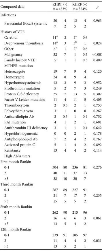
Table 2: Comparison of etiological factors and outcome according to the RHRF (-) group or the RHRF (+) group.
Compared data RHRF (-) RHRF (+) p
n = 453% n = 324%
Infections
Paracranial (focal) systemic 20 4 13 4 0.963
7 2 5 2
History of VTE
Cerebral 11a 2 2a 0.6
Deep venous thrombosis 14a 3 3b 1 0.024
Other 6a 1 2a 0.6
Malignancy 32 7 1 0.3 <0.001
Family history VTE 5 1 1 0.3 0.409
MTHFR mutation Heterozygote 19 7 9 4 0.120 Homozygote 24 8 9 4 Hyperhomocysteinemia 12 3 9 3 0.952 Prothrombin mutation 5 2 7 3 0.249 Protein C/S deficiency 25 7 13 5 0.302 Factor V Leiden mutation 11 4 11 5 0.405
Thrombocytosis 2 0.5 2 1 0.753
Polycythemia vera 3 1 0 0 0.267
Anticardiolipin Ab 2 0.5 1 0.4 0.752
PAI mutation 4 1 2 1 0.681
Antithrombin III deficiency 3 1 1 0.4 0.642
Hyperfibrinogenemia 0 0 2 1 0.178
Antiphospholipid Ab 7 2 4 2 0.767
Activated protein C 5 1 4 2 0.892
Resistance 13 4 4 2 0.114
High ANA titers First month Rankin
0-1 304 80 236 81 0.276
2 40 11 37 13
>3 38 10 20 7
Third month Rankin
0-1 287 89 227 91
2 21 7 17 7 0.235
>3 15 5 5 2
Sixth month Rankin
0-1 262 90 215 96 2 16 6 6 3 0.061 >3 13 5 4 2 12th month Rankin 0-1 239 91 185 97 2 11 4 4 2 0.031 >3 13 5 2 1
ANA: antinuclear antibody; MTHFR: methylenetetrahydrofolate reductase; PAI: plasminogen activator inhibitor; VTE: venous thromboembolism.
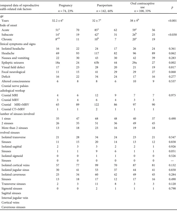
Table 3: Comparison of demographic and clinical characteristics of subgroups according to reproductive health-related risks. Compared data of reproductive
health-related risk factors
Pregnancy Puerperium Oral contraceptive
use p n = 74, 23% n = 142, 44% n = 108, 33% Age Years 32:2 ± 6a 32 ± 7a 38 ± 9b <0.001 Mode of onset Acute 51a 70 85a 62 59a 56 Subacute 14a 19 42a 31 26b 25 <0.030 Chronic 8a,b 11 10b 7 20a 19
Clinical symptoms and signs
Isolated headache 16 22 24 17 26 24 0.361
Headache 69 93 117 82 96 89 0.062
Nausea and vomiting 22 30 43 30 42 39 0.283
Epileptic seizures 18a 24 63b 44 29a 27 0.002
Visualfield defect 17 23 28 20 21 19 0.817
Focal neurological 11 15 41 29 29 27 0.068
Deficit 16 22 34 24 17 16 0.277
Altered consciousness 6 8 8 6 10 9 0.537
Cranial nerve palsies Radiological workup
Cranial MRI 4 6 12 9 7 7 0.975
Cranial MRV 3 4 6 4 3 3
Cranial MRI+MRV 65 89 122 86 97 90
Cranial CT+MRV 1 1 2 1 1 1
Number of sinuses involved
1 sinus 35 47 68 48 40 37 0.490
2 sinuses 26 35 51 36 49 45
More than 2 sinuses 13 18 23 16 19 18
Involved sinuses Isolated transverse 21 28 34 24 23 21 0.547 Sinuses 11 15 20 14 13 12 0.838 Isolated sagittal 2 3 3 2 2 1 0.926 Sinuses 1 1 9 6 1 1 0.051 Isolated sigmoid 0 0 1 1 0 0 0.526 Sinuses 0 0 0 0 0 0 —
Isolated cortical veins 57 77 99 70 87 81 0.132
Isolated jugular sinus 30 41 53 37 44 41 0.830
Isolated cavernous 25 34 60 42 49 45 0.284
Sinuses 13 18 17 12 17 16 0.490
Transverse sinuses 2 3 11 8 3 3 0.120
Sigmoid sinuses 0 0 2 1 1 1 0.798
Sagittal sinuses Internal jugular vein Cortical veins Cavernous sinuses
(systemic or paracranial infection
—otitis media, mastoiditis,
or sinusitis), systemic in
flammatory diseases, rheumatologic
or connective tissue disease, malignancies, and hematologic
diseases; and other specified causes were recorded.
The type of onset was considered to be acute if the
dura-tion of symptoms was less than 48 hours on admission,
subacute if the duration was between 48 hours and 1 month,
and chronic if the symptom duration was longer than 1 month.
The study was approved by the ethics committee of the
coordi-nating center (Acceptance No. 83045809/604/02-12333).
3. Results
In this study, 58% (
n = 453) of the total 777 female cases were
classified as RHRF (-) and 42% (n = 324) of them as RHRF
(+). The mean ages of the RHRF (+) group and the RHRF
(-) group were
34 ± 9 and 43:2 ± 13, respectively, and were
significantly different.
Acute onset is more frequent in the RHRF (+) group,
whereas a subacute chronic mode of onset is more common
in the RHRF (-) group. The most common symptoms were
headache, visual
field defects, and cranial neuropathies in
the RHRF (-) group and headache and epileptic seizure in
the RHRF (+) group. The comparison of these two groups
according to clinical symptoms and signs: epileptic seizures
(34%), nausea and vomiting (33%), and focal neurologic
def-icit (25%), was more common in the RHRF (+) group and
visual
field defect (29%) and cranial nerve palsies (13%) were
more common in the RHRF (-) group.
In the total female group investigations, CVST was
diagnosed with cranial MRI and MRV in 682 patients, with
cranial MRI in 46 patients, with only cranial MRV in 31
patients, and with cranial CT and MRV in 14 patients.
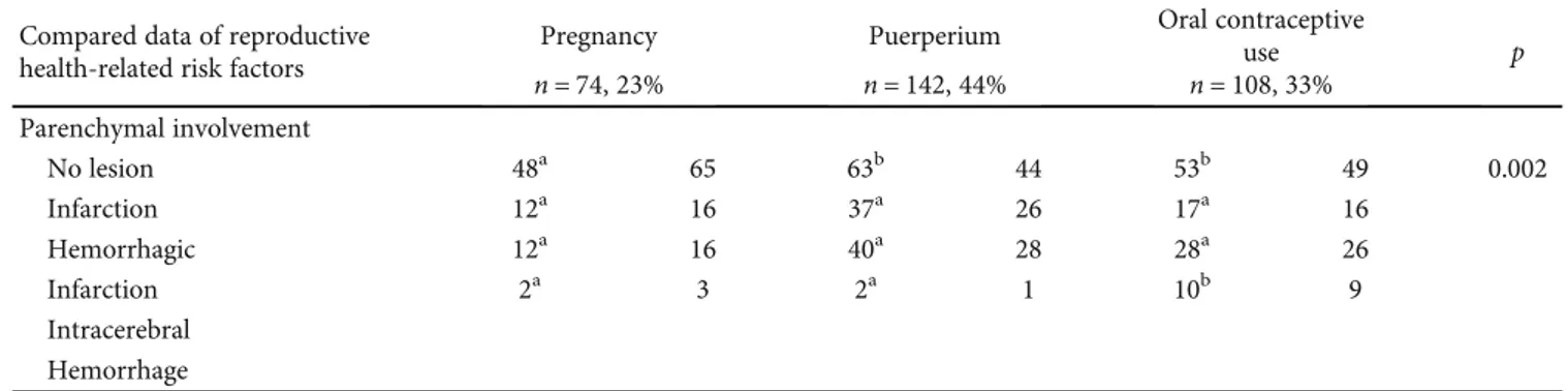
Paren-chymal lesions were detected in 333 (42.8%) female patients
including 160 (49.3%) in the RHRF (+) group and 173
(38.1%) in the RHRF (-) group (
p = 0:003). Parenchimal
lesion involvement, especially hemorrhagic transformation
(
n = 80, 25%), was more common in the RHRF (+) group.
Venous involvement was found in 1 sinus in 373 (48%)
female patients, in 2 sinuses in 274 (35%) patients, and in
more than 2 sinuses in 130 (17%) patients. In the comparison
of these two groups, there was no di
fference in intravenous
involvement. Transverse sinus involvement was the most
common site thrombosis within the total female group
(
n = 572, 73%), within the RHRF (+) group (n = 243, 75%),
and within the RHRF (-) group (
n = 329, 73%). The sigmoid
sinus and sagittal sinus involvements were followed by
trans-verse sinus in two groups.
Demographic aspects and comparative data of cases with
RHRF (+) and RHRF (-) are displayed in Table 1.
A positive previous history of venous thromboembolism
and malignancy was detected in 6% and 7% in the RHRF (-)
group. Hematological parameters were completed in 206
(26.5%) patients, and no differences were detected between
the two groups. When the RHRF (+) group was investigated,
it was found to be the largest group in the puerperium period
(43.8%) but the smallest group in the pregnancy period
(22.8%). Rankin scores, which suggested neurological
dis-ability after 12 months, were found signi
ficantly high in the
RHRF (-) group. Etiological factors and outcome according
to groups are presented in Table 2.
The mean age of OC users was higher than other groups.
The mode of acute clinical onset was high in all subgroups of
RHRF (+) cases. In addition, chronic onset and intracerebral
hemorrhage ratio were found more frequently in the OC user
group than in the other subgroups. Epileptic seizures were
found to be signi
ficantly higher in the puerperium group.
Demographic and clinical characteristics of subgroup
analy-ses are shown in Table 3.
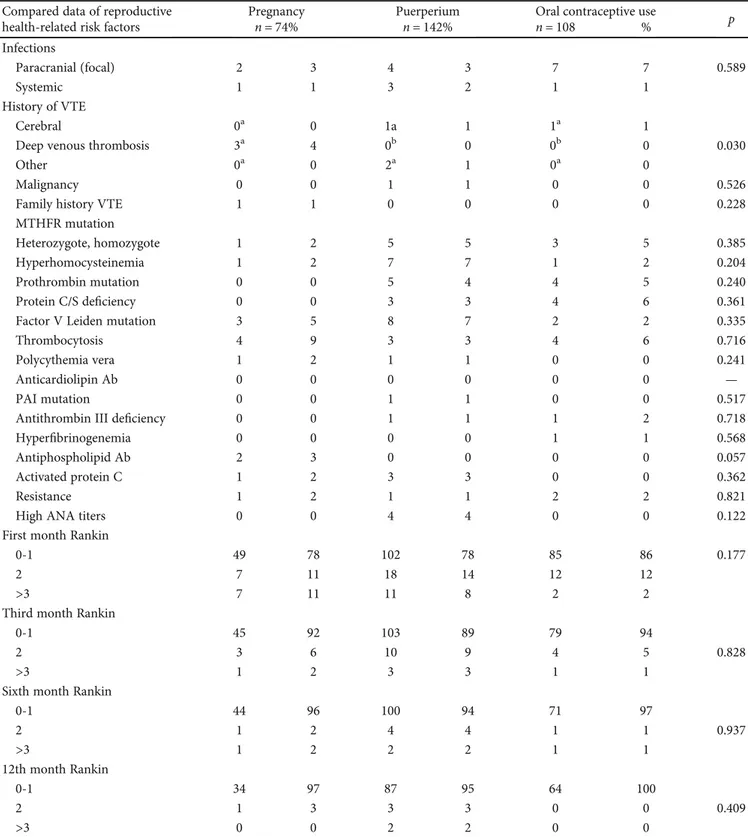
A history of deep venous thrombosis ratio was found
high in the pregnancy group. Hematologic and genetic tests
and Ranking scales were similar among the groups. A
com-parison of etiological factors and outcomes of subgroups is
seen in Table 4.
4. Discussion
Pregnancy, puerperium, and hormone replacement
treat-ment increase the tendency to cerebral venous sinus
throm-bosis (CVST) in women. CVST is much more frequently
seen in women than in men -a ratio of 3/1 [6]. In the study
of Coutinho et al., female ratio was found to be 75% and
female gender-specific risk factors at 65% [4]. In the
Interna-tional Study on Cerebral Venous and Dural Sinus
Thrombo-sis (ISCVST), the female ratio was found to be 75% of
patients. Gender-speci
fic risk factors such as OCs,
preg-nancy, puerperium, and hormone replacement therapy were
responsible [7]. The results of meta-analyses showed that
Table 3: Continued. Compared data of reproductive
health-related risk factors
Pregnancy Puerperium Oral contraceptive
use p n = 74, 23% n = 142, 44% n = 108, 33% Parenchymal involvement No lesion 48a 65 63b 44 53b 49 0.002 Infarction 12a 16 37a 26 17a 16 Hemorrhagic 12a 16 40a 28 28a 26 Infarction 2a 3 2a 1 10b 9 Intracerebral Hemorrhage
gender-specific risk factors were only not effective in children
and the elderly female groups and that the use of OCs
increased venous thrombosis development in reproductive
age females [8]. In our study, the female ratio was found to
be 68% and gender-speci
fic risks which were grouped as
RHRF (+) by us were found in 41% of women. Our
findings
are similar to the results of previous studies.
In our study, the mean age of women with reproductive
health-related risk factors (RHRF) was lower than that of
the RHRF (-) group. In subgroup analyses of RHRF (+) cases,
Table 4: Etiological factors and outcome of subgroups according to reproductive health-related risks. Compared data of reproductive
health-related risk factors
Pregnancy Puerperium Oral contraceptive use p
n = 74% n = 142% n = 108 % Infections Paracranial (focal) 2 3 4 3 7 7 0.589 Systemic 1 1 3 2 1 1 History of VTE Cerebral 0a 0 1a 1 1a 1
Deep venous thrombosis 3a 4 0b 0 0b 0 0.030
Other 0a 0 2a 1 0a 0
Malignancy 0 0 1 1 0 0 0.526
Family history VTE 1 1 0 0 0 0 0.228
MTHFR mutation
Heterozygote, homozygote 1 2 5 5 3 5 0.385
Hyperhomocysteinemia 1 2 7 7 1 2 0.204
Prothrombin mutation 0 0 5 4 4 5 0.240
Protein C/S deficiency 0 0 3 3 4 6 0.361
Factor V Leiden mutation 3 5 8 7 2 2 0.335
Thrombocytosis 4 9 3 3 4 6 0.716
Polycythemia vera 1 2 1 1 0 0 0.241
Anticardiolipin Ab 0 0 0 0 0 0 —
PAI mutation 0 0 1 1 0 0 0.517
Antithrombin III deficiency 0 0 1 1 1 2 0.718
Hyperfibrinogenemia 0 0 0 0 1 1 0.568
Antiphospholipid Ab 2 3 0 0 0 0 0.057
Activated protein C 1 2 3 3 0 0 0.362
Resistance 1 2 1 1 2 2 0.821
High ANA titers 0 0 4 4 0 0 0.122
First month Rankin
0-1 49 78 102 78 85 86 0.177
2 7 11 18 14 12 12
>3 7 11 11 8 2 2
Third month Rankin
0-1 45 92 103 89 79 94
2 3 6 10 9 4 5 0.828
>3 1 2 3 3 1 1
Sixth month Rankin
0-1 44 96 100 94 71 97 2 1 2 4 4 1 1 0.937 >3 1 2 2 2 1 1 12th month Rankin 0-1 34 97 87 95 64 100 2 1 3 3 3 0 0 0.409 >3 0 0 2 2 0 0
the mean age of OC users was higher than that of the other
groups. This difference may be related to planning of the
age of pregnancy [3].
Previous venous thrombosis history, thrombophilia,
cer-tain medical comorbidities, obesity, smoking, and
postpar-tum hemorrhage increase the risk of CVST [9, 10]. In our
study, the highest part of the RHRF (+) group consisted of
cases of puerperium. Puerperium often occurs in the sixth
to eighth week after delivery. In different population-based
case-control studies on venous thrombosis, it was explained
that risk increased 5-fold in the pregnancy period and a
60-fold in the puerperium [11]. Also, it has been reported that
it occurs more commonly after a cesarean birth than a
vagi-nal birth [12]. Infection, high matervagi-nal age and excessive
vomiting during pregnancy increase the development of
CVST [13]. All of the hormone levels, cardiovascular system,
and pregnancy-related hematologic changes return to the
baseline state within the slow process of puerperium. Human
chorionic gonadotropin (hCG) and sex steroids are at low
levels for the
first 2-3 weeks. These changes may cause the
tendency to thrombosis [14–16].
In our study, headache was the most frequent symptom
for all subgroups. However, epileptic seizures were higher
in the puerperium group. In the study of Kashkoush et al.,
the highest frequencies of symptom were found to be
head-ache (74%), seizure (50%), and an altered consciousness
(45%) in puerperium [17].
The inherited mutations in anticoagulant or
thrombo-lytic factors genes (the Factor V Leiden, the prothrombin
Factor II) and mutations in genes coding for proteins C and
S may increase the risk of developing venous thrombosis
[18]. We did not
find any relationship between inherited risk
factors and CVST.
Venous thrombosis risk increases with OC use.
Com-bined OCs containing estrogen and progesterone have higher
risk [19]. When the patient has a history of previous CVST,
the recurrence risk is increased by OC use [20, 21]. We did
not determine the content of the OCs. In the RHRF (-) group,
a previous history of CVST was high. In the subgroup
analy-sis, a previous history of CVST was high in the
“pregnancy
group.” Very little is known about the relapse rate during
pregnancy and puerperium in women with a history of CVST
[22]. The results of our study suggest that physicians must
keep in mind the possible recurrence of CVST in pregnancy.
In our study, malignancy was more frequent in the RHRF
(-) group. It has been reported that cancer patients have an
increased risk of tendency of venous thrombosis [23].
In the study by Lee et al., it has been reported that the
transverse sinus is involved in the majority of cases (75.6%).
Sigmoid sinus and superior sagittal sinus involvement
followed it at ratios of 58.5% and 29.3%, respectively [24]. In
our study, the
“transverse sinus” was affected more than other
venous sinuses. On the other hand, isolated cavernous sinus
involvement was significantly high in RHRF (-) group.
Cav-ernous sinus involvement is high in the presence of septicemia
and malignancy [25]. Therefore, in our study, this result was
expected to be more in the RHRF (-) group.
It has been reported that the prognosis in pregnant
patients is better than in nonpregnant patients with CVST
if they receive timely treatment [26]. In the VENOST main
study, the prognosis of CVST was found to be better in
women than men [1]. In our subgroup analysis, the
progno-sis was found to be worse in the RHRF (-) group.
5. Conclusions
Our results indicate that when CVST was detected in women
with RHFR (-), the existence of malignancy should be
inves-tigated. The previous history of CVST may be related to
recurrence in pregnancy. Clinical onset may present with
chronic headache in CSVT cases related to OC use. Epileptic
seizures may be a more frequent symptom in puerperal
CSVT cases. Physicians must keep these situations in mind.
Data Availability
All data will be available on request.
Conflicts of Interest
The authors declared that they have no con
flicts of interest
for this article.
Authors’ Contributions
Sevki Sahin and Taskin Duman have contributed equally.
References
[1] T. Duman, D. Uluduz, I. Midi et al.,“A Multicenter Study of 1144 Patients with Cerebral Venous Thrombosis: The VENOST Study,” Journal of Stroke and Cerebrovascular Dis-eases, vol. 26, no. 8, pp. 1848–1857, 2017.
[2] A. Ameri and M.-G. Bousser,“Cerebral venous thrombosis,” Neurologic Clinics, vol. 10, no. 1, pp. 87–111, 1992.
[3] M. Galarza and R. Gazzeri,“Cerebral venous sinus thrombosis associated with oral contraceptives: the case for neurosurgery,” Neurosurgical Focus, vol. 27, no. 5, article E5, 2009.
[4] J. M. Coutinho, J. M. Ferro, P. Canhão et al.,“Cerebral venous and sinus thrombosis in women,” Stroke, vol. 40, no. 7, pp. 2356–2361, 2009.
[5] C. Cantu and F. Barinagarrementeria, “Cerebral venous thrombosis associated with pregnancy and puerperium. Review of 67 cases,” Stroke, vol. 24, no. 12, pp. 1880–1884, 1993.
[6] J. Stam,“Thrombosis of the Cerebral Veins and Sinuses,” The New England Journal of Medicine, vol. 352, no. 17, pp. 1791– 1798, 2005.
[7] J. M. Ferro, P. Canhao, J. Stam, M. G. Bousser, F. Barinagarrementeria, and ISCVT Investigators,“Prognosis of cerebral vein and dural sinus thrombosis: results of the international study on cerebral vein and dural sinus thrombo-sis (ISCVST),” Stroke, vol. 35, no. 3, pp. 664–670, 2004. [8] J. M. Ferro, P. Canhao, M. G. Bousser, J. Stam, and
F. Barinagarrementeria,“Cerebral vein and dural sinus throm-bosis in elderly patients,” Stroke, vol. 36, no. 9, pp. 1927–1932, 2005.
[9] J. A. Heit, “Epidemiology of venous thromboembolism,” Nature Reviews Cardiology, vol. 12, no. 8, pp. 464–474, 2015.
[10] E. Jackson, K. M. Curtis, and M. E. Gaffield, “Risk of venous thromboembolism during the postpartum period,” Obstetrics and Gynecology, vol. 117, no. 3, pp. 691–703, 2011.
[11] E. R. Pomp, A. M. Lenselink, F. R. Rosendaal, and C. J. M. Doggen,“Pregnancy, the postpartum period and prothrombo-tic defects: risk of venous thrombosis in the MEGA study,” Journal of Thrombosis and Haemostasis, vol. 6, no. 4, pp. 632–637, 2008.
[12] C. Jaigobin and F. L. Silver,“Stroke and pregnancy,” Stroke, vol. 31, no. 12, pp. 2948–2951, 2000.
[13] D. J. Lanska and R. J. Kryscio,“Risk factors for peripartum and postpartum stroke and intracranial venous thrombosis,” Stroke, vol. 31, no. 6, pp. 1274–1282, 2000.
[14] C. L. Dennis, K. Fung, S. Grigoriadis, G. E. Robinson, S. Romans, and L. Ross, “Traditional postpartum practices and rituals: a qualitative systematic review,” Womens Health, vol. 3, no. 4, pp. 487–502, 2016.
[15] N. K. Tepper, S. L. Boulet, M. K. Whiteman et al.,“Postpartum venous thromboembolism: incidence and risk factors,” Obstet-rics and Gynecology, vol. 123, no. 5, pp. 987–996, 2014. [16] J. A. Heit, C. E. Kobbervig, A. H. James, T. M. Petterson,
K. R. Bailey, and L. J. Melton III,“Trends in the incidence of venous thromboembolism during pregnancy or postpartum: a 30-year population-based study,” Annals of Internal Medi-cine, vol. 143, no. 10, pp. 697–706, 2005.
[17] A. I. Kashkoush, H. Ma, N. Agarwal et al.,“Cerebral venous sinus thrombosis in pregnancy and puerperium: a pooled, systematic review,” Journal of Clinical Neuroscience, vol. 39, pp. 9–15, 2017. [18] G. J. Hankey, J. W. Eikelboom, F. M. van Bockxmeer, E. Lofthouse, N. Staples, and R. I. Baker,“Inherited thrombo-philia in ischemic stroke and its pathogenic subtypes,” Stroke, vol. 32, no. 8, pp. 1793–1799, 2001.
[19] W. H. W. Inman, M. P. Vessey, B. Westerholm, and A. Engelund,“Thromboembolic disease and the steroidal con-tent of Oral Contraceptives. A report to the Committee on Safety of Drugs,” BMJ, vol. 2, no. 5703, pp. 203–209, 1970. [20] C. Kolacki and V. Rocco,“The combined vaginal
Contracep-tive Ring, NuvaRing, and cerebral venous sinus thrombosis: a case report and review of the literature,” The Journal of Emer-gency Medicine, vol. 42, no. 4, pp. 413–416, 2012.
[21] A. McDaid, E. Logette, V. Buchillier et al.,“Risk prediction of developing venous thrombosis in combined oral contraceptive users,” PLOS ONE, vol. 12, no. 7, p. e0182041, 2017.
[22] S. Mehraein, H. Ortwein, M. Busch, M. Weih, K. Einhäupl, and F. Masuhr,“Risk of recurrence of cerebral venous and sinus thrombosis during subsequent pregnancy and puerperium,” Journal of Neurology, Neurosurgery, and Psychiatry, vol. 74, no. 6, pp. 814–816, 2003.
[23] J. W. Blom, J. P. M. Vanderschoot, M. J. Oostindier, S. Osanto, F. J. M. van der Meer, and F. R. Rosendaal,“Incidence of venous thrombosis in a large cohort of 66 329 cancer patients: results of a record linkage study,” Journal of Thrombosis and Haemostasis, vol. 4, no. 3, pp. 529–535, 2006.
[24] D. J. Lee, A. Ahmadpour, T. Binyamin, B. C. Dahlin, K. Shahlaie, and B. Waldau,“Management and outcome of spontaneous cerebral venous sinus thrombosis in a 5-year con-secutive single-institution cohort,” Journal of NeuroInterven-tional Surgery, vol. 9, no. 1, pp. 34–38, 2016.
[25] F. S. Southwick, E. P. Richardoson JR., and M. N. Swartz, “Septic thrombosis of the dural venous sinuses,” Medicine, vol. 65, no. 2, pp. 82–106, 1986.
[26] A. Razmara, K. Bakhadirov, A. Batra, and S. K. Feske, “Cere-brovascular complications of pregnancy and the postpartum period,” Current Cardiology Reports, vol. 16, no. 10, p. 532, 2014.