Introduction
Members of the phylum Myxozoa Grasse, 1970 mainly infect fish and about 1200 species of myxosporeans have been described so far (Lom and Dykova, 1992). Only seven species of myxosporean parasites, including Sphaerospora elegans Thelohan, 1892 and Myxobilatus gasterostei Davis, 1944, have been recorded from the three-spined stickleback worldwide (Bykovskaya-Pavlovskaya et al., 1964; Chappell, 1969; Lester, 1974; Wootton, 1976; Feist et al., 1991). The genera Sphaerospora and Myxobilatus are generally coelozoic parasites commonly found in the lumen of the kidney tubules or in the urinary bladder of freshwater and marine
fish, including Gasterosteus aculeatus L., 1758. Forty-eight species of Sphaerospora and 24 species of Myxobilatus are known, mostly from freshwater environments. Members of both genera show a strict host specificity as well as site specificity within the host (Arthur and Lom, 1985; El-Matbouli and Hoffmann, 1992). While Sphaerospora produces mono and disporous trophozoites which form one or two spores, Myxobilatus produces monosporous to polysporous trophozoites forming one or more spores in a pseudoplasmodium. Two developmental stages, extrasporogonic and sporogonic, have been shown to occur in S. elegans (Hedrick et al., 1988; Lom et al., 1991; Sultana, 1994).
Sphaerospora elegans Thelohan, 1892 and Myxobilatus gasterostei
Davis, 1944 (Phylum: Myxozoa) Infections in the Three-Spined
Stickleback, Gasterosteus aculeatus L., 1758 in Turkey
Ahmet ÖZER
Ondokuzmay›s University, Fisheries Faculty, 57000 Sinop - TURKEY
Received: 27.07.2002
Abstract:Sphaerospora elegans Thelohan, 1892 and Myxobilatus gasterostei Davis, 1944 infections in the three-spined stickleback (Gasterosteus aculeatus L., 1758) were studied from January to May 2000. Infection prevalence and intensity values were determined for mixed infections to be 26.5% and 3+ (0-5) parasites per fish, respectively. These values for each species were also determined. While infection prevalence and mean intensity values for S.elegans were 11.9% and 3+ (0-5), they were 17.9% and 3+ (0-5), respectively, for M. gasterostei. The existence of both parasite species in relation to the sex and length classes of the fish were determined and the infection interactions with these factors as well as some environmental factors are discussed. S.elegans and M. gasterostei represent two new parasite records for Turkey. This is also the first study on myxosporeans performed in brackish and freshwater conditions in the same environment.
Key Words:Sphaerospora elegans, Myxobilatus gasterostei, Gasterosteus aculeatus
Türkiye’deki Dikence Bal›¤›nda (Gasterosteus aculeatus L., 1758) Görülen Sphaerospora elegans Thelohan, 1892 ve Myxobilatus gasterostei Davis, 1944
(Phylum: Myxozoa) Enfeksiyonlar›
Özet: Dikence bal›klar›nda (Gasterosteus aculeatus L., 1758) bulunan Sphaerospora elegans Thelohan, 1892 ve Myxobilatus gasterostei Davis, 1944 enfeksiyonlar›, Ocak - May›s 2000 tarihleri aras›nda çal›fl›ld›. Enfeksiyon yüzdesi ve intensitesi de¤erleri her iki parazit türü beraber ele al›nd›¤›nda, s›ras›yla %26.5 ve enfekte bal›k bafl›na 3+ (0-5) parazit olarak kaydedildi. Bu de¤erler, her iki parazit türü için ayr› ayr› olarakta belirlendi. Sphaerospora elegans’›n enfeksiyon yüzdesi ve ortalama parazit say›s› s›ras›yla %11.9 ve 3+ (0-5) olarak tespit edilmiflken, bu de¤erler M. gasterostei için s›ras›yla %17.9 ve 3+ (0-5) olarak kaydedilmifltir. Her iki parazit türünün bal›klar›n cinsiyetleri ve boylar› ile iliflkileri belirlendi ve enfeksiyonlar›n bu faktörler ile beraber baz› çevresel faktörlerle olan etkileflimleri tart›fl›ld›. S.elegans ve M. gasterostei, Türkiye için iki yeni parazit türü olarak belirlendi. Bu çal›flma, ayr›ca mikzospor enfeksiyonlar› aç›s›ndan ac› su ve tatl› su karakterindeki bir ortamda yürütülen ilk araflt›rmad›r.
A knowledge of the epidemiology of myxozoan parasites is important in elucidating and understanding the two host life cycle of myxozoans, with actinosporean stages in oligochaetes, polychaetes and bryozoans and myxosporeans stages in fish hosts, of a given species (see Özer (1999) for detailed information). Following the breakthrough discovery of Markiw and Wolf (1983) that a tubificid oligochaete was an essential host in the life cycle of Myxobolus cerebralis Hofer, 1903, studies on the epidemiology of myxozoan parasites gained more attention, especially those of commercially important fish species (Wolf and Markiw, 1984, Foott and Hedrick, 1987; Sitja-Bobadilla and Alvarez-Pellitero, 1993; McGeorge, 1994; Özer and Wootten, 2000). Despite considerable information on the developmental stages of S. elegans and M. gasterostei, knowledge on the occurrence of these parasites in their host G. aculeatus is quite limited (Sultana, 1994).
In this study, infections of S. elegans and M. gasterostei in relation to the length class and sex of three-spined sticklebacks, as well as the environmental conditions, are investigated. Both S. elegans and M. gasterostei are new parasite records for Turkey. This study is also the first in which both myxosporean species were observed in brackish and freshwater conditions in the same environment.
Materials and Methods
Specimens of Gasterosteus aculeatus were collected by gill net and cast net from the Sırakırka¤açlar stream, which drains into the Black Sea at Sinop. The
Sırakırka¤açlar stream is characteristically brackish during winter and early spring (October to March) when the water level rises and the stream is connected with the Black Sea. Between late spring and early autumn, however, the water level drops, the waterway is no longer connected to the Black Sea and it becomes a freshwater stream.
Sampling was carried out on a monthly basis from January to May 2000. Following the May collection, as the water levels dropped, fish could no longer be caught as they moved upstream into deeper waters. For parasitological examination, fish were transported alive in local water directly to the Sinop Fisheries Faculty Laboratory. A total of 151 fish were examined (Table 1). Sticklebacks were weighed, their total length measured and sex determined at post-mortem. The body cavity of the fish was opened and the heart, liver, kidney, digestive tract, gall bladder, swim bladder, urinary bladder, reproductive organs and brain were dissected out and placed in separate Petri dishes in 0.8% physiological saline prior to examination.
All the organs used for the detection of myxosporeans were examined using squash preparations. At least three to four small pieces of tissue from each organ were compressed between a microscope slide and cover slip. Kidneys were examined by squash preparation or Giemsa-stained impression smears. These preparations were examined using standard light microscopy at x20 to x100 magnifications under oil immersion. The prevalence was calculated as the percentage of the total number of fish infected with parasitic stages after examination of the kidneys of all fish in a sample. Intensity of infection was
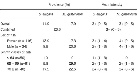
Prevalence (%) Mean Intensity
S. elegans M. gasterostei S. elegans M. gasterostei
Overall 11.9 17.9 3+ (0 - 5) 3+ (0 - 5)
Combined 26.5 3+ (0 - 5)
Sex of fish
Female (n = 116) 12.9 17.3 3+ (1 - 4) 4+ (0 - 5)
Male (n = 34) 8.9 20.5 2+ (1 - 3) 4+ (1 - 5)
Length classes of fish
≤64 (n=50) 10 0 1+ (1 - 3) 0
65 – 69 (n=61) 9.8 29.5 3+ (1 - 3) 3+ (1 - 3)
70 ≥(n=40) 17.5 22.5 2+ (0 - 4) 3+ (0 - 5)
Table 1. Infection prevalence (%) and mean intensity values in relation to the sex and length classes of G. aculeatus.
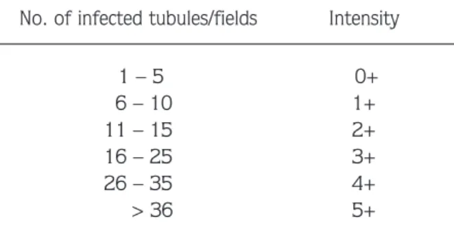
assessed from the presence of developing stages and mature spores in squash preparations. The intensity of infection was calculated semi-quantitatively and graded according to the number of infected kidney tubules per microscope field. For this purpose, squash preparations were standardised by examining the sections from the same part of the kidney. Twenty fields from each slide of the kidney of each fish were examined at x20 (two slides per fish) and the intensity of infection was categorised as follows:
No. of infected tubules/fields Intensity
1 – 5 0+ 6 – 10 1+ 11 – 15 2+ 16 – 25 3+ 26 – 35 4+ > 36 5+
Fifty and 70 mature (and developing) spores of M. gasterostei and S. elegans, respectively, from several fish specimens were measured (µm) according to the methods described by Lom and Arthur (1989). Measurement data are presented as mean ± SD, followed by ranges in parentheses.
Some water parameters measured with a digital analyser (Horiba U-10) and the number of fish sampled throughout the study period are also presented in Table 2.
The Kruskal-Wallis test (nonparametric ANOVA) was performed to find out the significant differences in the mean intensity values of both parasite species for length classes of fish as well as for the months in which this study was conducted. The difference between parasite loading on male and female sticklebacks was tested by the Mann-Whitney U-test. All the statistical tests were performed at the significance level of 5%.
Results
Myxobilatus gasterostei (Fig. 1A) and S. elegans (Fig. 1B) were the only myxosporean species identified in the kidney of G. aculeatus throughout the investigation period. The other organs examined were not infected. Pre-sporogonic and sporogonic stages of one or both species were found in individual fish.
In fresh kidney squash preparations, pre-sporogonic stages of S. elegans appeared as round- or oval bodies up to 25 µm in diameter. Mature spores, however, appeared to be spherical when viewed perpendicularly to the suture line. Monosporous and disporous plasmodia were observed. Mean spore size measured from 70 spores was 10.6 ± 0.8 µm (7.5 – 11.1) in length and 10.1 ± 0.6 µm (6.8 – 10.4) in width.
In fresh kidney squash preparations, monosporous to polysporous plasmodia of M. gasterostei were observed and pre-sporogonic stages appeared as round to elongated bodies up to 16.2 µm in diameter. Mature spores were, however, very elongated with two caudal processes and the measurements obtained from each part
Table 2. Some monthly water parameters measured and the number of G. aculeatus caught in the sampling site.
Months pH Temperature (°C) Dissolved O2(mg/l) Salinity (‰) G. aculeatus
October 7.0 17 6.3 1 0 November 7.1 11 7.1 3 0 December 6.4 6 7.1 3 0 January 6.4 4 6.8 4 16 February 6.8 4 6.6 4 39 March 6.2 8.6 6.3 3 65 April 6.9 15.4 6.1 0 20 May 7.3 24.8 6.6 0 11 June 8.1 26.4 6.0 0 0 July 8.0 30.6 5.8 0 0 August 7.6 31 5.1 0 0 September 7.6 23 6.1 0 0
were as follows: total length = 29.6 ± 0.9 µm (18.8 – 37.7), length of caudal processes = 17.2 ± 0.7 µm (10 – 25.5), length of spore body = 10.8 ± 0.08 µm (10.1 – 12.4), width of spore = 5.1 ± 0.1 µm (4.1 – 6.3), thickness of spore = 5.5 ± 0.1 µm (4.1 – 6.6), length of polar capsule= 5.7 ± 0.1 µm (4.2 – 7.1), and width of the polar capsule = 2.4 ± 0.08 µm (1.5 – 3) (based on 50 spores).
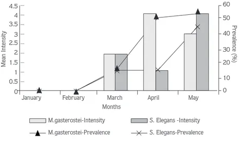
The overall infection prevalence rates of S. elegans and M. gasterostei were similar and the overall mean intensity values were the same for both parasite species (Table 1). Monthly prevalence and intensity values were also determined and are illustrated in Figure 2. Pre-sporogonic stages of both parasite species were first observed in March while sporogonic stages appeared in April. The occurrence of S. elegans and M. gasterostei in relation to the sex and length class of G. aculeatus is shown in Table 1. No statistically significant differences in the mean intensity values were determined for the loading of each parasite species between the months and length classes as well as the sex of fish.
Discussion
Sphaerospora elegans and M. gasterostei were identified in the kidney of G. aculeatus for the first time in Turkey and also represent two new parasite records for the country. The light microscopic structure of pre-sporogonic and pre-sporogonic stages of S. elegans and M. gasterostei were similar to those described previously (Davis, 1944; Feist et al., 1991; Sultana, 1994) and the spore dimensions of both parasite species obtained in this study are in the range given by other authors (Lester, 1974; Hedrick et al., 1988; Feist et al., 1991; Sultana, 1994).
Seasonal patterns of occurrence have been shown in myxosporeans, some being present in the fish all year round (Molnar, 1979), while others show a single peak in a one-year life cycle (Molnar, 1982; Molnar and Kovacs-Gayer, 1985; Baska, 1990) and some others show two A
B
Fig. 1. Hand drawings of M. gasterostei (A) and S. elegans (B). (Scale bar = 5 µm). 0 0.5 1 1.5 2 2.5 3 3.5 4 4.5
January February March April May
Months 0 10 20 30 40 50 60
M.gasterostei-Intensity S. Elegans -Intensity M.gasterostei-Prevalence S. Elegans-Prevalence Mean Intensity Prevalence (%) — — — — — — — — —
Figure 2. Monthly infection prevalence (%) and mean intensity values of M. gasterostei and S. elegans.
peaks of infectivity each year (Kovacs-Gayer and Molnar, 1983). Mixed infections of some myxosporeans, including S. elegans and M. gasterostei in the three-spined stickleback have also been recorded (Feist, 1991; Sultana, 1994).
Seasonal studies on Sphaerospora and Myxobilatus are limited (Booker and Current, 1981; Grupcheva et al., 1985; Molnar, 1988; Sitja-Bobadilla and Alvarez-Pellitero, 1993; Sultana, 1994). The combined and single prevalence values of S. elegans and M. gasterostei found in the present study were much lower than those given by Sultana (1994), possibly because fish samples could be collected only from January to May rather than all year round. Mean intensity values, however, were similar in both studies. Neither developing nor spore stages of either parasite species were observed in January and February. The developing stages of both species were seen in March and the spore stages were first observed in April. This data partially agrees with Sultana (1994) and could be explained by several factors: first, the extrasporogonic stages of S. elegans were reported in the choroidal rete mirabile of the eye, the circulating blood and in the blood sinuses of the interstitial spaces of the kidney of G. aculeatus in January and February and in June by Sultana (1994). A 15- to 35-day pre-patent period between the point at which fish become infected and at which infections become detectable in the population have also been shown in myxosporeans which have extrasporogonic stages (Kent and Hedrick, 1984; McGeorge, 1994). The extrasporogonic stages of S. elegans were not studied here. It might be that January and February represent a pre-patent period of infection of S. elegans and M. gasterostei with developing stages becoming apparent in March and spore stages in April. Alternatively, fish might have become infected in the previous year in June in freshwater conditions, followed by development and sporogony in the following months. It was shown for Myxidium salvelini that sporogony was recommenced following the transfer of infected fish to sea water and their return to fresh water (Higgins et al., 1993). McGeorge (1994) also recorded the loss of sporogonic stages and mature spores of Sphaerospora truttae in Salmo salar L., 1758 after 4 months in sea water. Due to the brackish character of the sampling site, both S. elegans and M. gasterostei may have remained dormant at developing and spore stages in the kidney. Following the return to fresh water in April, these stages
became apparent with increasing prevalence and intensity. Third, pre-sporogonic and sporogonic stages could not be observed in January and February due to the small number of fish examined or the techniques used in this study. Considering that the length of the examined fish indicated that they might be about 1 year old, it seems more likely they would have picked it up the previous summer. On the other hand, it is not known whether an extrasporogonic form survived over the saline winter conditions. Histological studies could be useful in determining exact starting date of infection and its intensity.
It has been suggested that environmental and host-related factors have important impacts on the pattern of myxosporean infections (Wolf and Markiw, 1984; Foott and Hedrick, 1987; Lom and Dykova, 1992). In general, temperature has been considered to be the major factor (Mitchell, 1977). While some myxosporeans, such as Myxobolus dujardini, have maximum infection during cold months (Mitchell, 1977), others, such as Ceratomyxa shasta, show generally higher infection frequencies in summer (Udey et al., 1975). It was also suggested that low temperatures inhibit proliferative kidney disease infections (Ferguson, 1981). Sultana (1994) determined seasonal infection patterns of S. elegans and M. gasterostei and noted that their prevalence was lowest in spring and summer in a freshwater lake. The results obtained in the present study also show low level infections. It is not yet known whether S. elegans and M. gasterostei have actinosporean stages in alternate hosts in their life cycle. If one can presume that it is so, low demand by fish for consumption of alternate hosts or low number presence of these hosts may be responsible for the lower infection prevalences of both parasite species.
Studies on the effects of host sex on myxosporean infection are quite rare and the data available show considerable variations (Janovy and Hardin, 1987; Alvarez-Pellitero and Sitja-Bobadilla, 1993; Sultana, 1994). Both infection prevalence and mean intensity values of S. elegans and M. gasterostei in male and female G. aculeatus were similar in the present study. This data partly agrees with Sultana (1994), although she noted a significantly higher prevalence of M. gasterostei in female fish. However, it was noted by Alvarez-Pellitero and Sitja-Bobadilla (1993) that in most cases there was no correlation between myxosporean infections and host sex.
Some of the major influences on infection appear to be the size and age of fish. Some reports show a higher prevalence and intensity of myxosporeans as a result of the increasing probability of the fish encountering infective stages over time (Molnar and Kovacs-Gayer, 1985; Sitja-Bobadilla and Alvarez-Pellitero, 1993). A number of myxosporean species, however, appear to infect very young hosts in their first year (Mitchell, 1977; Molnar, 1982; Molnar and Kovacs-Gayer, 1985; Ferguson, 1987; McGeorge, 1994). Infection with M.
gasterostei was not determined in the smallest length class of G. aculeatus, but it was present in larger length classes of fish. S. elegans, however, was present in all length classes. Similar results for both parasite species were also recorded by Sultana (1994).
Acknowledgement
This study was financed by Ondokuzmayıs Üniversity under project number S-076.
References
Alvarez-Pellitero, P. and Sitja-Bobadilla, A.S. 1993. Ceratomyxa spp. (Protozoa: Myxosporea) infections in wild and cultured sea bass Dicentrarchus labrax from the Spanish Mediterranean area. J. Fish Biol. 42: 889-901.
Arthur, J.R. and Lom, J. 1985. Sphaerospora araii n. sp. (Myxosporea: Sphaerosporidae) from the kidney of a long nose skate (Raja rhina, Jordan and Gilbert) from the Pacific Ocean of Canada. Can. J. Zool. 63: 2902 – 2906.
Baska, F. 1990. Chloromyxum inexpectatum n. sp. and Sphaerospora colomani (Myxozoa: Myxosporea), parasites of the urinary system of the sterlet, Acipencer ruthenus. Syst. Parasitol. 16: 185 – 193. Booker, O.J. and Current, W.L. 1981. Myxobilatus mictospora (Kudo, 1920) (Myxozoa: Myxosporea) in the largemouth bass Micropterus salmoides Lacepe: plasmodium morphology and fine structure. J. Parasitol. 67: 859 – 865.
Bykovskaya-Pavlovskaya, I.E., Gusev, A.V., Dubinina, N.A., Izyumova, N.A., Smirnova, T.S., Sokolovskaya, I.L., Shtein, G.A., Shulman, S.S. and Epshtein, V.M. 1964. Key to Parasites of Freshwater fish of the USSR. Israel Program for Scientific Translations, Jerusalem. Chappell, L.H. 1969. The parasites of the three-spined stickleback Gasterosteus aculeatus L., from a Yorkshire pond. 1. Seasonal variation of parasite fauna. J. Fish Biol. 1: 137 – 152.
Davis, H.S. 1944. A revision of the genus Henneguya (Myxosporidia) with description of two new species. Trans. American Micr. Soc. 63: 311 – 320.
El-Matbouli, M. and Hoffmann, R. W. 1992. Sphaerospora scardinii n.sp. (Myxosporea: Sphaerosporidae) observed in the kidney of rudd, Scardinius erythropthalmus. Dis. Aquat. Org. 14: 23 – 29. Feist, S.W., Chilmonczyk, S. and Pike, A.W. 1991. Structure and
development of Sphaerospora elegans Thelohan, 1892 (Myxozoa: Myxosporea) in the sticklebacks Gasterosteus aculeatus L. and Pungitius pungitius L. (Gasterosteidae). J. Fish Dis. 4: 175 –177. Ferguson, H.W. 1981. The effects of water temperature on the
development of proliferative kidney disease in rainbow trout, Salmo gairdneri Richardson. J. Fish Dis. 4: 175 – 177.
Foott, J.S. and Hedrick, R.P. 1987. Seasonal occurrence of the infectious stage of proliferative kidney disease (PKD) and resistance of rainbow trout, Salmo gairdneri Richardson, to reinfection. J. Fish Biol. 30: 477 – 484.
Grupcheva, G., Dykova, I. and Lom, J. 1985. Seasonal fluctuation in the prevalence of Sphaerospora renicola and myxosporean blood stream stages in carp fingerlings in Bulgaria. Folia Parasitol. 32: 193 – 203.
Hedrick, R.P., Kent, M.L., Toth, R.J. and Morrison, J.K. 1988. Fish infected with Sphaerospora spp. Thelohan (Myxosporea) from fish in water enzootic for proliferative kidney disease of salmonids. J. Protozool. 35: 13 – 18.
Higgins, M.J., Margolis, L. and Kent, M.L. 1993. Arrested development in a freshwater myxosporeans Myxidium salvelini, following transfer of its host, the sockeye salmon (Onchorhynchus nerka), to sea water. J. Parasitol. 79: 403 – 407.
Janovy, J and Hardin, E. 1987. Population dynamics of the parasites in Fundulus zebrinus in the Platte River of Nebraska. J. Parasitol. 73: 689 – 696.
Kent, M.L. and Hedrick R.P. 1985. PKX, the causative agent of proliferative kidney disease (PKD) in Pacific salmonid fishes and its affinities with the Myxozoa. J. Protozool. 32(2): 257-260. Kovacs-Gayer, E. and Molnar, K. 1983. Studies on the biology and
pathology of the common carp parasite Myxobolus basilamellaris Lom et Molnar, 1983 (Myxozoa: Myxosporea). Acta Vet. Hung. 31: 91 – 102.
Lester, R.J.G. 1974. Parasites of Gasterosteus aculeatus near Vancouver British Columbia. Sysis 7: 195 – 200.
Lom, J. and Arthur, J.R. 1989. A guideline for the preparation of species description in Myxosporea. J. Fish Dis. 12: 151 – 156. Lom, J. and Dykova, I. 1992. Protozoan Parasites of Fishes.
Development in Aquaculture and Freshwater Science. Volume 26. Elsevier Science Publishers, Amsterdam.
Lom. J., Pike, A.W. and Feist. S.W. 1991. Myxosporean vegetative stages in the choroidal rete mirabile of Gasterosteus aculeatus infected with Myxobilatus gasterostei and Sphaerospora elegans. Dis. Aquat. Org. 11: 67 – 72.
Markiw, M. E. and Wolf, K. 1983. Myxosoma cerebralis (Myxozoa: Myxosporea) etiological agent of salmonid whirling disease requires tubificid worm (Annelida: Oligochaete) in its life cycle. J. Protozool. 30: 561-564.
McGeorge, J. 1994. Studies on the biology of Sphaerospora sp. (Myxozoa: Myxosporea) from farmed Atlantic salmon, Salmo salar L., in Scotland. PhD Thesis. University of Stirling, 353 pp. Mitchell, L.G. 1977. Myxosporidia. In: Parasitic Protozoa, Vol. 4. (Ed.
Kreieer, J.P). Academic Press, New York.
Molnar, K. 1979. Myxobolus pavlovskii (Achmerov, 1954) (Myxosporida) infection in the silver carp and bighead. Acta Vet. Hung. 27: 207 – 216.
Molnar, K. 1982. Biology and histopathology of Thelohanellus nikolskii Achmerov, 1955 (Myxosporea: Myxozoa), a protozoan parasite of common carp (Cyprinus carpio). Parasitol. Res. 1982: 269 – 277. Molnar, K. 1988. Development of Myxobilatus legeri in cyprinid fishes.
Dis. Aquat. Org. 4: 181 – 187.
Molnar, K. and Kovacs-Gayer, E. 1985. The pathogenicity and development within the host fish of Myxobolus cyprini Doflein, 1898. Parasitol. 90: 549 – 555.
Özer, A. 1999. Studies on actinosporeans (Phylum: Myxozoa) from a salmon farm in Northern Scotland, with special reference to the actinosporean and myxosporean stages of Sphaerospora truttae Fischer-Scherl, El-Matbouli and Hoffmann, 1986. PhD Thesis, University of Stirling, 333 pp.
Özer, A. and Wootten., R. 2000. The life cycle ofSphaerospora truttae (Myxozoa: Myxosporea) and some features of the biology of both the actinosporean and myxosporean stages. Dis. Aquat. Org. 40: 33-39.
Sitja-Bobadilla, A. S. and Alvarez-Pellitero, P. 1993. Population dynamics of Sphaerospora dicentrarchi Sitja-Bobadilla et Pellitero, 1992 and S. testicularis Sitja-Bobadilla and Alvarez-Pellitero, 1990 (Myxosporea: Bivalvulda) infections in wild and cultured Mediterranean sea bass (Dicentrarchus labrax L.). Parasitol. 106: 39 – 45.
Sultana, Q. 1994. Studies on the biology of some parasites of the three-spined stickleback, Gasterosteus aculeatus L. with special reference to the myxosporea. PhD Thesis. University of Stirling. 287 pp.
Udey, L.R., Fryer, J.L. and Pilcher, K.S. 1975. Relation of water temperature to ceratomyxosis in rainbow trout (Salmo gairdneri) and coho salmon (Oncorhynchus kisutch). J. Fish. Res. Board of Can. 32: 1545 – 1551.
Wolf, K. and Markiw, M. E. 1984. Biology contravenes taxonomy in the Myxozoa: new discoveries show alternation of invertebrate and vertebrete hosts. Science, 225: 1449-1459.
Wootton, R.J. 1976. The biology of the sticklebacks. Academic Press. London.