Original Article
Kuwait Medical Journal 2020; 52 (1): 43 - 49
Expressions of p53, KAI1, PTEN and their use as prognostic
markers in urothelial carcinoma of the bladder
ABSTRACT
KEYWORDS: KAI1, PTEN, p53, urinary bladder, urothelial carcinoma
Ozgur Ilhan Celik1, Serkan Yasar Celik1, Unsal Han2, Binnur Onal3
1Department of Pathology, Mugla Sitki Kocman University, Faculty of Medicine, Mugla, Turkey
2Department of Pathology, Ankara Diskapi Yildirim Beyazit Education and Research Hospital, Ankara, Turkey
3 Department of Pathology and Cytology, Düzce University, Faculty of Medicine, Düzce, Turkey
Objectives: To determine the expression levels of p53,
KAI1, PTEN and their relations with the clinicopathologic parameters in patients with urothelial carcinoma of the bladder
Design: Retrospective study
Setting: Department of Pathology, Ankara Diskapi Yildirim
Beyazit Education and Research Hospital, Ankara, Turkey
Subjects: Seventy-eight patients with a diagnosis of
urothelial carcinoma of the bladder at the Pathology Department of Ankara Diskapi Yildirim Beyazit Education and Research Hospital and who were followed up for two years
Intervention: Archived pathology materials of all patients
were reviewed and clinical data were collected from medical database records retrospectively.
Main outcome measures: We considered the following
parameters: age, gender, survivals and disease-free survivals of the patients, tumor-stage, lymphovascular-angiovascular invasions, expressions of KAI1, PTEN and p53 in tumorous tissues.
Results: In non-metastatic-patients, 55.2% were p53
negative; however, 45.8% of the metastatic-patients had diffuse-strong p53-expressions. Hence, p53 positivity indicates that the tumor will metastase.
In transurethral resection (TUR) materials, patients with positive KAI1-expression had better survival (p=0.0285). The tumor seemed to be more aggressive and invasive in patients with decreased KAI1-expressions because there was no KAI1 expression in 50% of the metastatic-patients, although it wasn’t statistically significant (p=0.550). For PTEN expression, no statistically-significant relation was found with the tumor-stage (p=0.34) and survival/ disease-free-survival (TUR:p=0.9599/0.7576, radical
cystectomy:p=0.8219/0.5790).
Conclusions: The presence of metastasis dramatically
decreases the overall survival in patients with urothelial carcinoma. Identification of these patients as early as possible would effect their therapies, follow-up intervals and surveys. p53 and KAI1 seem to be especially better indicators of the prognosis than PTEN.
INTRODUCTION
Urothelial malignancy of the bladder is the fifth most common malignancy occurring worldwide. In the genitourinary tract, it is the second most common malignancy. Approximately 90% of bladder tumors are of epithelial origin and the majority of them are the urothelial carcinomas of the bladder (UCB)[1,2]. The
ratio of men to women is approximately 3:1[3,4].
About 25% of patients with UCB present with advanced (metastatic or muscle-invasive) disease[3].
The gold standard of treatment for patients with
advanced and refractory non-muscle-invasive UCB is radical cystectomy (RC) with bilateral pelvic lymphadenectomy (BPL) with or without perioperative chemotherapy and radiotherapy. In approximately 50% of patients with muscle-invasive UCB, the recurrence of disease and even death from the disease can be seen, in spite of advances in surgical techniques and perioperative chemotherapy[3,5,6].
The standard prognostic, predictive factors and the risk factors for recurrence and progression of UCB can be listed as; multifocality, tumor size of >3 cm, Address correspondence to:
Ozgur Ilhan Celik, Department of Pathology, Mugla Sitki Kocman University, Faculty of Medicine, Kotekli-Mugla, Turkey. Tel: +90 506 305 83 43; Fax: +90 252 211 13 45; E-mail: [email protected]
concurrent carcinoma in situ, tumor extension beyond the bladder on bimanual examination, infiltration of the ureteral orifice, lymph node metastases, presence of systemic dissemination, histological grade, stage, lymphatic and/or vascular invasion, specific subtypes or histological variants of urothelial carcinomas (such as small cell carcinoma, sarcomatoid carcinoma, nested variant, micropapillary carcinoma and lymphoepithelioma-like carcinoma have worse prognosis), margin status after cystectomy and the pattern of tumor growth (pushing or infiltrative)[7].
However, all these factors always do not adequately show the individual biological potential and clinical behaviour of the tumor.
Therefore, it is important to find out the molecular events to explain the clinical heterogeneity of these tumors in order to help clinicians apply individualized adjuvant therapy according to individualized prognostic and predictive factors[8-10]. Some of these
controversial molecular events are antibodies like PTEN, KAI1 and p53.
PTEN
PTEN (MMACI-The phosphatase and tensin homolog mutated on chromosome ten) is a tumor suppressor gene located on chromosome 10 that inhibits cell proliferation and tumorogenesis by inducing apoptosis and by arresting G1 phase of cell cycle[4]. PTEN is known to be mutated or deleted
in many human cancers, including gliomas, breast, prostate, lung, thyroid and endometrial carcinomas. This gene is also reported to show loss of heterozygosity in bladder cancer and more frequently in muscle-invasive urothelial carcinomas[11-14].
KAI1
KAI1 protein (also named as R2, C33, IA4, 4F9, CD82), located on chromosome 11, is a cell surface glycoprotein initially detected as a specific tumor metastasis suppressor gene in prostate cancer[14].
KAI1 inhibits cell migration and attenuates epidermal growth factor signaling[15-16]. Many experimental
evidences have shown that expression of KAI1 is down-regulated or lost in the advanced stages of many different human cancers (breast, stomach, colon, lung cancers, melanoma), indicating that a loss of KAI1 function may be an important step in disease progression, invasiveness and metastasis[17-20].
p53
p53 is a tumor suppressor gene located on chromosome 17 that plays a key role in regulating cell cycle progression and apoptosis. It blocks the damaged cell entrance to the S phase of cell cycle until its DNA is repaired and it sends specific damaged cells to apoptosis. p53 is the most commonly mutated gene in human cancers[3,21,22].
In the present study, we analyzed the expressions of PTEN, KAI1 and p53 in materials of the patients with UCB and compared with pathological parameters to determine the prognostic significance of these proteins in the UCB.
SUBJECTS AND METHODS Patients and tissue samples
We analyzed 78 patients (69 males and 9 females) retrospectively who underwent RC with BPL (53 patients) and/or transurethral resection (TUR) (49 patients) who were diagnosed as urothelial carcinoma
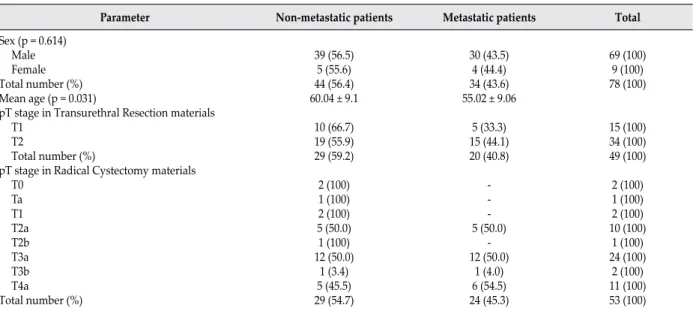
Parameter Non-metastatic patients Metastatic patients Total
Sex (p = 0.614) Male Female Total number (%) Mean age (p = 0.031)
pT stage in Transurethral Resection materials T1
T2
Total number (%)
pT stage in Radical Cystectomy materials T0 Ta T1 T2a T2b T3a T3b T4a Total number (%) 39 (56.5) 5 (55.6) 44 (56.4) 60.04 ± 9.1 10 (66.7) 19 (55.9) 29 (59.2) 2 (100) 1 (100) 2 (100) 5 (50.0) 1 (100) 12 (50.0) 1 (3.4) 5 (45.5) 29 (54.7) 30 (43.5) 4 (44.4) 34 (43.6) 55.02 ± 9.06 5 (33.3) 15 (44.1) 20 (40.8) -5 (-50.0) -12 (50.0) 1 (4.0) 6 (54.5) 24 (45.3) 69 (100) 9 (100) 78 (100) 15 (100) 34 (100) 49 (100) 2 (100) 1 (100) 2 (100) 10 (100) 1 (100) 24 (100) 2 (100) 11 (100) 53 (100)
Table 1: The sex, mean age and pathological stage (pTstage) distribution of the patients without lymph node/distant metastasis and with
between 2007 and 2008 and followed up for 2 years (Table 1). We obtained informed consents from all patients and certification from Ankara Dışkapı Yıldırım Beyazıt Education and Research Hospital Ethics Committee about the relevance of the study (Ethics Committee no: 17/19-03.11). We followed up all the patients after the 3rd month control with intervals
of 4 months for 2 years.
The pathology slides of RC and TUR materials of the patients were reviewed and representative sections and paraffin blocks of each tumor were selected.
The histological grade of the bladder tumors were determined according to the 2004 World Health Organization classification[7]. Tumor staging was done
using Tumor Node Metastasis system according to the American Joint Committee on Cancer[23].
The patients were seperated into two groups as;
Group 1 (non-metastatic group) : Composed of
the patients (n = 44) with no lymph node metastasis present at the time of cystectomy and no distant organ metastasis determined on the follow-up.
Group 2 (metastatic group): Composed of the
patients (n = 23) who had lymph node metastasis present at the time of cystectomy and the patients (n = 11) who did not have any lymph node metastasis present at the time of cystectomy, but distant organ metastasis occured on the follow-up.
The age, gender, survivals and disease free survivals of the patients, stage of the tumors, lymphovascular-angiovascular invasions, expressions of KAI1, PTEN and p53 in TUR and RC materials were statistically evaluated in two groups (non-metastatic and metastatic).
Immunohistochemistry
For immunohistochemical staining, 4µm sections of selected tumor blocks were cut and mounted on poly-L-lysine–coated slides. Following deparaffinization
and rehydration, antigen retrieval was performed using citrate buffer (pH 6.0) at 121 ˚C for 10 minutes. Endogenous peroxidase activity was blocked with 3% hydrogen peroxide for five minutes. Later, the sections were incubated with antibodies against KAI1 (Santa Cruz, sc-17752, G-2), p53 (Novocastra, NCL-L-p53-D-07) and PTEN (Neomarker, ser370, RB-10620-P1). 3,3’-diaminobenzidine and 3-amino-9-ethylcarbazole were performed as chromogenes and finally slides were counterstained with hematoxylin. Breast carcinoma tissue was used as positive controls for p53 and PTEN. Prostate carcinoma tissue was used as a positive control for KAI1. Cases not treated with primary antibodies served as negative controls.
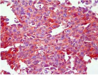
Immunostaining was evaluated by two pathologists. Nuclear staining for p53 (Figure 1) and cytoplasmic staining for KAI1 (Figure 2) and PTEN (Figure 3) was accepted as positive. The ‘expression rate’, the extent of staining for each antibody, was scored according to the number of cytoplasmic or nuclear stained carcinoma cells in 100 tumor cells. Less
Fig 1: Nuclear p53 positive staining (p53 x 100) Fig 2: Cytoplasmic KAI1 positive staining (KAI1 x 200)
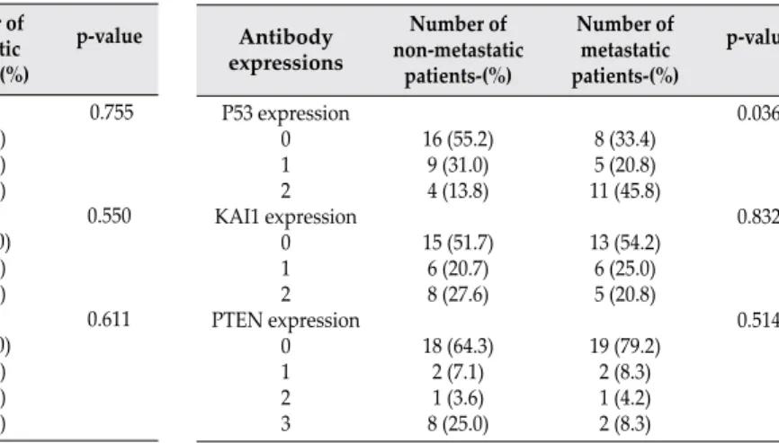
Antibody expressions Number of non-metastatic patients-(%) Number of metastatic patients-(%) p-value P53 expression 0 1 2 KAI1 expression 0 1 2 PTEN expression 0 1 2 3 9 (31.0) 10 (34.5) 10 (34.5) 16 (55.2) 5 (17.2) 8 (27.6) 12 (41.4) 3 (10.4) 9 (31.0) 5 (17.2) 5 (25.0) 6 (30.0) 9 (45.0) 10 (50.0) 6 (30.0) 4 (20.0) 12 (60.0) 2 (10.0) 4 (20.0) 2 (10.0) 0.755 0.550 0.611 Antibody expressions Number of non-metastatic patients-(%) Number of metastatic patients-(%) p-value P53 expression 0 1 2 KAI1 expression 0 1 2 PTEN expression 0 1 2 3 16 (55.2) 9 (31.0) 4 (13.8) 15 (51.7) 6 (20.7) 8 (27.6) 18 (64.3) 2 (7.1) 1 (3.6) 8 (25.0) 8 (33.4) 5 (20.8) 11 (45.8) 13 (54.2) 6 (25.0) 5 (20.8) 19 (79.2) 2 (8.3) 1 (4.2) 2 (8.3) 0.036 0.832 0.514
Table 2: p53, KAI1 and PTEN expressions of the urothelial carcinoma
of the bladder in transurethral resection materials. Table 3: p53, KAI1 and PTEN expressions of the urothelial carcinoma of the bladder in radical cystectomy materials.
than 5% positive cells were accepted as negative, while ≥5% positive cells were being accepted as positive.
Scoring for p53 and KAI1:
0 (negative): <5% positive cells; 1+ (positive): 5-50% positive cells; 2+ (positive): >50% positive cells.
Scoring for PTEN:
0 (negative): <5% positive cells; 1+ (positive): 5-25% positive cells; 2+ (positive): 26-50% positive cells; 3+ (positive): >50% positive cells.
Statistical analysis
Statistical evaluation was performed by using Chi-square test, Mann-Whitney test or Fisher’s Exact test. The survival and the factors that have effects on disease free survival were evaluated with Log Rank survival analysis. The patients who died within the first month after the operation were excluded during the survival analysis. P-values less than 0.05 were considered to be statistically significant.
RESULTS
Age and gender of the patients
Patients included 69 males and 9 females. The relation of sex with the status of metastasis was not statistically significant. The mean age was lower in metastatic group and this was statistically significant (Table 1).
TUR materials
In TUR materials, there was no statistically significant relation between p53, KAI1, PTEN expressions and the metastatic, non-metastatic groups (Table 2). Also, there were no statistically significant relations between p53, KAI1, PTEN expressions and pT stages of the tumors in metastatic and non-metastatic groups (p = 0.248, p = 0.755, p = 0.059 respectively).
Radical cystectomy materials
In RC materials, there was a statistically significant relation between p53 expressions and the groups (non-metastatic and (non-metastatic, Table 3). However, this relation was not significant with KAI1, PTEN. Also, there was no statistically significant relation between
Antibody
expressions Survival Transurethral Resection Materialsp-value Disease free survival p-value Survival p-valueRadical Cystectomy materialsDisease free survival p-value
P53 - (<5%) +(≥ 5%) KAI1 -(<5%) +(≥ 5%) PTEN - (<5%) +(≥ 5%) 50.00 44.12 27.27 53.33 45.79 54.48 0.9378 0.0285 0.9599 78.57 70.59 69.23 77.27 69.13 75.34 0.6581 0.7461 0.7576 40.00 38.77 37.50 36.66 41.89 42.75 0.7771 0.9808 0.8219 71.43 69.23 65.22 75.00 66.67 73.91 0.7923 0.5081 0.5790
Table 4: Survivals-disease free survivals of the patients with the urothelial carcinoma of the bladder and p53, KAI1 and PTEN
p53, KAI1, PTEN expressions and pT stages of the tumors in metastatic and non-metastatic groups (p = 0.185, p = 0.723, p = 0.34 respectively).
However, when the lymphovascular vessel invasion was evaluated, it was seen positive only in 2 patients (6.8%) in non-metastatic group and in 11 patients (45.8%) in metastatic group. These rates were statistically quite significant (p <0.001). As the angiovascular invasion was evaluated, it was seen positive in 2 patients (6.8%) in non-metastatic group and in 12 patients (50.0%) in metastatic group. These rates were also statistically quite significant (p <0.001).
When the survival, the disease free survival and p53, KAI1 and PTEN expressions were evaluated in TUR and RC materials; according to TUR materials, there was a statistically significant relation in survival only between the patients with positive and negative KAI1 expressions (Table 4).
DISCUSSION
Bladder carcinoma is the second most common cancer of the genitourinary tract and the incidence of it has been increasing in the last 20 years. UCB is a progressive disease typically seen in older ages. The depth of invasion, grade of the tumor and lymph node metastasis are the major factors used in determining clinical treatment. About 70% of bladder tumors are superficial, 25% are invasive and 5% are metastatic. Also, 30% of the superficial tumors become invasive in the follow-up. Long term survivals are better in patients with organ-confined disease[24-25].
BPL helps to understand the local spread of the disease. The risk of pelvic lymph node metastasis increases with the stage of the disease. The risk of pelvic lymph node metastasis in patients with stage pT3 and pT4 is 30 – 60% respectively, while the risk of lymph node positivity at the time of surgery in patients with stage pT2 is about 10 - 30%. Generally, the patients with positive lymph node metastasis determined postoperatively show simultaneous distant organ micrometastasis. Hence, it is important to predict the tumors that tend to metastise before the surgery in order to determine the apropriate clinical treatment and aproach. Still, there is no reliable indicator to use in predicting the metastatic disease[26-27].
The first notable finding in our study was that, the average age of the metastatic group was lower than the non-metastatic group. This shows that the tumors seen in younger ages are more aggressive.
p53 located in the nucleus blocks the cell cycle in G1 phase and prevents the cell passing to the S phase. Then it activates the genes that repair DNA and sends the damaged and unrepaired cells to apoptosis. p53 mutations can be seen in many tumors like lung, colon and breast[26]. It was shown that p53 can be used in
diagnosing urothelial carcinoma in situ of the urinary bladder and in predicting the progression of the non-invasive pT1 lesions[28]. In a study, Uygur et al evaluated
p53 expressions in TUR materials of 31 patients with RC. In 17 patients with p53 positivity (nuclear staining 20% and higher was accepted as positive), 11 had lymph node metastasis. Therefore, patients who had pT2 and pT3a disease with p53 positivity were accepted as high risk group and concluded that early aggressive therapy had to be started as soon as possible[29]. In another study, Esrig et al reported that
there was a positive relation between p53 staining and the pathological stage. In that study, they also reported that in p53 positive patients, recurrence of the disease was higher and the survival was lower. Hence, p53 was determined as a prognostic factor independent from the stage and the grade[22]. On the other hand, there
are studies reporting that there is no relation between p53 expression and the stage[30]. Also Puzio-Kuter et
al reported similar results; that p53 overexpression was seen in invasive bladder carcinoma compared to non-invasive papillary tumors[1]. Goebell et al showed
in their international study that p53 positivity was significantly correlated with tumor progression in pT1 disease and advanced bladder cancer and p53 appeared to be predictive in high grade bladder cancer[21].
In our study, we found out that in RC materials; there was a statistically significant relation between p53 expressions and the groups (non-metastatic and metastatic). p53 positivity in metastatic group was higher. We thought that the patients with p53 expression of 50% or higher had more risk to develop metastasis. So, p53 positivity may be a bad prognostic factor and the patients expressing 50% or more p53 may be treated more radically and followed up carefully. However, we could not find this relation in TUR materials and we thought that p53 is not sufficient in predicting lymph node metastasis of the tumor in TUR materials. The reason for this is thought to be that the material obtained by TUR is inadequate and also cautery artefact can limit adequate examination of all material. We also showed that the lymphovascular vessel and the angiovascular invasions were seen more frequently in metastatic group than the non-metastatic group as known before[7]. When we looked
at the survivals and disease-free survivals, the increase of p53 expression had negative effects on the survival and disease-free survival. However, this effect was not statistically significant. We think that this effect would be statistically significant in new studies with more patients.
KAI1 was detected in tumors like prostate, stomach, colon, breast carcinomas and the invasiveness and the metastatic potentials of the tumors were shown to increase with the loss of KAI1 expressions[18-20]. In a
study, Su et al reported that decreased KAI1 expression was associated with the degree of invasiveness and progression of cancer and was an independent prognostic factor for tumor recurrence in primary pTa and pT1 UCB[16]. We found that according to TUR
materials, patients with positive KAI1 expression had better survival than patients with negative KAI1 expression. The tumor was more aggressive and invasive in TUR materials with lower KAI1 expressions. We thought that this was not statistically significant because of the low number of patients.
PTEN inhibites cell proliferation and tumorogenesis by inducing apoptosis and by arresting G1 phase of cell cycle. PTEN was detected in many human cancers, like gliomas, breast, prostate, lung, thyroid and endometrial carcinomas. This gene is also reported to show loss of heterozygosity in bladder cancer, more frequently in muscle-invasive urothelial carcinomas[11-15]. In their
study, Lee et al reported that loss of PTEN expression was associated with non-papillary histology, high grade and invasive urothelial carcinomas[13].
We only found that the loss of PTEN expression in metastatic group was more than the loss of PTEN expression in non-metastatic group. However, this correlation was not statistically significant.
In UCB, the lymphovascular vessel and the angiovascular invasions are very important as they are indicators of the lymph node or distant metastasis. The presence of lymph node or distant metastasis shortens the surveys. We need more reliable indicators to use at the beginning of the disease before the radical operation to predict the tumors which will be more aggressive, in order to decide the correct therapy, neoadjuvant or early adjuvant therapy. p53, KAI1 and PTEN seem to be good indicators, although we could not reach perfect results. We think that the reason is the limited number of our patients as indicated before. New studies with large series seem to support the usefulness of p53, KAI1 and PTEN.
CONCLUSION
In UCB, it is very important to predict the progression of the disease before the surgery as it frequently relaps and shortens the lives of the patients. It is very important to find new indicators showing the aggressiveness of the disease. The results of this study give the impressions that p53 positivity, KAI1 negativity and PTEN negativity in UCB are good indicators of worse prognosis and aggressiveness.
ACKNOWLEDGMENT
The authors wish to thank Dr Alper Aygün from Ankara Dışkapı Yıldırım Beyazıt Education and Research Hospital, Department of Urology, Ankara, Turkey for his clinical support.
REFERENCES
1. Puzio-Kuter AM, Castillo-Martin M, Kinkade CW, Wang X, Shen TH, Matos T, et al. Inactivation of p53 and Pten promotes invasive bladder cancer. Genes Dev 2009; 23(6):675-680.
2. Lee K, Jung ES, Choi YJ, Lee KY, Lee A. Expression of pRb, p53, p16 and Cyclin D1 and their clinical implications in urothelial carcinoma. J Korean Med Sci 2010; 25(10):1449-1455.
3. Matsushita K, Cha EK, Matsumoto K, Baba S, Chromecki TF, Fajkovic H, et al. Immunohistochemical biomarkers for bladder cancer prognosis. Int J Urol 2011; 18(9):616-629.
4. Jemal A, Siegel R, Xu J, Ward E. Cancer statistics, 2010. CA Cancer J Clin 2010; 60(5):277-300.
5. Shariat SF, Karakiewicz PI, Palapattu GS, Lotan Y, Rogers CG, Amiel GE, et al. Outcomes of radical cystectomy for transitional cell carcinoma of the bladder: a contemporary series from the Bladder Cancer Research Consortium. J Urol 2006; 176(6 Pt 1):2414-2422.
6. Hautmann RE, Gschwend JE, de Petriconi RC, Kron M, Volkmer BG. Cystectomy for transitional cell carcinoma of the bladder: results of a surgery only series in the neobladder era. J Urol 2006; 176(2):486-492.
7. Eble JN, Sauter G, Epstein JI, Sesterhenn IA. Pathology and genetics of tumours of the urinary system and male genital organs. World Health Organization Classification of Tumours. IARC Press, Lyon, 2004. 8. Shariat SF, Karam JA, Lerner SP. Molecular markers in
bladder cancer. Curr Opin Urol 2008; 18(1):1-8.
9. Vrooman OP, Witjes JA. Molecular markers for detection, surveillance and prognostication of bladder cancer. Int J Urol 2009; 16(3):234-243.
10. Kim YK, Kim WJ. Epigenetic markers as promising prognosticators for bladder cancer. Int J Urol 2009; 16(1):17-22.
11. Litlekalsoy J, Hostmark JG, Costea DE, Illemann M, Laerum OD. Time-dependent biological differences in molecular markers of high-grade urothelial cancer over 7 decades (ras proteins, pTEN, uPAR, PAI-1 and MMP-9). Virchows Arch 2012; 461(5):541-551.
12. Herlevsen M, Oxford G, Ptak C, Shabanowitz J, Hunt DF, Conaway M, et al. A novel model to identify interaction partners of the PTEN tumor suppressor gene in human bladder cancer. Biochem Biophys Res Commun 2007; 352(2):549-555.
13. Lee H, Choı SK, Ro JY. Overexpression of DJ-1 and HSP90α, and loss of PTEN associated with invasive urothelial carcinoma of urinary bladder: Possible prognostic markers. Oncol Lett 2012; 3(3):507-512. 14. Røtterud R, Fosså SD, Nesland JM. Protein networking
in bladder cancer: Immunoreactivity for FGFR3, EGFR, ERBB2, KAI1, PTEN, and RAS in normal and malignant urothelium. Histol Histopathol 2007; 22(4):349-363. 15. Zhang XA, He B, Zhou B, Liu L. Requirement of the
p130CAS-Crk coupling for metastasis suppressor KAI1/CD82-mediated inhibition of cell migration. J Biol Chem 2003; 278(29):27319-27328.
16. Su JS, Arima K, Hasegawa M, Franco OE, Umeda Y, Yanagawa M, et al. Decreased expression of KAI1 metastasis suppressor gene is a recurrence predictor in primary pTa and pT1 urothelial bladder carcinoma. Int J Urol 2004; 11(2):74-82.
17. You JJ, Madigan MC, Rowe A, et al. An inverse relationship between KAI1 expression, invasive ability and MMP-2 expression and activity in bladder cancer cell lines. Urologic Oncology: Seminars and Original Investigations 30. 2012:502-508.
18. Ilhan O, Celik SY, Han U, Onal B. Use of KAI1 as a prognostic factor in gastric carcinoma. Eur J Gastroenterol Hepatol 2009; 21(12):1369-1372.
19. Takaoka A, Hinoda Y, Satoh S, Adachi Y, Itoh F, Adachi M, et al. Suppression of invasive properties of colon cancer cells by a metastasis suppressor KAI1 gene. Oncogene 1998; 16(11):1443-1453.
20. Yang X, Wei LL, Tang C, Slack R, Mueller S, Lippman ME. Overexpression of KAI1 suppresses in vitro invasiveness and in vivo metastasis in breast cancer cells. Cancer Res 2001; 61(13):5284-5288.
21. Goebell PJ, Groshen SG, Schmitz-Dräger BJ; ISBC. p53 immunohistochemistry in bladder cancer—a new approach to an old question. Urol Oncol 2010; 28(4):377-388.
22. Esrig D, Elmajian D, Groshen S, Freeman JA, Stein JP, Chen SC, et al. Accumulation of nuclear p53 and tumor progression in bladder cancer. N Engl J Med 1994; 331(19):1259-1264.
23. Edge S, Byrd DR, Compton CC, Fritz AG, Greene
FL, Trotti A. American Joint Committee on Cancer-AJCC. Cancer Staging Manual. Springer, Chicago, 2010.
24. Skinner DG. Management of invasive bladder cancer. A meticulous pelvic node dissection can make a difference. J Urol 1982; 128(1):34-36.
25. Schoenberg MP, Walsh PC, Breazeale DR, Marshall FF, Mostwin JL, Brendler CB. Local recurrence and survival following nerve sparing radical cystoprostatectomy for bladder cancer: 10-year followup. J Urol 1996; 155(2):490-494.
26. Walsh PC, Retik ED, Wein AJ. Campbell’s Urology. Saunders, Philadelphia, 2002.
27. Smith JA Jr, Whitmore WF Jr. Regional lymph node metastases from bladder cancer. J Urol 1981; 126(5):591-593.
28. Bernardini S, Billerey C, Martin M, Adessi GL, Wallerand H, Bittard H. The predictive value of muscularis mucosae invasion and p53 over expression on progression of stage T1 bladder carcinoma. J Urol 2001; 165(1):42-46.
29. Uygur MC, Yaman I, Kutluay L, Altug U, Erol D. The relation between p53 overexpression and lymph node metastases in clinical stage T2 and T3a transitional cell bladder carcinoma. J Exp Clin Cancer Res 1999; 18(3):391-395.
30. Sınık Alkıbay T, Ataoğlu Ö, Akyol G, et al. Correlation of nuclear p53 over-ekspession with clinical and histopathological features of transitional cell bladder cancer. Int Urol Nephrol 1997; 29:25-31.