99ournalof Neurology,Neurosurgery, and Psychiatry 1994;57:998-1001
SHORT REPORT
Apomorphine induced cognitive changes
in
Parkinson's disease
EvzenRilizka, Jan Roth, Natasa Spa6kova, PetrMecir, RobertJech
Abstract
Auditory eventrelated potentials (ERPs)
andvisual evokedpotentials (VEPs)were
recorded from eight patients with
Parkinson's disease, before and after a singledose ofapomorphine. Toassessthe
treatment effects, the patients' motor
state, Benton visual retention test (BVRT), and digit span tests were also examined. After apomorphine, although
motor performance improved, the ERP latencies were delayed and the N2-P3
ERP amplitude was significantly
dimin-ished by comparison with pretreatment values. These data suggestthat
apomor-phineinduces,besides itsmotoreffects in
patientswithParkinson's disease,a
slow-ing down of cognitive processing.
Preferential stimulation of dopamine
autoreceptors in mesocortical and
mesolimbic systems may represent a
neuralmechanismforthese effects. Also,
the posttreatment BVRT rotation errors
significantly increased, suggesting an
apomorphine induced impairment of
visuospatial perception.
(7 Neurol Neurosurg Psychiatry 1994;57:998-1001)
ClinicofNeurology, 1stMedicalFaculty, CharlesUniversity, Prague, Czech Republic ERiizika J Roth NSpa6kovA PMeAf RJech Correspondenceto:
DrEvzenRuzicka,Clinic of Neurology,Katehnska 30,
120 00 Praha2, Czech
Republic.
Received1June1993
andinfinal revised form 28January1994.
Accepted11February 1994
Dopaminergicdeficiency is knownas a
funda-mental mechanism of motor impairment in
idiopathic Parkinson's disease and may also playan important partinparkinsonian
cogni-tivedeficits.' The benefit fromlevodopa treat-ment on cognition is usuallyless pronounced
than that onmotor symptoms. Some specific cognitive deficits may recover whereas others
remain unimproved.2 Several studies even
found a degradation ofcognitive functions in patients with Parkinson's disease under lev-odopa.3
The evaluation of cognitive deficits in
Parkinson's disease is often complicated by
the interference with motor disability. Electrophysiologicalmethods ofinvestigation, namely late "event related" auditory evoked
potentials (ERPs) have thus beenproposed as
objective indicators of neural function in Parkinson's disease.4 Recently, ERPs were
used to assess the effects of dopaminergic treatment in Parkinson's disease. After lev-odopa, the latency of the major cognitive component P3 (P300) was shortened,
sug-gestingapost-treatment improvement of cog-nitiveprocessing,5 but another study, showed
a delay of the P3 ERP after levodopa.6
Similarly, inhealthysubjects, a single dose of levodopa provoked a prolongation of the P3
wavelatency in comparison with the
pretreat-mentvalues.7Visual evokedpotentials (VEPs) have also been assessed in patients with
Parkinson's disease. The delay of the P100 wave, the major component of VEPs, have beenproposed as acorrelate of dopaminergic dysfunction in the retina and visual
path-ways.8
To improve our understanding of
dopaminergic involvement in Parkinsonian
cognitive deficits, we aimed to assess the
behavioural effects ofapomorphine, a potent direct agonist ofD, and D2 dopamine
recep-tors with a rapid but short term effect.
Transient sedation and sleepiness have often beenseeninpatientswithParkinson's disease after isolated doses ofapomorphinebutmore profoundanalysis of this phenomenon and of itsunderlyingmechanisms isnotavailable.
Materials and methods
Eight patients with Parkinson's disease (five men and three women, mean age 59 4 (SD 8-3) years) were studied aftergivinginformed
consent. The mean duration of disease was 8 3 (64) years, withtwo patients classified as being in each ofHoehn and Yahr stagesI,
iI,
III, and IV. All of the patients were on
dopaminergicmedication withmean duration of5 4(48) years, andameanlevodopadose of
422 (SD 294) mg. Only non-demented
patients with mean mini mental status (MMS) score 28-6 (SD 2 2), with normal or
corrected to normalvision, and with nosigns or history ofconcurrent neurological or psy-chiatric diseases were included. Nine healthy volunteers (six men and three women, mean
age 61 9 (SD 2 7) years) were taken as
con-trols for ERP and VEP testing. A detailed interview, standardneurological examination, and MMStestingwere doneto includein the control group only subjects without previous history of neurological or psychiatric
dis-orders, with normal neurological state, and MMS>28.
The ERPs were examined according to an auditory oddballparadigm requiring amental
count ofrare target tones (2 kHz) randomly 998
Apomorphine induced cognitive changesinParkinson's disease
occurring (p= 02) in asequence offrequent indifferent tones (1 kHz, p = 0 8), carried binaurally byheadphones, and with an
inten-sity of 75 dB. Evoked potentials were
recorded from the vertex (Cz) with a
refer-ence tolinkedmastoids. Electrode impedance
was maintained below 5 kfQ. The EEG data
were amplified (filter bandpass 05-50 Hz), digitised, and averaged separately for target
and indifferent tones by a Dantec E-4000.
Trials with excessive eye movement were
automatically rejected. At least two averages
of20 artifact free trials in response to target
stimuli were obtained. The ERPs were
analysed and the N1, P2, N2, and P3 waves were identified on the basis of sequence,
polarity, and latencyrange, onthe waveforms elicited by target stimuli. Latencies ofERPs weremeasured in ms from stimulus onset to wavepeaks, amplitude valuesN1-P2and
N2-P3 were determined as the difference in ,uV between therespective peaks.
The VEPs wereexamined monocularly, by
successive stimulation of the right and left
eyes. Pattern reversal vertical grating stimuli with square wave luminance profile, spatial frequency of4cpd, and temporal frequencyof
1 Hz were generated on a TV screen placed
150 cm from theobserver's eye and subtend-ing 90 of his or her visual field. The mean
luminance of the screen was 619 cd/M2n, the
contrast was 95%. The VEPs were recorded from Ozreferencedto Fz.Filterbandpasswas set at 1 and 100 Hzand analysis timewas400 ms. At least two averages of200 artifact free trials were performed to ensure reproducibil-ity. The latencies of N70, P100, and N140 VEPcomponents weremeasuredin ms, from stimulusonset(pattern reversal) topeak;VEP
amplitude was determined by measuring the difference in,uV between the peaks of N70 andP100.
A short battery ofneuropsychological tests
included the Benton visual retention test,
administration A (BVRT), and forward and backwarddigitspan tests(DS). On theBVRT
examination, after a 10 second exposure to
each of10 cards ofoneorthreefigure design,
an immediate recall by drawing was
demanded. To minimise practice effects, two
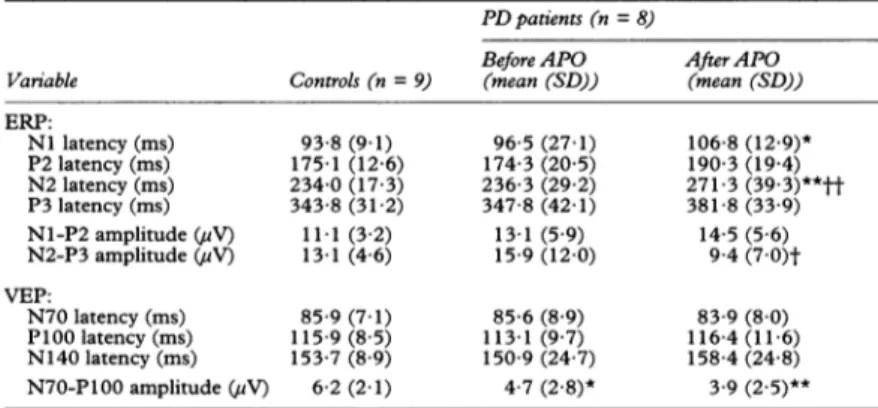
Table1 Event relatedpotentials and visual evoked potentials in eight patients with Parkinson's disease
PDpatients(n= 8)
Before APO AfterAPO
Variable Controls(n=9) (mean(SD)) (mean(SD))
ERP: NIlatency (ms) 93-8(9 1) 96-5 (27-1) 106-8(12-9)* P2latency (ms) 175-1 (12-6) 174-3(20 5) 190-3(19-4) N2latency (ms) 234-0(17-3) 236-3 (29-2) 271-3(39-3)**tt P3latency (ms) 343-8(31-2) 347-8(42-1) 381-8 (33-9) N1-P2amplitude (uV) 11 1(32) 13-1 (5 9) 14-5 (5 6) N2-P3amplitude(uV) 13-1(4 6) 15-9(12-0) 9-4(70)t VEP: N70latency(ms) 85-9(7-1) 85-6 (8 9) 83-9 (8 0) P100latency(ms) 115-9(85) 113-1 (97) 116-4 (11-6) N140latency(ms) 153-7(8 9) 150-9 (24 7) 158-4 (24 8) N70-P100amplitude(jV) 6-2(2-1) 4-7(28)* 3 9(2.5)** *p<0-05; **p <0-01;patientswith Parkinson's disease v controls (Mann-Whitney U test)
tP<005; tp<001; patientswith Parkinson's disease beforevafter apomorphine (paired
Wilcoxontest).
equivalent forms of the BVRT (C and D) were made before and after apomorphine. The numbers ofcorrect reproductions and of
errors wererecorded (more thanoneerror per
cardwas counted if differenterrors were
com-bined). The DS tests required an immediate
recallofdigit series (from 1 to 9)of increasing length. The forward and backward DS repre-sent the largest numbers of digits correctly repeatedin agivenorder.
Atleast 48 hoursbefore tests, 20 mg dom-peridone given three times daily. All medica-tion except for domperidone was withdrawn
12-14 hours before the testing. The patients
weretested twice during the same experimen-tal session, before and after apomorphine (a dose ofapomorphine hydrochloride in water
correspondingto 0-05 mgofapomorphine/kg of weight, injected subcutaneously). Both
parts of the testingwere identical in arrange-mentand duration (40-45 minutes); the
sec-ond part started 15 minutes after apomorphine. Examination of ERPs and VEPs and neuropsychological testing were
conducted as described. The patients' motor status was evaluated according to the Columbia University rating scale (CURS) in
which the maximumdisabilityscore = 100.
Results
After apomorphine, motor performance in
eight patients was greatly improved in
com-parison withthe pretreatment condition. The
average CURS score decreased from 32-5
(SD 24 6) before apomorphine to 14-5 (9-7)
after apomorphine (p< 005 on paired
Wilcoxon test).No furtherchanges exceeding 10% of post-treatment CURS values were
noted until the end of the examination. All patients and control subjects were able
toperform theERPtask and the target count errors did not exceed ±2. The electrophysio-logical data obtained from patients with
Parkinson's disease and from controls were
compared by Mann-Whitney U tests. The
indices of the ERP recorded in patients with
Parkinson's disease before apomorphine did
00 01) 0. en LO Nl N2 P2
~~~~~~~~~~~~~~~~1
.,,2vo_N
1-
/,
Ip3^ II2 I%I
~~~~~~~3
0 200 400 600 800 1000 msFigure1 Eventrelatedpotentials recorded at Cz from a controlsubject (trace 1) and fromapatient with Parkinson'sdiseasebefore (trace 2) and after (trace 3) a single doseofapomorphine.Eachtrace represents the averageof40responsestothetargettone.Note the relative delays ofthe N2 andP3 wavesafterapomorphine compared with the control and with the pretreatment waveform.
Rzicika, Roth,Spackova,Meeif,3'ech c 0 a, CN 0 80 160 240 ms 320 400
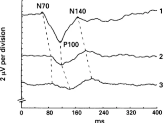
Figure2 Visualevoked potentials recorded fromOz in a
control subject(trace 1) and in apatientwithParkinson's diseasebefore(trace 2)and after (trace3) asingle dose of
apomorphine. Eachtracing represents an average of 400 responses elicitedby rightandlefteyestimulation. Notethe
post-treatmentmodificationsofwaveformshape with
slightly delayed Pl00and Nl40latenciesincomparison with thepotentials recorded before apomorphine.
not differ from control values but the post-treatmentNI and N2 ERPlatencieswere sig-nificantly delayed in comparison with control data.
Latencies in ERPsgenerally increased after apomorphine compared with the
pretreat-mentvalues. This increase was significant for
the N2 ERPlatency andjustunder the
signif-icancethresholds for the P2 and P3 latencies, asassessed by Wilcoxonpairedtests.Also, the N2-P3 ERP interpeak amplitude was signifi-cantly diminished after apomorphine com-pared with the pre-treatment results (table 1, fig 1).
The latencies of the VEP recorded in
patients with Parkinson's disease did not dif-fer from control data. The N70-P100 VEP
amplitudesweresignificantlylower inpatients
with Parkinson's disease than in controls,
both before and after apomorphine. Slight and non-significant delays of the P100 and N140 wave latencies were found after
apo-morphine compared with the pretreatment values (table 1,fig 2).
The pretreatment numbers of correct
BVRTreproductionswerewithin normal lim-its and remained unchanged after
apomor-phine. Total BVRT error scores were somewhat higher after apomorphine than
before.Thiswas mainlydueto the significant
Table2 Neuropsychologicaltestsineightpatientswith Parkinson's disease
Before After apomorphine apomorphine
Variable (mnean(SD)) (mean(SD))
Benton visualretentiontest:
Correctreproductions 5 0(2-1) 4-8 (20) Total number oferrors 70(2-7) 8-5 (49) Rotationerrors 1-1 (08) 25(09)t Omissions 0-5(0*8) 1*5(1*8) Distortions 25 (20) 1-6(1 9) Misplacements 13(1-0) 1.9(1-6) Sizeerrors 1.1 (17) 0-9(08) Digit span: Forward 59(0-8) 6-3(1-0) Backward 3 5(0-9) 4-1(1-8)
p <005;patientswith Parkinson's disease beforevafter
apo-morphine(pairedWilcoxontest).
rise of figure rotation errors and, to a lesser extent, of omissions. Scores for DS were within normal limits and did not significantly change after apomorphine (table 2).
Spearman rank correlations calculated between electrophysiological and
neuropsy-chological data (both before and after apo-morphine) and the age of the patients, duration ofParkinson's disease, duration and dose of levodopa treatment, MMS, and CURS scores, were not significant.
Discussion
In eight non-demented patients with
Parkinson's disease, motor disability as
assessed by CURS was improved after
apo-morphine, whereas cognitive performance as indexed by ERP was worse after than before
apomorphine.
These results are similar to previous find-ings of improved motor performance and
delayed ERP after levodopa in patients with
Parkinson's disease.6 The delays in the ERP components seem to represent a slowing down ofneural events underlyingperception,
discrimination, and categorisation of stimuli,9 the post-treatment reduction ofthe P3 ERP
amplitude corresponding to a decrease of the
stimulus information amounts processed.'0
Also, on BVRT which provides measures of
visuospatial cognitive functions, our patients
had significant post-treatment increases in
rotation errors, indicating an apomorphine-inducedimpairment ofvisualperception.
The contrasting effects of dopaminergic
treatment, improving overall motor
perfor-mance but impairing some aspects of cogni-tive processing, might be interpreted in the
light of several earlier studies. After
apomor-phine treatment in low doses, laboratory ani-mals showed decreased motor activity and
yawning behaviour,whereashigherdoses
pro-duced hypermobility and stereotypy." These
biphasic behavioural responses were attrib-uted to a selective drug action at presynaptic and postsynaptic dopamine receptors.'2 The
presynaptic dopamine "autoreceptors" were
shown to be more sensitive to the effects elicitedbydopamineagoniststhan the postsy-naptic receptors in the striatum. Low doses of
dopamine agoniststhus produceaninhibition ofdopaminergic activity,and larger doses act to stimulate postsynaptic receptors directly elicitingthe expected response. Furthermore, presynaptic dopamine receptors in the meso-corticalsystemseemedtobemoresensitiveto dopamine agonists than the autoreceptors on
the terminals of the nigrostriatal neurons.'2
Preferential stimulation ofdopamine
autore-ceptors in the mesocortico-limbic system might, therefore, explain the post-treatment ERP delays in patients with Parkinson's
dis-ease. Denervation supersensitivity of
postsy-naptic dopamine receptors in the striatum may account for the parallel improvement of
motor performances induced by
apomor-phine. Of course, more complex interactions within distinct subpopulations of
post-synaptic dopamine receptors or within
Apomorphine induced cognitive changesinParkinson's disease
dopaminergic neurotransmitter systems may occur.
On the otherhand, taking intoaccountthe fixedorderof testing in thesame session, the findings might be interpreted as non-specific
effects of apomorphine on arousal. Such an
interpretation, however, seemsunlikely, given
that the DS test performances remained unchanged with the treatment, indicating a
preserved level of attention all through the testing.
Finally, the pretreatment latencies of the
ERPandVEPrecordedin ournon-demented patients with Parkinson's diseasewere not dif-ferent from the control data. ForERP, this is in accordance with the fact that definite
latency delays have generally been shown in demented but not in non-demented patients with Parkinson's disease, reflecting probably cognitive slowing down associated with dementia.4' On VEP testing, normal laten-cies and reduced amplitudes were found in the present study similarto, for example, the non-demented patients (mean MMS =
28&2)
of Hansch etal.14 Several other studies,how-ever,showeddelayedVEP inParkinson's
dis-ease,theP100VEPbeingmoredelayed "off'
than "on" levodopa.8Without elaborating on
dopaminergic involvement ofthe visual sys-teminParkinson'sdisease,onthebasisof this discussion, perhaps the definite VEP delays might also be related to the presence of dementiainpatients with Parkinson's disease,
as ERPs are. In most previous VEP studies, the patients' mental state was not, unfortu-nately, specified. Interestingly, a significant prolongation of VEP latencies was recently reported in demented as distinct from
non-dementedpatients with Parkinson'sdisease.'5
Inconclusion, thepresentresults show that apomorphine modifiessomeaspects of cogni-tivefunctioning innon-demented parkinson-ian patients. Preferential stimulation of the presynaptic dopamine receptors in the
meso-cortical and mesolimbic system may at least partly explain these findings. Further studies should be undertaken inboth demented and non-demented patients with Parkinson's
disease, employing more specific test
para-digms focused on isolated cognitive deficits,
to shed light on complex subcorticocortical
interactions supposedly occurring under dopaminergic treatment in Parkinson's disease.
This research was supported by a grant from the Czech Ministry of Health (IGA 1039-2). We thank Olga Kucerova for hertechnicalassistance, to Farid El Massioui andBernard
Pillon for theirhelpfulcomments, and toSylvie Margulesfor herlanguage revision.
1 Agid Y,Javoy-AgidF, Ruberg M.Biochemistryof neuro-transmitters in Parkinson's disease. In: Marsden CD,
Fahn S, eds. Movement disorders. New York: Butterworth's, 1987:166-230.
2 DuboisB,Boller F, Pillon B, Agid Y. Cognitivedeficitsin Parkinson's disease. In: Boller F and GrafmanJ, eds. Handbook of neuropsychology. Vol 5. Amsterdam: Elsevier,1991:195-240.
3 PoeweW,Berger W, Benke T, Schelosky L. High-speed
memoryscanning in Parkinson's disease: Adverse effects oflevodopa.Ann Neurol 1991;29:670-3.
4 RiiN&a E, El Massioui F. Event-related potentials in
Parkinson's disease: a review. Behav Neurol 1993;6: 15-26.
5 Starkstein SE, Esteguy M, Berthier ML, Garcia H,
LeiguardaR:Evokedpotentials,reactiontime and cog-nitiveperformancein onandoffphasesof Parkinson's
disease.JNeurol NeurosurgPsychiatry1989;52:338-40.
6 PrasherD, FindleyL.Dopaminergic inducedchangesin
cognitive and motorprocessing in Parkinson'sdisease: Anelectrophysiological investigation.JfNeurol Neurosurg
Psychiatry1991;54:603-9.
7 StanzioneP,FattapostaF,GiuntiP,etal. P300 variations in
parkinsonian patients before and duringdopaminergic
monotherapy: A suggested dopamine component in P300. Electroencephalogr Clin Neurophysiol 1991;80: 446-53.
8 Bodis-Wollner I. Visual deficits related to dopamine deficiency in experimental animals and Parkinson's diseasepatients.TrendsNeurosci 1990;13:296-302. 9 KutasM, McCarthy G,Donchin E: Augmentingmental
chronometry: The P300 as a measure of stimulus evaluation time. Science1977;197:792-5.
10 Johnson R. A triarchic model of P300 amplitude. Psychophysiology1986;23:367-84.
11 Strombom U. Catecholamine receptor agonists. Effects on motor activity and rate of tyrosine hydroxylation in mouse brain. Naunyn Schmiedeberg's Arch Pharmacol
1976;292: 167-72.
12 Roth RH.Dopamineautoreceptors: Pharmacology,
func-tionand comparison with postsynaptic dopamine recep-tors.CommunPsychopharmacol1979;3:429-45. 13 GoodinDS,AminoffMJ. Electrophysiologicaldifferences
between demented and nondemented patients with
Parkinson'sdisease. Ann Neurol 1987;21:90-4. 14 HanschEC,Syndulko K, Cohen SN, Goldberg ZI, Potvin
AR,TourtellotteWW.Cognition in Parkinson's disease: Anevent-relatedpotentialperspective. Ann Neurol 1982; 11:599-607.
15 Okuda B, Tachibana H, Kawabata K, Takeda M, Toda K,
SugitaM. Correlationof visual evoked potentials with dementia inParkinson's disease.NipponRonenIgakkai
Zasshi1992;29:475-9.