http://journals.tubitak.gov.tr/medical/ © TÜBİTAK
doi:10.3906/sag-1507-90
Segmental epidural anesthesia for percutaneous kyphoplasty:
comparison with general anesthesia
Alparslan APAN1, Özgün CUVAŞ APAN1,*, Emine Arzu KÖSE2
1Department of Anesthesiology and Intensive Care Medicine, Faculty of Medicine, Giresun University, Giresun, Turkey
2Department of Anesthesiology and Intensive Care Medicine, Faculty of Medicine, İstanbul Medipol University, İstanbul, Turkey
1. Introduction
Vertebroplasty, kyphoplasty, and lordoplasty are minimally invasive procedures mainly performed for refractory vertebral body fracture pain due to osteoporosis, trauma, hemangioma, or metastasis. Fluoroscopic-guided injection of bone cement is applied into a compressed vertebral body in these procedures. Kyphoplasty is a more advanced technique. Beside pain relief, the goal of kyphoplasty is height restoration of a fractured vertebra. A cavity is created with insertion and inflation of a balloon into the vertebral corpus, and then is filled with cement (1,2).
Local anesthetic infiltration combined with monitored anesthesia care is an option for pain relief during vertebroplasty and kyphoplasty (3–6). On the other hand, especially while in the prone position, the level of sedation can easily shift from consciousness sedation to deep sedation and cause airway-related problems. Local infiltration anesthesia alone is not sufficient for pain relief during these procedures (7,8). Because of severe pain,
although kyphoplasty is recommended to be performed under general anesthesia, especially in elderly patients with comorbidities, general anesthesia can be associated with life-threatening problems (1,9).
There are a few case reports about spinal anesthesia for percutaneous kyphoplasty in the literature. In one of these reports, spinal anesthesia combined with conscious sedation was successfully performed in an elderly patient undergoing percutaneous kyphoplasty (10). Another report is a case series, and the authors conclude that subarachnoid anesthesia may be an adequate technique for kyphoplasty. On the other hand, they stated that additional conscious sedation may be required during the procedure (11).
When we take into account the disadvantages of spinal and general anesthesia techniques, segmental epidural anesthesia might be used for kyphoplasty procedures and may offer advantages for elderly patients with pulmonary disease, not only during the intraoperative period, but Background/aim: This is a feasibility study evaluating whether segmental epidural anesthesia is an alternative anesthetic approach to general anesthesia for percutaneous kyphoplasty.
Materials and methods: After ethics committee approval was obtained, 52 ASA class I–III patients scheduled for elective, single-level percutaneous kyphoplasty were recruited. The patients were divided into two equal groups. In Group E (Group Epidural) segmental epidural anesthesia was performed using the loss of resistance technique with saline. In Group G (Group Control) general anesthesia was performed. Hemodynamic parameters, intraoperative and postoperative analgesic requirements, visual analogue scale (VAS) scores, length of stay in the postanesthesia care unit (PACU), and complications were recorded.
Results: Hemodynamics were similar between the two groups. Postoperative analgesic requirement was significantly higher in Group G than in Group E (P < 0.004). VAS scores were significantly lower in Group E than in Group G (P < 0.05). Time to first pain experience at the first postoperative 4 h was significantly longer and length of stay in the PACU was significantly shorter in Group E than in Group G (P < 0.001).
Conclusion: Segmental epidural anesthesia is a safe anesthetic technique for percutaneous kyphoplasty. This technique offered advantages over general anesthesia in terms of postoperative analgesia, analgesic consumption, early recovery, and short PACU stay. Therefore, it should be considered a suitable anesthetic technique in patients undergoing single level percutaneous kyphoplasty. Key words: General anesthesia, epidural anesthesia, kyphoplasty, neurosurgery
Received: 12.07.2015 Accepted/Published Online: 22.03.2016 Final Version: 20.12.2016 Research Article
also during the postoperative period. To the best of our knowledge, there is no published study evaluating the efficacy of segmental epidural anesthesia for elective kyphoplasty and comparing it to general anesthesia. Therefore, we aimed to assess the effectiveness of segmental epidural anesthesia for elective kyphoplasty and compared it to general anesthesia, as to postoperative analgesic requirement, pain scores, and length of PACU stay.
2. Materials and methods
The Institutional Ethics Committee approved the study and it was conducted in accordance with the Declaration of Helsinki. Written informed consent was also obtained from all the patients. Fifty-two ASA physical status class I–III patients, selected between June 2012 and September 2013, aged between 18 and 85 years, scheduled for elective, single-level percutaneous kyphoplasty were enrolled in this prospective, randomized study. Patients with a severe systemic disease such as myelomatous disease, ASA IV patients, and those with a pathology in cervical vertebra, allergic history to the study drugs, any contraindication to epidural anesthesia, pregnancy, and cognitive disorders preventing cooperation were excluded. Patients requiring surgical intervention at more than one level were also excluded.
A preoperative neurologic examination, conventional radiography of the affected spinal segment, and magnetic resonance imaging with fat-saturated T2-weighted imaging were performed in all patients. Midazolam 1–1.5 mg was given intravenously as premedication. The patients were monitored (Infinity Kappa, Drager, Lubeck, Germany) for standard electrocardiography (ECG), blood pressure noninvasively, peripheral oxygen saturation (SpO2), and end-tidal CO2 monitoring in the operating room.
The patients were randomly allocated into one of two groups according to numbers inserted into sealed opaque envelopes. In Group E (Group Epidural, n = 26), the patients were placed in a sitting position. After skin preparation, local anesthesia was performed with 2 mL of lidocaine 2%. A Tuohy needle 18 gauge (Perifix, B. Braun, Melsungen, Germany) was introduced into the intervertebral space one segment lower than the affected one. Fluoro guidance was used to confirm the injured vertebral level. The aperture of the needle was directed cranially and 0.5% concentration of levobupivacaine of 1.25 mL per segment for thoracal and 1.5 mL per segment for lumbar segments was administered. The patients were then placed in the prone position. Analgesia was assessed using a sharp pinprick test. Surgery was allowed when epidural analgesia (VAS 0) was determined at least two upper and two lower dermatomal levels of the surgical area. The patients received oxygen 5 L min–1 via a facial
mask. During trocar insertion, in patients who complained
of pain (VAS > 30 mm) iv 25 µg of fentanyl was added for supplemental analgesia. In cases of insufficient anesthesia/ analgesia despite iv fentanyl, surgery would be switched to general anesthesia and the patient would be excluded from the study.
In Group G (Group Control, n = 26), after preoxygenation, propofol 2 mg kg–1, fentanyl 1 µg kg–1,
and rocuronium bromide 0.6 mg kg–1 were given for
induction of anesthesia. Endotracheal intubation was applied and anesthesia was maintained with end tidal 1%–2% concentration of sevoflurane in a nitrous oxide/ oxygen mixture (FiO2 = 35%). The respiratory rate was set to maintain an end-tidal CO2 between 4 and 4.5 kPa. Following intervention, neuromuscular block was antagonized with 15 µg kg–1 atropine sulfate and 40 µg
kg–1 neostigmine. If adequate spontaneous ventilation
(more than 6 mL kg–1) was achieved, the endotracheal
tube was removed. A neurologic examination (motor functions of the lower extremities) was performed in all patients, whenever they were awake. The patients were observed until cooperation was achieved and then they were transferred to the postanesthetic care unit (PACU).
Kyphoplasty was performed by an experienced neurosurgical team under biplane fluoroscopic guidance (Phillips BV 3000, Bent, the Netherlands) by using a transpedicular approach for lumbar vertebrae and a transpedicular or extrapedicular approach for thoracal vertebrae. Four to six milliliters of cement was injected into each vertebra, and the injection was stopped if cement leakage was observed. Cement leakage was detected by intraoperative biplane radiographs. Hemodynamic parameters were recorded every 5 min during the intraoperative and postoperative period. A heart rate less than 50 beats per minute was evaluated as bradycardia and atropine sulfate 0.5 mg was administered intravenously. A 20% decrease from baseline mean arterial pressure (MAP) or MAP < 60 mmHg was considered hypotension and treated with intravenous fluid loading and ephedrine 5 mg intravenously. In cases of hypertension defined as an increase in MAP more than 20% of the baseline value, antihypertensive therapy was individualized according to the patient’s characteristics. IV 10 mg of metoclopramide was given for the treatment of postoperative nausea and vomiting (PONV). Patients were transferred to the ward when Modified Aldrete Score was 9 or greater (12). In Group E, patients with Modified Aldrete Score 9 or greater at the end of surgery bypassed the PACU and were directly discharged to the surgical ward; otherwise they were transferred to the PACU. The time between arrival and discharge from the PACU was regarded as length of stay in the PACU. Pain was assessed by an investigator who was blinded to the study groups at 1, 2, 3, and 4 h postoperatively. Pain was assessed by using a standard
two-sided plastic millimetric scale (VAS = visual analogue scale), ranging from 0 mm = no pain to 100 mm = worst pain imaginable. When VAS scores were higher than 30 mm, dexketoprofen 50 mg was given intravenously for the treatment of pain. Despite iv dexketoprofen treatment, if VAS score was persistently higher than 30 mm in the following visit, paracetamol 1 g was added intravenously. If these treatments were inefficient, then tramadol hydrochloride 25 mg was started intravenously and repeated as required. Postoperative analgesic requirements, VAS scores, length of stay in the PACU, and complications were recorded. Number of patients complaining of pain, number of patients requesting analgesic at the first postoperative 4 h, time to first pain experience, time to first analgesic administration, and time to home discharge were noted. The patients were observed routinely for 4 h postoperatively and discharged from wards on the day of surgery when they met the discharge criteria. The discharge criteria for home were awake and alert patient, stable vital signs, no intractable side effects, and ambulating without assistance (13). Patients and surgeons were asked to comment on their satisfaction about the anesthetic technique for their operation as satisfied or unsatisfied. Dexketoprofen 25 mg tablet twice a day, and tramadol hydrochloride 50 mg capsule orally were prescribed to the patients before discharge. A telephone interview was performed 24 h after the operation and the patients were asked to assess their pain scores with a numerical rating scale (NRS) ranging from 0 = no pain to 10 = worst pain imaginable. Analgesic consumption was questioned and all analgesic consumptions were also noted 24 h after the operation.
2.1. Statistical analysis
As the aim of this study was to assess the effectiveness of segmental epidural anesthesia, primary outcomes were regarded as analgesic requirement, pain scores, and length of PACU stay. Secondary outcomes were patient and surgeon satisfaction, hemodynamic parameters, and side effects.A total sample size of 44 (22 per group) was required to detect at least 25% difference in analgesic requirement ratio between the groups with a power of 90% at the 5% significance level (analgesic requirement ratio: ratio of the number of patients requesting analgesic at the first postoperative 4 h to the number of patients enrolled in each group). Sample size estimation was performed by using NCSS and PASS 2000 (Hintze J. 2001. NCSS and PASS. Number Cruncher Statistical Systems. Kaysville, UT, USA) software. This study was designed to enroll 26 patients in each group to allow for potential dropout of subjects.
Data analysis was performed by using SPSS for Windows, version 11.5 (SPSS Inc., Chicago, IL, USA). While the differences between groups for normally distributed data
were compared by Student’s t test, the Mann–Whitney U test was used for not normally distributed data (e.g., length of stay in the PACU). Categorical data were analyzed by Fisher’s exact or likelihood ratio test, where applicable (number of patients complaining of pain, number of patients requesting analgesic). While the differences among repeated hemodynamic measurements (at 10-min interval) were analyzed by repeated measurements of ANOVA, Friedman’s test was applied for comparisons of VAS levels. When the P-values from the repeated measurements of ANOVA or Friedman’s test were statistically significant, the Bonferroni adjusted multiple comparison test or Wilcoxon sign rank test were used to determine which measurement time differed from which others. A P-value less than 0.05 was considered statistically significant.
3. Results
The study population consisted of patients with refractory vertebral fracture pain due to osteoporosis (44 patients, 84.6%), trauma (5 patients, 9.6%), and hemangioma (3 patients, 5.8%). A total of 52 patients were enrolled in the study. One patient in Group E was excluded because of the failure of epidural anesthesia and it was switched to general anesthesia. Another patient in group G was found to have upper respiratory tract infection and was excluded from the study. Therefore 50 patients (25 in each group) were presented. The distribution of the level of the pathologic vertebra in the two groups is shown in Figure 1. Cement leakage was found in five patients (3 patients in Group E, 2 patients in Group G) during the procedure.
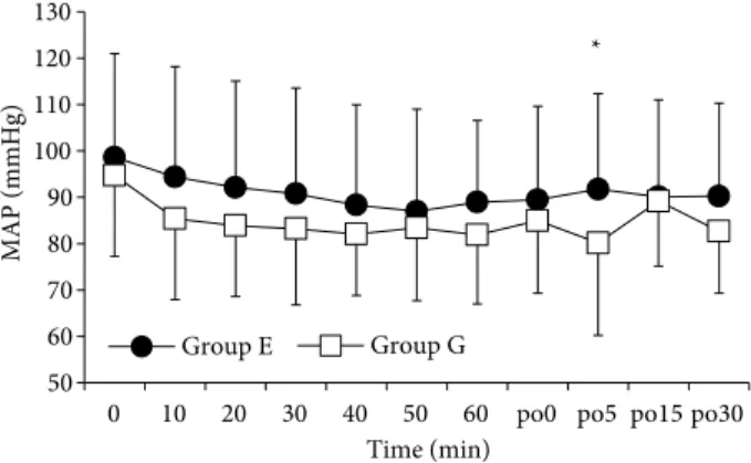
Patients’ characteristics, ASA physical status, and duration of surgery and anesthesia were comparable between the two groups (Table 1). Vital signs including heart rates and mean arterial blood pressure variations were also similar and are presented in Figures 2 and 3.
0 1 2 3 4 5 6 T4 T7 T8 T9 T10 T11 T12 L1 L2 L3 L4 Number of patients Group E Group G
Figure 1. Distribution of the level of the pathologic vertebra in the study groups.
No hemodynamic fluctuation was observed in group E and G patients during the intervention. However, in the PACU, mean blood pressure were higher than 20% of baseline values (P = 0.490) in two patients (8%) in Group G and they were treated with 0.5 mg kg–1 esmolol bolus
over 1 min followed by 50 µg kg–1 min–1 infusion. Oxygen
saturation was almost always over 94% throughout the study periods in group E.
Four patients needed additional intraoperative iv bolus doses of fentanyl (total dose in each patient: 25, 50, 50, and 75 µg) for rescue analgesia in Group E. Fentanyl 1 µg kg–1
was given in all patients during induction of anesthesia in Group G. All the analgesic doses used for intraoperative analgesia are shown in Table 1.
Although VAS scores were lower than 30 mm, 15 patients experienced pain in Group E, but no patient requested analgesic at the first postoperative 4 h. However, all patients in Group G complained of pain and only 8 out of the 25 patients were treated with dexketoprofen. Four patients requested supplement paracetamol due to pain (>30 mm VAS score) despite dexketoprofen treatment. No patient requested tramadol in either group before discharge. VAS scores were significantly lower in Group E than in Group G at the first postoperative 4 h (Figure 4). All the analgesic doses used for postoperative analgesia are shown in Table 2.
The number of patients complaining of pain at the first postoperative 4 h was significantly lower in Group E than Table 1. Patients’ characteristics, duration of surgery, and anesthesia.
Group E (n= 25) Group G(n= 25) P-value Age (years) 67.0 ± 10.4 62.7 ± 14.3 0.232 Weight (kg) 72.7 ± 12.6 70.5 ± 9.3 0.478 Height (cm) 162.8 ± 11.2 159.8 ± 8.6 0.310 Sex (F/M) (%) 20/5 (80/20) 21/4 (84/16) 1.0 ASA I/II/III (%) 2/16/7 (8/64/28) 2/14/9 (8/56/36) 0.825
Duration of surgery (min) 50.9 ± 14.6 46.7±12.6 0.277
Duration of anesthesia (min) 69.3 ± 16.8 64.2±13.4 0.239
Number of patients receiving
intraoperative fentanyl (%) 4 (16) 25 (100)
NA
Total dose (µg) 50.00 ± 20.41 70.60 ± 9.38
Data presented as mean ± SD, n (%). NA: not available. Student’s t test and Fisher’s exact test were used.
50 60 70 80 90 100 110
0 10 20 30 40 50 60 po0 po5 po15 po30
HR (beats/min) Time (min) Group E Group G * * 50 60 70 80 90 100 110 120 130
0 10 20 30 40 50 60 po0 po5 po15 po30
MAP (mmHg)
Time (min)
Group E Group G
*
Figure 2. Heart rate (HR) variables in the two groups. po0, po5, po15, po30: Postoperative 0, 5, 15, 30 min. *: P < 0.05, a significant difference between groups. Data were analyzed by repeated measurements of ANOVA.
Figure 3. Mean arterial blood pressure (MAP) variables in the two groups. po0, po5, po15, po30: Postoperative 0, 5, 15, 30 min. *: P < 0.05, a significant difference between groups. Data were analyzed by repeated measurements of ANOVA.
in Group G (Table 2). Time to first pain experience in the postoperative period was significantly longer in Group E than in Group G (Table 2). Analgesic requirements in the first postoperative 4 h were significantly lower in Group E than in Group G (Table 2).
Patients in Group E were awake and oriented at the end of the operation. Length of stay in the PACU was significantly shorter in Group E than in Group G (Table 2). Thirteen patients were directly transferred to the surgical ward in Group E and the rest stayed in the PACU. Motor functions of the lower extremities were similar and no neurological deficit nor urinary retention was observed in
any patient in Group E. All patients were discharged from the ward uneventfully after 4 h observation.
On call, pain (NRS) scores after 24 h were not statistically significant between the two groups (Group E: 1.5 ± 0.8 vs. Group G: 1.6 ± 0.9, P = 0.67). All patients in the two groups received dexketoprofen 25 mg tablet twice daily on the first day after discharge. Four patients in Group G and two patients in Group E used rescue 50 mg of oral tramadol after discharge (P = 0.66) (Table 2).
No patient experienced postoperative nausea and vomiting (PONV) in Group E, whereas 6 (24%) patients in Group G experienced PONV and were treated with 10 mg metoclopramide intravenously. Incidence of PONV was significantly higher in group G than in group E (P = 0.022).
All procedures were completed successfully and the patients were discharged without any complication related to surgery and anesthesia. All surgeons agreed that there was no difference between the epidural anesthesia and general anesthesia with respect to satisfaction. There was also no significant difference in patient satisfaction between the two groups (number of patients satisfied/ unsatisfied = 22/3 in Group E, and 24/1 in Group G, P = 0.297).
4. Discussion
This study showed that segmental epidural anesthesia offered advantages over general anesthesia in terms of postoperative analgesia and recovery. Therefore, it should be considered a suitable anesthetic technique in patients undergoing single-level percutaneous kyphoplasty.
0 5 10 15 20 25 30 35 40 45 0 1 2 3 4 V isu al an al og ue sc al e ( m m) Time (h) Group E Group G * * * * *
Figure 4. Distribution of the visual analogue scales (VAS) scores in the postoperative period, *: P < 0.05, a significant difference between groups. Friedman’s test was applied for comparisons of VAS levels.
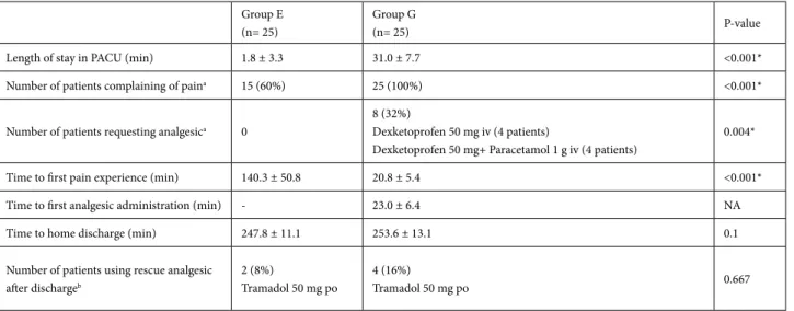
Table 2. Recovery characteristics in the two groups.
Group E (n= 25)
Group G
(n= 25) P-value
Length of stay in PACU (min) 1.8 ± 3.3 31.0 ± 7.7 <0.001*
Number of patients complaining of paina 15 (60%) 25 (100%) <0.001*
Number of patients requesting analgesica 0
8 (32%)
Dexketoprofen 50 mg iv (4 patients)
Dexketoprofen 50 mg+ Paracetamol 1 g iv (4 patients)
0.004*
Time to first pain experience (min) 140.3 ± 50.8 20.8 ± 5.4 <0.001*
Time to first analgesic administration (min) - 23.0 ± 6.4 NA
Time to home discharge (min) 247.8 ± 11.1 253.6 ± 13.1 0.1
Number of patients using rescue analgesic after dischargeb
2 (8%)
Tramadol 50 mg po
4 (16%)
Tramadol 50 mg po 0.667
Data presented as mean ± SD, n (%).*: P < 0.05, a significant difference between groups. Student’s t test, Mann–Whitney U test, Fisher’s exact, or likelihood ratio test were used. PACU: Postanesthetic care unit. NA: not available. a: during the postoperative 4 h. b: All the patients in the two groups received dexketoprofen 25 mg tablet twice daily on the first day after discharge.
Kyphoplasty is a painful procedure and so some clinicians prefer to perform surgery under general anesthesia (1,9). However, especially in elderly patients, general anesthesia may cause postoperative pulmonary complications. Most patients undergoing kyphoplasty are elderly and need longer PACU stay. The mortality rate from pulmonary disease is increased in the elderly and a neuroaxial block may offer advantage for this kind of patient population (2). This was the main reason why we compared segmental epidural anesthesia with general anesthesia.
Segmental epidural anesthesia offers some benefits over lumbar spinal anesthesia in terms of daytime surgery (14). However, spinal block might be related to problems such as high or low level of spinal anesthesia,hypotension, and nausea and vomiting. Additionally, local anesthetic may pool in the sacral region due to lumbar lordosis, and the level of anesthesia may not reach an adequate high segmental level. An unpredictable increase in anesthetic level may cause cardiac and respiratory depression (15). Furthermore, especially in elderly populations, the prone position may significantly exacerbate these complications. Therefore, in the present study, this was the other reason to choose segmental epidural anesthesia.
Kyphoplasty is associated with 8% to 9% risk of cement leakage (16).The neuroaxial block does not cover signs of cement embolism, and most leaks are clinically asymptomatic (17). In the literature, at least 1 mL segment–1 of local anesthetic solution is required
to provide sufficient epidural anesthesia, and 1.5 mL segment–1 of local anesthetic solution for each segment is
advised for thoracal epidural anesthesia in nonpregnant patients (18,19). Considering the advanced age of the study population, 1.25 mL segment–1 of local anesthetic
solution for each thoracal and 1.5 mL segment–1 of local
anesthetic solution for each lumbar segment were used to produce epidural anesthesia.
An epidural catheter was not inserted because the placement of the epidural catheter would be in the surgical area due to the characteristics of the procedure. A low volume of local anesthetic solution was sufficient to offer anesthesia for surgery on a vertebral lesion affecting one segment level. Therefore, local anesthetic solution was given using the single-shot technique.
Segmental epidural anesthesia offers limited distribution of the solution, and prevents hemodynamic fluctuation. In the present study, no serious hemodynamic adverse event was observed in the two groups. Although epidural block extended to T4–5 dermatomal levels, no hemodynamic disturbances were recorded.
Another advantage of segmental epidural anesthesia was that it also allowed us to perform a neurologic examination at the end of the procedure. Patients were awake and oriented at the end of the operation. Additionally, postoperative pain scores and incidence of PONV were significantly lower in Group E patients than in Group G patients. Therefore, segmental epidural anesthesia allowed us to discharge the patients earlier from the PACU.
Percutaneous kyphoplasty is a minimally invasive technique and mostly performed on an outpatient basis (20). In the present study, all patients were discharged uneventfully on the day of surgery. Additionally, patients in the epidural group stayed less in the PACU and had lower VAS scores, less PONV, and less analgesic requirement during the first 4 h postoperatively. These findings supported the benefits of this technique in patients undergoing percutaneous kyphoplasty compared to general anesthesia.
Patient and surgeon satisfaction in this study suggested that segmental epidural anesthesia offered adequate analgesia at the surgical side, and established a good surgical condition. Only four patients needed additional intraoperative iv bolus doses of fentanyl (mean 50 µg) for rescue analgesia in Group E. It was also planned that if adequate analgesia (VAS < 30 mm) had not been achieved in group E patients, we would have increased the dose of local anesthetic. However, no patient needed additional local anesthetic doses after iv fentanyl for rescue analgesia.
One limitation of this study was that patients whose ASA classification was grade I, II, or III were enrolled in the study. Although patients undergoing percutaneous kyphoplasty were generally elderly and had co-morbid disease, patients with ASA class IV or more were not evaluated in this study. Even though there was no significant difference between the two groups in terms of perioperative complications, except PONV, patients having comorbid diseases may benefit from regional anesthesia.
Another limitation was that patients requiring surgical intervention at only one segment were included in the study. Segmental epidural anesthesia may be suitable for these patients. It is obvious that the duration of the intervention will be longer and the complication rate higher as the number of segments is increased.
In conclusion, segmental epidural anesthesia is a safe anesthetic technique for percutaneous kyphoplasty. This technique offers advantages over general anesthesia in terms of postoperative analgesia, analgesic consumption, early recovery, and short PACU stay. Therefore, it should be considered a suitable anesthetic technique in patients undergoing single-level percutaneous kyphoplasty.
References
1. Luginbuhl M. Percutaneous vertebroplasty, kyphoplasty and lordoplasty: implications for the anesthesiologist. Curr Opin Anaesthesiol 2008; 21: 504-513.
2. Frost EAM, Johnson DM. Anesthetic considerations during vertebroplasty, kyphoplasty, and intradiscal electrothermal therapy. Int Anesthesiol Clin 2009; 47: 45-55.
3. Sesay M, Tauzin-Fin P, Jeannin A, Vignes JR, Dousset V, Maurette P. Median effective infusion dose (ED50) of alfentanil for monitored anesthesia care of percutaneous vertebroplasty of osteoporotic fractures. J Neurosurg Anesthesiol 2009; 21: 165-169.
4. Della Puppa A, Andreula C, Frass M. Assisted sedation: a safe
and easy method for pain-free percutaneous vertebroplasty. Minerva Anestesiol 2008; 74: 57-62.
5. Lin BF, Huang YS, Kuo CP, Ju DT, Lu CH, Cherng CH, Wu CT. Comparison of A-line autoregressive index and observer assessment of alertness/sedation scale for monitored anesthesia care with target-controlled infusion of propofol in patients undergoing percutaneous vertebroplasty. J Neurosurg Anesthesiol 2011; 23: 6-11.
6. Mohr M, Pillich D, Kirsch M, Mueller JU, Fleck S, Hosten N, Langner S. Percutaneous balloon kyphoplasty with the patient under intravenous analgesia and sedation: a feasibility study. AJNR Am J Neuroradiol 2011; 32: 649-653.
7. Brinjikji W, Comstock BA, Gray L, Kallmes DF. Local anesthesia with bupivacaine and lidocaine for vertebral fracture trial (LABEL). A report of outcomes and comparison with the investigational vertebroplasty efficacy and safety trial (INVEST). AJNR Am J Neuroradiol 2010; 31: 1631-1634. 8. Venmans A, Klazen CA, Lohle PN, van Rooij WJ. Percutaneous
vertebroplasty and procedural pain. AJNR Am J Neuroradiol 2010; 31: 830-831.
9. Schofer MD, Illian CH, Illian JB, Kortmann HR. Balloon kyphoplasty for recent vertebral fractures in the elderly. Orthopade 2008; 37: 462-469 (in German).
10. Hannallah M, Gibby E, Watson V. Fluoroscopy-guided, small-dose spinal anesthesia for kyphoplasty: a collaborative effort between the anesthesiologist and interventional radiologist. Anesth Analg 2008; 106: 1329-1330.
11. Souvatzis X, Katonis PG, Licoudis SA, Marouli DG, Askitopoulou H. Subarachnoid anesthesia for kyphoplasty: is anesthesia adequate? Anesth Analg 2010; 111: 238-240.
12. Aldrete JA. The post-anesthesia recovery score revisited. J Clin Anesth 1995; 7: 89-91.
13. Chung F. Recovery pattern and home-readiness after ambulatory surgery. Anesth Analg 1995; 80: 896-902.
14. Tzovaras G, Pratsas K, Georgopoulou S. Laparoscopic cholecystectomy using spinal anaesthesia. Br J Anaesth 2007; 99: 744; author reply 745.
15. Kim JT, Shim JK, Kim SH, Jung CW, Bahk JH. Trendelenburg position with hip flexion as a rescue strategy to increase spinal anaesthetic level after spinal block. Br J Anaesth 2007; 98: 396-400.
16. Hadjipavlou AG, Tzermiadianos MN, Katonis PG, Szpalski M. Percutaneous vertebroplasty and balloon kyphoplasty for the treatment of osteoporotic vertebral compression fractures and osteolytic tumours. J Bone Joint Surg Br 2005; 87: 1595-1604.
17. Hulme PA, Krebs J, Ferguson SJ, Berlemann U. Vertebroplasty and kyphoplasty: a systematic review of 69 clinical studies. Spine 2006; 31: 1983-2001.
18. Kleinmen W. Spinal, epidural, & caudal blocks. In: Morgan GE, Mikhail MS, Murray MJ, Larson CP, editors. Clinical Anesthesiology. 3rd ed. New York, NY, USA: Lange Medical Books/McGraw-Hill; 2002. pp: 253-282.
19. Selvan RB, Veliath DG, Rao PB, Ramachandran, Ranjan RV. Cholecystectomy under segmental thoracic epidural block in a patient with twin gestation. Saudi J Anaesth 2012; 6: 73-75. 20. Burton AW, Hamid B. Kyphoplasty and vertebroplasty. Curr