Comparison of L-FABP, ALT and AST Levels in Chronic
Hepatitis C
[Kronik Hepatit C’de L-FABP, ALT ve AST Düzeylerinin Karşılaştırılması]
Research Article [Araştırma Makalesi]
Türk Biyokimya Dergisi [Turkish Journal of Biochemistry–Turk J Biochem] 2011; 36 (2) ; 102–106.
Yayın tarihi 15 Haziran, 2011 © TurkJBiochem.com [Published online 15 June, 2011]
Seren Ozenirler1, Gulbanu Erkan1, Aycan Erkan4, Ceyla Konca1 , Sehri Elbeg2, Gulen Akyol3
Gazi UniversityFaculty of Medicine,Departments of 1Gastroenterology, 2Medical Biochemistry 3Pathology, Ankara, Turkey.
4Ufuk University, Faculty of Medicine, Department
of Cardiology, Ankara, Turkey.
Yazışma Adresi
[Correspondence Address]
Aycan Erkan, MD
Department of Cardiology, Faculty of Medicine, Ufuk University, Dr. Rıdvan Ege Hospital, 86-88 Mevlana Boulevard, Ankara, Turkey Phone: +90(312)2044184
Fax: +90(312)2044055 e-mail: [email protected]
Registered: 1 July 2010; Accepted: 17 February 2011 [Kayıt Tarihi: 01 Temmuz 2010; Kabul Tarihi: 17 Şubat 2011]
ABSTRACT
Purpose: To determine the plasma level of liver- type fatty acid binding protein (L-FABP)
in chronic hepatitis C (CHC), and to compare this to Alanine Aminotransferase (ALT) and Aspartate Aminotransferase (AST) levels.
Methods: We tested 38 biopsy-proven CHC patients and 33 age- and sex- matched healthy
controls in whom biopsy was not performed for ethical considerations. Patients with persis-tently elevated plasma aminotransferase levels (ALT and/or AST), and positive anti-HCV antibody and HCV-RNA results were included, but not those with liver cirrhosis, chronic alcohol consumption, severe and/or acute cardiac, renal, cerebrovascular or intestinal disea-se, seropositive for Hepatitis B virus or HIV. In addition to routine biochemical assessment, L-FABP was determined using a solid phase specific sandwich ELISA.
Results: Plasma ALT and AST level were significantly higher in the patient group than in the
control group (57.7±27.9 IU/L vs 19.0± 7.7 IU/L, p<0.01 and 44.5±19.2 IU/L vs 18.3± 4.2 IU/L, p<0.01, respectively). Plasma L-FABP was significantly higher in the patient group than in the control group (5480.3± 4387.8 ng/mL vs 1710.5± 911.6 ng/mL, p<0.01). No correlation was found between L-FABP and either of the enzyme levels. ROC analysis produced cut-off values above which the likelihood of CHC increased. The cut-off value was 27.5 IU/L for ALT and 23.5 IU/L for AST. The cut-off value for L-FABP was 2600 ng/mL.
Conclusion: L-FABP is elevated significantly in CHC when compared to healthy controls,
independent of aminotranferase levels. Further studies are warranted to evaluate the poten-tial use of L-FABP in the setting of CHC.
Conflict of interest: The authors have no conflict of interest to declare.
Keywords: Chronic Hepatitis C, Hepatocellular Damage, Liver-Type Fatty Acid Binding
Protein, AST, ALT
ÖZET
Amaç: Kronik Hepatit C hastalarında Karaciğer Tipi Yağ Asidi Bağlayıcı Protein (L-FABP)
düzeylerinin belirlenmesi ve bu düzeylerin aspartat aminotransferaz (AST) ve alanin ami-notransferaz (ALT) düzeyleri ile ilişkisinin tanımlanması amaçlanmıştır.
Metotlar: Tanısı biyopsiyle doğrulanmış 38 kronik Hepatit C hastası ve etik nedenlerden
ötürü biyopsi yapılmamış olan 33 sağlıklı kontrol çalışmaya alınmıştır. Süregelen aminot-ransferaz (ALT ve/veya AST) yüksekliği, pozitif anti-HCV antikoru ve pozitif HCV-RNA sonucu diğer ön koşullardır. Karaciğer sirozu, kronik alkol kullanımı, ciddi ve/veya akut kardiyak, renal, serebral veya intestinal hastalığı olanlar, Hepatit B veya HIV için seropozitif hastalar çalışmaya dahil edilmemiştir. Hastaların rutin biyokimyasal değerlendirmelerinin yanı sıra, solid faz özgül sandviç ELISA yöntemi ile plazma L-FABP düzeyleri ölçülmüştür.
Bulgular: Kronik Hepatit C grubunda plazma ALT ve AST değerleri kontrol grubu ile
kar-şılaştırıldığında anlamlı olarak daha yüksek bulunmuştur (sırasıyla 57.7±27.9 IU/L’e karşılık 19.0± 7.7 IU/L, p<0.01 ve 44.5±19.2 IU/L’e karşılık 18.3± 4.2 IU/L, p<0.01). Hasta grubun-da plazma L-FABP düzeyleri kontrol grubuna göre anlamlı derecede yüksek bulunmuştur (5480.3± 4387.8’ ng/mL’ye karşılık 1710.5± 911.6 ng/mL, p<0.01). L-FABP ve aminotransfe-raz düzeyleri arasında korelasyon olmadığı görülmüştür. ROC analizinde, aşıldığında kronik Hepatit C olasılığının arttığı kesim noktaları ALT için 27,5 IU/L, AST için 23,5 IU/L ve L-FABP için 2600,0 ng/mL olarak belirlenmiştir.
Sonuçlar: Sağlıklı kontrollerle karşılaştırıldığında, kronik Hepatit C hastalarında plazma
L-FABP düzeyleri, karaciğer aminotransferaz düzeylerinden bağımsız olarak, anlamlı dere-cede yüksek bulunmuştur. Bulgularımız L-FABP’ın kronik Hepatit C hastalarında hepatosit hasarının bağımsız bir göstergesi olabileceğini düşündürmektedir. Bu hipotez geniş ölçekli çalışmalarla doğrulanmalıdır.
Anahtar Kelimeler: Kronik Hepatit C, Hepatosellüler hasar, Karaciğer Tipi Yağ Asidi
Introduction
Hepatitis C virus (HCV) is one of the most common ca-usative agents of chronic viral hepatitis. Chronic hepati-tis C (CHC) can progress to cirrhosis and eventually to hepatocellular carcinoma, and thus constitutes a major public health problem. Biochemical markers of hepato-cellular injury, especially the liver aminotransferases, have a very important role in the diagnosis, follow-up, and risk stratification of CHC [1-4].
Release of cytoplasmic proteins from damaged hepa-tocytes into the vascular system occurs as a result of vi-ral or toxic hepatitis, ischemia or congestion of the liver, shock, trauma, or rejection after transplantation. Alanine aminotransferase (ALT) and aspartate aminotransferase (AST) are routinely employed for the initial assessment and monitoring of hepatic disease [3]. Currently, ALT is the only marker with adequate specificity for liver dise-ase, but its rather large size (96 kDa) may retard the rise of its concentration in the plasma, thus rendering its use in detection of early stages of liver damage [3]. Alpha glutathione S-transferase (α-GST), which is normally present in liver, kidney and intestine, has been shown to be a useful marker for detection of hepatocellular injury due to rejection after liver transplantation [5].
Markers that can be detected earlier and that are more sensitive than ALT are needed [3]. Fatty acid-binding proteins (FABPs) are a family of intracellular proteins of molecular mass 15 kDa that function in the intracellu-lar transport of long-chain fatty acids. Nine FABPs have been identified of which liver-type (L-FABP) is mainly present in mammalian hepatocytes and enterocytes. It constitutes 2–5% of cytosolic protein content, and very rapidly leaks out of damaged cells, leading to an early rise in plasma level [6-9]. L-FABP has effective endoge-nous protectant, including antioxidant, properties [10-12]. L-FABP binds long-chain fatty acids and other ligands such as fibrates [13] and endogenous PPARα [14]. It ri-ses significantly after bile duct ligation in rats [15], and during episodes of acute rejection of liver transplant [16]. This study was designed to compare plasma L-FABP, AST and ALT levels in CHC patients in view of no pub-lished data concerning L-FABP levels in chronic Hepa-titis C infection.
Materials and Methods
We enrolled 38 biopsy-proven CHC patients (14 males and 24 females) referred to the Gazi University Gastroentero-logy Department and 33 age- and sex- matched healthy controls (15 males and 18 females). All of the patients were newly diagnosed with CHC. The control group con-sisted of individuals without any systemic disease whose biochemical, hematological, virological serum markers (HbsAg, anti-HBc total, anti-HCV, antiHIV) and abdomi-nal ultrasonographies were normal. Liver biopsy was not performed in the control group for ethical considerations. Informed consent was obtained from all subjects. The study protocol was approved by the local ethics committee.
The patient group included those who showed persis-tently elevated serum transaminase levels for 6 months, who were positive for both anti-HCV antibody and HCV-RNA, and had liver biopsy results compatible with CHC. Excluded were patients with liver cirrhosis, those in whom liver biopsy was contraindicated, with positive serological markers for hepatitis B virus or for anti-HIV antibody, with chronic alcohol consumption in excess of 20 g per day, with signs of autoimmune or metabolic li-ver disease, with seli-vere cardiac disease, with acute renal damage or failure, with acute cerebrovascular disease, with skeletal muscle injury or myositis, or with any acu-te inacu-testinal condition.
Plasma samples were assayed for AST and ALT as well as gamma-glutamyl transferase (GGT), bilirubin, and L-FABP levels.
AST was determined using a standard UV absorption technique [17]. L-FABP levels were determined using a specific sandwich ELISA method (Hycult Biotechno-logy B.V., Uden, Netherlands).
Liver biopsies were performed and all were evaluated by the same pathologist using the Metavir scoring system [18].
Serum HCV-RNA level was determined by reverse transcriptase – PCR using a commercial kit (MagAtt-ract Virus Mini M48 Kit, RealTime™ HCV Amplifica-tion Reagent Kit, Abbott) and Anti-HCV antibody was determined by ELISA (chemiluminescence).
Genotype analysis of all subjects was performed by the Line Assay (innolipa) strip method and all were determi-ned as genotype 1b [19].
Results were evaluated using the SPSS 12.0 software (SPSS Inc., Chicago,Illinois, USA). Comparisons were made using the Mann-Whitney U test between two gro-ups [20]. The Pearson Chi-square test [21] and Fisher’s exact test [22] were used for categorical data. ROC analysis was performed for AST, ALT, and L-FABP; area under curve and confidence interval for each of the-se parameters were calculated. Cut-off values determi-ning the likelihood of CHC were also determined. The relation between variables was examined with correlati-on analysis. P values of less than 0.05 were ccorrelati-onsidered to be statistically significant.
Results
Mean age at enrollment was 51.1± 11.1 years in the CHC group and 49.8± 12.8 years in the control group (p=0.556) (Table 1). The distribution of individuals according to body mass index (BMI) did not significantly differ bet-ween the CHC and control groups (p=0.173) (Table 2). Plasma ALT and AST levels were significantly higher in the CHC group than in the control group (57.7±27.9 IU/L vs 19.0± 7.7 IU/L, p<0.01 and 44.5±19.2 IU/L vs 18.3± 4.2 IU/L, p<0.01, respectively). The levels of GGT were higher in the CHC group (48.3±34.3 vs 20.3±9.0, p<0.01). Total and direct bilirubin levels were signifi-cantly higher in the CHC group (see Table 3).
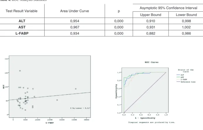
Plasma level of L-FABP was significantly higher in the CHC group than in the control group (5480.3± 4387.8 ng/ mL vs 1710.5± 911.6 ng/mL, p< 0.01). We did not obser-ve any correlation between L-FABP leobser-vels and neither aminotransferase levels (Figure 1).
L-FABP/ALT ratio was not significantly different in the CHC and control groups (94.97 vs 90.02, p=NS). Subgroup analysis of patients with normal (≤40 IU/mL) against those with elevated (≥40 IU/mL) ALT levels showed that the L-FABP levels were not significantly different. However, L-FABP level in patients with nor-mal ALT level tended to be higher than that in controls, but the difference did not reach statistical significance. ROC analysis for ALT, AST, and L-FABP was performed. None of these parameters showed any superiority to the others in terms of area under curve. It was noted that the area under curve approximated the numerical value 1 for all three parameters studied in ROC analysis (Table 4). ROC curves for ALT, AST, and L-FABP are given in Figure 2.
ROC analysis also produced cut-off values above which the likelihood of CHC increased. The cut-off value was 27.5 IU/L for ALT and 23,5 IU/L for AST. The cut-off value for L-FABP was 2600 ng/mL.
Discussion
CHC is a serious condition that leads to debilitating liver cirrhosis and fatal hepatocellular carcinoma. Sensitive detection of hepatocellular injury can aid in diagnosis and in monitoring of CHC. Of the markers routinely used for these purposes, only ALT has adequate sensiti-vity but rises slowly. Thus, a specific marker the levels of which rise rapidly after hepatocellular damage would be ideal.
L-FABP, owing to its small molecular weight (15 kDa), leaks out of damaged hepatocytes very early and ra-pidly following injury. L-FABP levels rise earlier than the levels of ALT, making earlier detection of acute liver transplant rejection possible. The high intracellular
he-Table 1. Age and gender characteristics of CHC and control groups
CHC Control Male, n (%) 14 (36,8) 15 (45,5) Female, n (%) 24 (63,2) 18 (54,5) Age, Mean ± SD 51.1 ± 11.0 49.8 ± 12.8 Numbers in brackets denote percentages
CHC: Chronic Hepatitis C
Table 2. The distribution of individuals according to Body Mass
Index in CHC and control groups
CHC Control n % n % Normal1 13 34,2 14 42,4 Overweight2 15 39,5 16 48,5 Obese3 10 26,3 3 9,1 CHC: Chronic Hepatitis C SD: Standard Deviation
1Body Mass Index between 18.5 and 24.9 kg/m2 2Body Mass Index between 25 and 29.9 kg/m2 3Body Mass Index equal to or greater than 30 kg/m2
Table 3. Biochemical findings
N Mean Median SD Min Max P
ALT CHC 38 57,7 51,5 27,9 16 157 P=0,0001 Control 33 19,0 17 7,7 8 43 AST CHC 38 44,5 42 19,2 19 102 P=0,0001 Control 33 18,3 18 4,2 6 25 GGT CHC 38 48,3 37,5 34,3 10 150 P=0,0001 Control 33 20,3 18 9,0 9 50 T. bil CHC 38 0,8 0,645 0,6 0,19 3,31 P=0,053 Control 33 0,6 0,53 0,2 0,28 1,38 D. bil CHC 38 0,3 0,285 0,2 0,1 0,93 P=0,055 Control 33 0,2 0,21 0,1 0,1 0,46 L-FABP CHC 38 5480,3 5000 4387,8 1700 25600 P=0,0001 Control 33 1710,5 1500 911,6 640 4000 *p<0.05, Statistically Significant
CHC: Chronic Hepatitis C, ALT: Alanine Aminotransferase, AST: Aspartate Aminotransferase, GGT: Gamma-Glutamyl Transferase, T. bil: Total bilirubin, D. bil: Direct bilirubin , L-FABP: Liver Type Fatty Acid Binding Protein
patic concentration of L-FABP increases its sensitivity for hepatocellular damage. L-FABP is normally a cytop-lasmic protein and hence its serum levels are neglectably low. For the sandwich ELISA method, which is used for the detection of L-FABP, the detection limit of the assay is 0.1ng/mL, and the intra- and interassay CVs are <5% and <15%,respectively.
The fact that L-FABP has cysteine and methionine gro-ups implies that this protein may also have antioxidant properties. In an in vitro experimental model of hepatic oxidative stress, it was demonstrated that L-FABP acts as an endogenous cytoprotectant [10]. In our study, we found elevated plasma levels of L-FABP as a result of le-akage from damaged hepatocytes in CHC. Considering that oxidative stress is a major mechanism of hepatocel-lular damage in CHC, the role of cytoplasmic L-FABP against oxidative stress remains to be elucidated in this clinical setting.
To the best of our knowledge, this is the first report on the plasma levels of L-FABP in CHC. We also determi-ned a cut-off value for L-FABP above which the likeliho-od of CHC increases. This cut-off value, which is 2600 ng/mL, may be valuable in aiding the diagnosis and risk stratification of CHC. In ROC analysis, L-FABP was not superior to AST or ALT in terms of area under curve.
However, the knowledge from previous studies that its levels raise earlier than those of AST and ALT may con-fer an advantage on L-FABP.
The main limitations of L-FABP were that our sample size was small and lacking a follow-up, so we could not observe changes of L-FABP levels over time in the pa-tient and control groups.
ALT level may be normal in CHC and marked fibrosis and even cirrhosis can be encountered in patients with such levels [23-25]. This may explain why we found no correlation between ALT and L-FABP levels. However, L-FABP level may reflect hepatocellular damage in presence of normal ALT levels.
Our results suggest that L-FABP may be a sensitive mar-ker of hepatocellular damage in the setting of CHC. This needs to be tested in larger studies. This sensitive mar-ker of hepatocyte injury may potentially be useful for the detection of hepatocyte damage, possibly including flare ups in the course of CHC.
Acknowledgements
We would like to thank Mr. Ahmet Gül for the statistical analysis.
Figure 1. The correlation graphic of ALT and L-FABP. No correlation
was observed between ALT and L-FABP.
Table 4. ROC Analysis Statistics
Test Result Variable Area Under Curve p Asymptotic 95% Confidence Interval Upper Bound Lower Bound
ALT 0,954 0,000 0,910 0,998
AST 0,967 0,000 0,931 1,002
L-FABP 0,934 0,000 0,882 0,986
[17] Rej R. (1984) Measurement of aminotransferases: Part 1. Aspartate aminotransferase. Crit Rev Clin Lab Sci.21 (2):99-186. [18] Bedossa P, Poynard T (1996) An algorithm for the grading of
activity in Chronic Hepatitis C. Hepatology. 24:289-293. [19] Bouchardeau F, Cantaloube JF, Chevaliez S, Portal C,
Ra-zer A, Lefrere JJ, Pawlotsky JM, De Micco P, Laperche S. (2007) Improvement of Hepatitis C Virus (HCV) genotype determinati-on with the new versideterminati-on of the INNO-LIPA HCV assay. Journal of Clinical Microbiology. 45:1140-1145.
[20] Mann HB, Whitney DR. (1947) On a test of whether one of two random variables is stochastically larger than the other. The Annals of Mathematical Statistics.18 (1):50-60.
[21] Pearson K. (1950) On the criterion that a given system of devia-tions from the probable in the case of a correlated system of va-riables is such that it can be reasonably supposed to have arisen from random sampling. Philosophical Magazine.302:157-175. [22] Fisher R.A. (1922) On the interpretation of χ2 from contingency
tables, and the calculation of P. Journal of the Royal Statistical Society.85 (1):87-94.
[23] Puoti C, Castellacci R, Montagnese F. (2000) Hepatitis C virus carriers with persistently normal aminotransferase levels: he-althy people or true patients? Dig Liver Dis.32:634-643. [24] Puoti C, Guido M, Mangia A, Persico M, Prati D. (2003)
Clini-cal management of HCV carriers with normal aminotransferase levels. Dig Liver Dis. 35:362-369.
[25] Puoti C, Magrini A, Stati T, Rigato P, Montagnese F, Rossi P, et al (1997) Clinical, histological, and virological features of hepatitis C virus carriers with persistently normal or abnormal alanine transaminase levels. Hepatology. 26:1393-1398.
References
[1] Lee MH, Yang HI, Lu SN, Jen CL, Yeh SH, Liu CJ, Chen PJ, You SL, Wang LY, Chen WJ, Chen CJ. (2010) Hepatitis C virus
seromarkers and subsequent risk of hepatocellular carcinoma: long term predictors from a community-based cohort study. J Clin Oncol. 28(30): 4587-4593.
[2] Liu CH, Liang CC, Liu CJ, Hsu SJ, Lin JW, Chen SI, Hung PH, Tsai HB, Lai MY, Chen PJ, Chen JH, Chen DS, Kao JH. (2010) The ratio of aminotransferaseto platelets is a useful index for predicting hepatic fibrosis in hemodialysis patients with chronic hepatitis C. Kidney Int. 78(1):103-109.
[3] Dufour DR, Lott JA, Nolte FS, Gretch DR, Koff RS, Seeff LB. (2000) Diagnosis and monitoring of hepatic injury: I. Performan-ce characteristics of laboratory tests. Clin Chem. 46(12):2027– 2049.
[4] Trull AK. (2001) The clinical validation of novel strategies for monitoring transplant recipients. Clin Biochem. 34:3–7. [5] Hughes VF, Trull AK, Gimson A, Friend PJ, Jamieson N,
Dun-can A, et al. (1997) Randomized trial to evaluate the clinical benefits of serum alpha-glutathione S-transferase concentra-tion monitoring after liver transplantaconcentra-tion. Transplantaconcentra-tion. 64:1446–1452.
[6] Glatz JF, van der Vusse GJ. (1996) Cellular fatty acid-binding proteins: their function and physiological significance. Prog Li-pid Res. 35:243–282.
[7] Bass NM, Barker ME, Manning JA, Jones AL, Ockner RK. (1989) Acinar heterogeneity of fatty acid-binding protein exp-ression in the livers of male, female and clofibrate-treated rats. Hepatology 9:12–21.
[8] Ockner RK, Manning JA, Kane JP. (1982). Fatty acid-binding protein. Isolation from rat liver, characterization, and immunoc-hemical quantification. Biol Chem. 257:7872–7878.
[9] Gordon JI, Alpers DH, Ockner RK, Strauss AW. (1983) The nuc-leotide sequence of rat liver fatty acid-binding protein mRNA. J Biol Chem. 258:3356–3363.
[10] Rajaraman G, Wang GQ, Yan J, Jiang P, Gong Y, Burczynski FJ. (2007) Role of cytosolic liver fatty acid binding protein in hepatocellular oxidative stress: effect of dexamethasone and clo-fibrate treatment. Mol Cell Biochem. 295(1-2):27-34.
[11] Wang G, Gong Y, Anderson J, Sun D, Minuk G, Roberts MS, Burczynski FJ. (2005) Antioxidative function of L-FABP in L-FABP stably transfected Chang liver cells. Hepatology. 42(4):871-879.
[12] Liu Y, Wang J, Wei Y, Zhang H, Xu M, Dai J. (2008) Induc-tion of time-dependent oxidative stress and related transcriptio-nal effects of perfluorododecanoic acid in zebrafish liver. Aquat Toxicol. 89(4):242-250.
[13] Chuang S, Velkov T, Horne J, Wielens J, Chalmers DK, Porter CJ, Scanlon MJ. (2009) Probing the fibrate binding specifity of rat liver fatty acid binding protein. J Med Chem. 52(17):5344-5355.
[14] Hostetler HA, McIntosh AL, Atshaves BP, Storey SM, Payne HR, Kier AB, Schroeder F. (2009) L-FABP directly interacts with PPARα in cultured primary hepatocytes. J Lipid Res. 50(8):1663-1675.
[15] Wang G, Shen H, Rajaraman G, Roberts MS, Gong Y, Ji-ang P, Burczynski F. (2007) Expression and antioxidant function of liver fatty acid binding protein in normal and bile-duct ligated rats. Eur J Pharmacol. 560(1):61-68.
[16] Pelsers MAL, Morovat A, Alexander GJM, Hermens WT, Trull AK, Glatz JFC. (2002) Liver Fatty Acid Binding Protein as a Sensitive Serum Marker of Acute Hepatocellular Damage in Liver Transplant Recipients. Clinical Chemistry 48:2055-2057.