HEADACHE & FACIAL PAIN SECTION
Original Research Article
Duration of Migraine Is Associated with Cardiac
Diastolic Dysfunction
Berkay Ekici, MD,* Isin Unal-Cevik, MD, PhD,†
Ebru Akgul-Ercan, MD,* Gulin Morkavuk, MD,†
Yavuz Yakut, PhD,‡and Aycan F. Erkan, MD, PhD* Departments of *Cardiology and
†Neurology, Faculty of Medicine, Ufuk University,
Ankara;
‡Department of Physiotherapy and Rehabilitation,
Hacettepe University, Faculty of Health Sciences, Ankara, Turkey
Reprint requests to: Isin Unal-Cevik, MD, PhD, Department of Neurology, Faculty of Medicine, Ufuk University, Balgat, 06520-Ankara, Turkey. Tel:+90(312) 204-4000; Fax:+90(312) 287-2390; E-mail:
Disclosure: The authors report no conflicts of interest. Authors’ Contributions: BE has contributed in
conception and design, acquisition of data, analysis and interpretation of data, and involved in drafting and revising the manuscript; IUC has contributed in conception and design, coordination, acquisition analysis, and interpretation of data, and involved in drafting and revising the manuscript critically for important intellectual content; EAE has contributed in acquisition of data, analysis and interpretation of data, and involved in revising the manuscript; GM has contributed in acquisition of data, analysis, and interpretation of data; YY has contributed in statistical analysis and interpretation of data, AFE has
contributed in design of the manuscript. All authors read and approved the final manuscript.
Authors’ Information: BE is staff cardiologist in Ufuk University Faculty of Medicine, Department of Cardiology; IUC is a neurologist, neuroscientist, and pain specialist, currently Professor and the
chairperson in Ufuk University Faculty of Medicine Department of Neurology; EAE is Associate Professor and cardiologist in Ufuk University Faculty of
Medicine, Department of Cardiology; GM is the chief neurology resident in Ufuk University Faculty of Medicine Department of Neurology; YY is currently Professor in Hacettepe University Faculty of Health
Sciences, Department of Physiotherapy and Rehabilitation; AFE is Assistant Professor and cardiologist in Ufuk University Faculty of Medicine, Department of Cardiology.
Abstract
Objective. Migraine is a common type of headache accompanied or preceded by signs of central and autonomic nervous system dysfunction. Autonomic dysfunction has been suggested to be a potential contributor to impaired cardiac diastolic function. Cardiac diastolic dysfunction is characterized by normal left ventricular contractility but impaired ventricular relaxation. It is a growing clinical entity implicated in morbidity and mortality due to heart failure. The aim of this study was to determine if any relationship exists between migraine and diasto-lic dysfunction.
Methods. Migraineurs (N=55), and age- and sex-matched healthy controls (N=52) were evaluated by conventional and tissue Doppler echocardio-graphy. Migraine-related disability in the previous 3 months was assessed by the Migraine Disability Assessment questionnaire. Baseline characteris-tics were recorded, and blood samples were collected.
Results. The groups did not differ in terms of sex or age. The migraine group had higher lipid levels compared with the control group. Diastolic dys-function was significantly higher among the 30 migraineurs with a history of migraine of 10 years or more compared with the 25 migraineurs with a history of less than 10 years, (P=0.003). In logistic regression analysis, migraine duration was shown to be an independent predictor of diastolic dys-function (odds ratio 1.130, 95% confidence interval,
P=0.044).
Conclusions. Cardiac diastolic dysfunction is asso-ciated with migraine. A long history of migraine is an independent predictor of diastolic dysfunction. Key Words. Headache; Risk Factor; Hyperlipi-demia; Quality of Life; Burden of Disease
Pain Medicine 2013; 14: 988–993 Wiley Periodicals, Inc.
Introduction
A headache of usually unilateral and throbbing type, lasting 4–72 hours, accompanied by nausea, vomiting, photophobia, and phonophobia, worsening with physical activity, and with aura (reported in 20% patients) not attrib-uted to another disorder is called migraine [1]. Migraine is a common primary headache with a prevalence of 15–17% in females and 6% in males [2]. It may be trig-gered by a variety of environmental factors all thought to activate meningeal nociceptors, and is further associated with a local neurogenic inflammatory response [3]. More-over, skipping a meal, stress, and sleep disturbances are thought to activate the hypothalamus, perifornical areas, and stria terminalis, which converge on the superior sali-vatory nucleus and parasympathetic sphenopalatine gan-glion, leading to increased sensitivity of the meningeal nociceptors [4]. Brainstem activation is observed during a migraine attack, mainly in the dorsal pons [5]. Autonomic nervous system (ANS) symptoms characterized by nausea, vomiting, flushing, pallor, diarrhea, diaphoresis, or polyuria may accompany acute migraine attacks due to activation of the hypothalamus and brainstem autonomic nuclei [6]. Autonomic dysfunction in migraine is shown by many studies based on sudomotor axon reflex, changes in blood pressure measurements and heart rate variability during resting and active standing, deep breathing, the Valsalva maneuver, and the tilt-table test [7,8]. Results of pupillometry or sweating tests are controversial; they report either sympathetic hypofunction or hyperfunction in migraine [9,10]. However, there is overall agreement that an autonomic dysregulation is present in migraine [8]. ANS regulates heart rate and cardiac contractility by dual effects of the sympathetic and parasympathetic nerves. Comprehensive electrocardiographic analyses have pro-vided more information in terms of the detection of abnor-malities in heart rate variability related to the ANS dysfunction in migraineurs [11].
Cardiac diastolic dysfunction is characterized by normal left ventricular contractility but impaired ventricular relax-ation [12]. Numerous conditions such as diabetes mellitus, obesity, hypertension, advanced age, cardiomyopathy, constrictive pericarditis, coronary heart diseases, and sleep apnea are associated with diastolic dysfunction [13]. To the best of our knowledge, the relationship between migraine and diastolic dysfunction has not been studied. In this age- and sex-matched, case-control study, we aimed to determine if any relationship exists between migraine and diastolic dysfunction.
Methods
The subjects enrolled had an adequate level of under-standing of the questionnaires, were aged between 18 and 60 years, and had given written consent. The study was approved by the Local Ethical Committee. Migraine was diagnosed by two neurologists according to The International Classification of Headache Disorders: 2nd edition criteria [1]. Subjects who were older than 60 years;
who had a history of diabetes, renal disease, cardiomy-opathy, coronary heart disease, valvular heart disease, hypertension, or obesity; or were taking any migraine pro-phylactic medications such as antihypertensives were excluded due to their effects on cardiac diastolic function. The control group consisted of age- and sex-matched healthy volunteers, who were admitted to the neurology outpatient clinic due to symptoms unrelated to headache, syncope, stroke, or cardiovascular disease. A detailed form that included demographic characteristics, the diag-nosis, clinical characteristics, and Migraine Disability Assessment questionnaires (MIDAS) [14] were assessed by the neurologists. MIDAS is a quickly applied, 5-item questionnaire that evaluates the disability and impact of migraine headaches on daily living in the past 3-month period. All subjects gave baseline blood samples. Conventional and tissue Doppler echocardiography were performed by cardiologists who were blind to the clinical status of the subjects. The echocardiographic assess-ments were performed during headache-free intervals in migraineurs. Cardiac diastolic dysfunction is characterized by normal left ventricular contractility but impaired ventricu-lar relaxation [12]. There are four basic echocardiographic patterns of diastolic dysfunction graded from I to IV in which the mildest form is grade I. Grade II diastolic dysfunction is called pseudonormal filling dynamics and is considered a moderate diastolic dysfunction. Grade III and IV diastolic dysfunctions represent restrictive filling dynamics [15,16]. In the present study, a Vingmed System echocardiography unit (Vivid 7 Pro, General Electric, Horten, Norway) was used. A sample volume of 2 mm was placed between the mitral leaflet tips, E and A velocities were measured, and the E/A ratio was calculated. Using continuous-wave Doppler tracings, isovolumetric contraction time (ICT), isovolumet-ric relaxation time (IVRT), and aortic ejection time (ET) were measured. The Doppler-derived myocardial performance index (MPI) (also denoted as the Tei-index) is an index of combined cardiac systolic and diastolic function [17]. It is defined as the sum of ICT and IVRT divided by the ET. Tissue Doppler was employed to measure systolic (S) and diastolic (E′ and A′) mitral annular velocities.
Statistical Analysis
To show a statistically significant difference between migraine and diastolic dysfunction, the required sample size of a minimum of 32 volunteers in each group was calculated (Alpha= 0.05 and Beta = 0.20 [Power 80%]). The data were analyzed with the PASW Statistics version 18 software package (SPSS Hong Kong Headquarters, Quarry Bay, Hong Kong). The normal distribution of vari-ables was verified by the Kolmogorov–Smirnov test. Spearman’s rho correlation was used when one or both of the variables were not normally distributed. Comparisons between the groups were made with either Student’s t-test or the Mann–Whitney U-test. A chi-square (c2) test
was used to investigate whether distributions of categori-cal variables differed within the groups. Logistic regression analyses were conducted according to body mass index (BMI), total cholesterol, and history of migraine duration.
Data are shown as mean⫾ standard deviation for con-tinuous variables and absolute numbers (%) for dichoto-mous variables. P values less than 0.05 were considered statistically significant.
Results
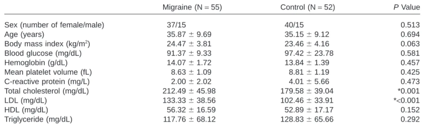
In this study, 107 subjects were included. Demographic characteristics, and hematological and biochemical parameters of the migraineurs (N= 55) and healthy con-trols (N= 52) are displayed in Table 1. Age, sex, and BMI did not differ between the groups (all P> 0.05). Mean total cholesterol and low-density lipoprotein (LDL)-cholesterol values were higher in the migraineurs compared with the control group as well (212.49⫾ 45.98 vs 179.58 ⫾ 39.04, P= 0.001 and 133.33⫾ 38.56 vs 102.46 ⫾ 33.91, P< 0.001, respectively). Blood glucose, C-reactive protein, hemogram, and MPV values did not differ between the groups.
According to the ICHD-II criteria, among the 55 migraineurs, 23 were diagnosed with episodic migraine with aura, 27 with episodic migraine without aura, and 5 with chronic migraine. The frequency of migraine attacks in episodic migraine and chronic migraine patients were
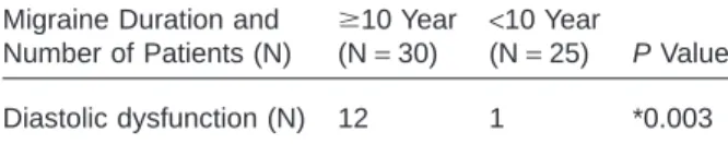
4.22⫾ 3.17 and 17.60 ⫾ 2.30 days/month, respectively. On echocardiographic examinations, atrial and ventricular dimensions were found to be within normal limits. We did not observe any echocardiographic findings of concentric hypertrophy, left atrial enlargement, etc., but two of the migraine with aura patients had patent foramen ovale. Diastolic dysfunction was found in 23.6% of the migraine group (eight had Grade II and five had Grade I) and 3.8% of the control group (one had Grade I and the other had Grade II diastolic dysfunction); the difference between the groups was statistically significant (P< 0.05) (Table 2). Among the 13 migraineurs with diastolic dysfunction, 5 had episodic migraine with aura. The mean MPI scores were also significantly higher in migraineurs (0.41⫾ 0.10) compared with the control group (0.35⫾ 0.05) (P= 0.014) (Table 2). In order to assess the effect of migraine burden on diastolic dysfunction, we divided the patients into two groups according to history of migraine of less than 10 years or of 10 years or more. The mean migraine durations of the migraine groups, 10 years or more vs less than 10 years, were 16.60⫾ 5.90 and 4.80⫾ 2.22 years, respectively. The difference was statistically significant (t= 9.440, P< 0.001). Migraine patients with a history of 10 years or more were signifi-cantly more prone to have diastolic dysfunction as well
Table 1 Comparisons of baseline characteristics and blood values between the groups
Migraine (N=55) Control (N=52) P Value
Sex (number of female/male) 37/15 40/15 0.513 Age (years) 35.87⫾9.69 35.15⫾9.12 0.694 Body mass index (kg/m2) 24.47⫾3.81 23.46⫾4.16 0.063
Blood glucose (mg/dL) 91.37⫾9.33 97.42⫾23.78 0.581 Hemoglobin (g/dL) 14.07⫾1.72 13.84⫾1.39 0.457 Mean platelet volume (fL) 8.63⫾1.09 8.81⫾1.19 0.425 C-reactive protein (mg/L) 2.00⫾2.02 4.01⫾5.66 0.473 Total cholesterol (mg/dL) 212.49⫾45.98 179.58⫾39.04 *0.001 LDL (mg/dL) 133.33⫾38.56 102.46⫾33.91 *<0.001 HDL (mg/dL) 56.32⫾16.59 52.89⫾17.17 0.152 Triglyceride (mg/dL) 117.76⫾68.12 128.83⫾65.66 0.292 * P<0.05.
HDL=high-density lipoprotein; LDL=low-density lipoprotein.
Data are shown as mean⫾standard deviation for continuous variables.
Table 2 Continuous wave Doppler-derived parameters and comparisons between the groups
Migraine (N=55) Control (N=52) P Value
Diastolic dysfunction, N (%) 13 (23.6%) 2 (3.8%) *0.004 AET (milliseconds) 300.44⫾30.43 325.58⫾33.46 *<0.001 IVRT (milliseconds) 73.27⫾13.41 70.69⫾9.91 0.317 ICT (milliseconds) 47.49⫾15.15 43.50⫾9.25 0.463 MPI 0.41⫾0.10 0.35⫾0.05 *0.010 * P<0.05.
AET=aortic ejection time; IVRT=isovolumetric relaxation time; ICT=isovolumetric contraction time; MPI=myocardial perfor-mance index; N=number of patients.
(P= 0.003) (Table 3). There was no significant relationship between MIDAS groups and presence of diastolic dys-function (Table 4). In the logistic regression analysis, the relationship between diastolic dysfunction and migraine duration was independent of BMI and total cholesterol levels (P= 0.044, odds ratio = 1.130, 95% confidence interval= 1.003–1.272) (Table 5).
Discussion
Migraine is associated with ANS symptoms and signs. Diastolic dysfunction is a chronic process associated with autonomic neuropathy or dysfunction [18,19]. To our knowledge, this is the first study designed to assess the relationship between migraine and cardiac diastolic
func-tion. Here, we report that the prevalence of diastolic dys-function was significantly higher in migraineurs compared with healthy controls. Compared with the healthy controls, lipid levels were also increased in migraineurs. In the logis-tic regression analysis, a migraine history of 10 years or more was found to be an independent predictor for devel-oping diastolic dysfunction.
There is growing evidence of an association between migraine and an increased risk for ischemic stroke and other cardiovascular events [20]. Although migraine is reported to be a risk factor for stroke and coronary heart disease, the risk is low in the general population, and it is important to identify which migraineurs will develop these events [21]. The identification of comorbidities in migraineurs is essential in our understanding of the patho-physiology of migraine and assessing the therapeutic options [22]. In a case-control study, the aortic stiffness measured by aortic pulse wave velocity was found to be higher in young migraineurs without significant cardiovas-cular risk factors [23]. In another study investigating the vascular dysfunction associated with migraine, migraine with aura patients and healthy controls were assessed by carotid intima-media thickness, and endothelial function by using peripheral arterial tonometry [24]. Although the peripheral endothelial function was not impaired, arterial stiffness was found to be greater in migraineurs [24]. The authors concluded that these findings might contribute to the increased stroke risk in migraineurs [24]. Cardiac dias-tolic dysfunction with preserved ejection fraction is a growing clinical entity implicated in morbidity and mortality due to heart failure [25]. The role of specific treatments for diastolic dysfunction per se is unclear. Therapy is directed to reduce associated risk factors such as obesity, hypertension, and high blood glucose levels. In diabetic patients, decreased myocardial 123I-metaiodobenzylguanidine uptake was shown to be asso-ciated with diastolic dysfunction [26]. Recently, in a rat model of insulin resistance with hyperglycemia, sympa-thetic nervous dysregulation was proposed in the devel-opment of diastolic dysfunction in the absence of systolic left ventricular dysfunction as well [27]. However, our migraine patients did not have a history of diabetes, and there was no significant difference in glucose levels com-pared with the control group. Age is an important factor that influences the prevalence of diastolic dysfunction [28]; thus, we limited our study population to age 60 and under. Hypertension, diabetes, and obesity are implicated in the development of symptomatic myocardial dysfunction in individuals with normal coronary arteries [29]. In our study, we also did not include subjects with a history of hyper-tension, coronary heart disease, or arrhythmia. Another risk factor for diastolic dysfunction is obesity [30]. Inter-estingly, migraine is also associated with obesity [31]. It is reported that increased inflammatory mediators, vascular hyperreactivity, adipocytokines, and sympathetic tone are associated with an increase in headache frequency [31]. In our study, migraine patients’ BMI values were less than 30 kg/m2, and there were no significant differences
between the migraineurs and the controls. Dyslipidemia, a component of metabolic syndrome, has also been
Table 3 The correlation between migraine duration and diastolic dysfunction
Migraine Duration and Number of Patients (N) ⱖ10 Year (N=30) <10 Year (N=25) P Value Diastolic dysfunction (N) 12 1 *0.003 * P<0.05.
Table 4 Distribution of the migraineurs’ MIDAS grades and diastolic dysfunction
MIDAS (N) Number of Migraineurs with Diastolic Dysfunction Number of Migraineurs without Diastolic Dysfunction Grade 1 2 5 Grade 2 4 6 Grade 3 4 13 Grade 4 3 18
MIDAS=Migraine Disability Assessment Questionnaire;
MIDAS Grade 1=little or no disability; Grade 2=mild disability; Grade 3=moderate disability; Grade 4=severe disability.
Table 5 Results of multivariate analysis of migraine and diastolic dysfunction (logistic regression model without interaction)
OR (95% CI) P Value
Migraine duration (year) 1.130 (1.003–1.272) *0.044 BMI (kg/m2) 1.157 (0.928–1.444) 0.195
Total cholesterol (mg/dL) 1.015 (0.997–1.033) 0.097
* P<0.05.
BMI=body mass index.
Odds ratio (OR) is statistically significant if confidence interval (CI) does not include 1.
associated with migraine [32]. In our study, similar to the literature, our migraine group had significantly higher total and LDL cholesterol levels compared with the controls.
In order to support evidence for migraine’s impact over the patient’s lifetime, we divided our patients into two groups according to migraine history of less than 10 years or of 10 years or more. Diastolic dysfunction was significantly higher in the migraineurs with a history of 10 years or more. Furthermore, in the logistic regression analysis, migraine duration was shown to be an independent predictor of diastolic dysfunction. MIDAS assesses migraine-related disability in the past 3 months. In our study, we did not find a significant relationship between MIDAS scores and dias-tolic dysfunction. However, being a university hospital, the migraine patients who were referred to us had higher MIDAS scores. Only one patient, reporting a history of migraine of less than 10 years and MIDAS grade 4, had diastolic dysfunction. These findings may further strengthen our hypothesis that a long migraine history may cumulatively contribute to the susceptibility of the migraine sufferers to develop diastolic dysfunction. Still, we think that large-scale prospective studies would provide greater statistical power. In future, new studies investigating the effect of migraine prophylactic medications on diastolic dysfunction might further clarify several issues.
Limitations of the Study
In this study, we did not include tests to evaluate auto-nomic dysfunction such as heart rate variability, changes in blood pressure measurements during resting and active standing, deep breathing, the Valsalva maneuver, the tilt-table test, sudomotor axon reflex, or sweating tests in our study population. However, based on previous studies, there is overall agreement that an autonomic dysregula-tion is present in migraine. Our study populadysregula-tion consisted of 13 migraineurs with diastolic dysfunction. Large-scale prospective studies are needed to obtain further informa-tion if cardiac diastolic dysfuncinforma-tion is a cause or conse-quence, or simply an epiphenomenon of a persistent autonomic dysregulatory process that is promoted by migraine or is a constitutional factor that promotes persis-tent migraine.
Conclusion
Migraine has a negative impact on general well-being. Cardiac diastolic dysfunction might represent a footprint of autonomic imbalance in migraineurs with a more than 10-year history. Echocardiography is a practical and noninvasive additional method to define the risk of cardiac disease in patients with a long history of migraine.
Acknowledgments
The authors would like to thank Drs. Utku Kutuk, MD; Gunhan Gultekin Demir, MD; Ferda Ince, MD; and Aslihan Alhan, PhD, for their technical assistance.
References
1 Headache Classification Subcommittee of the Interna-tional Headache Society. The internaInterna-tional classifica-tion of headache disorders, 2nd ediclassifica-tion. Cephalalgia 2004;24(suppl 1):9–160.
2 Stewart WF, Shechter A, Rasmussen BK. Migraine prevalence. A review of population-based studies. Neurology 1994;44:S17–23.
3 Levy D, Strassman AM, Burstein R. A critical view on the role of migraine triggers in the genesis of migraine pain. Headache 2009;49:953–7.
4 Burstein R, Jakubowski M. Unitary hypothesis for mul-tiple triggers of the pain and strain of migraine. J Comp Neurol 2005;493:9–14.
5 May A. New insights into headache: An update on functional and structural imaging findings. Nat Rev Neurol 2009;5:199–209.
6 Goadsby PJ, Charbit AR, Andreou AP, Akerman S, Holland PR. Neurobiology of migraine. Neuroscience 2009;161:327–41.
7 Mosek A, Novak V, Opfer-Gehrking TL, Swanson JW, Low PA. Autonomic dysfunction in migraineurs. Head-ache 1999;39:108–17.
8 Shechter A, Stewart WF, Silberstein SD, Lipton RB. Migraine and autonomic nervous system function: A population-based, case-control study. Neurology 2002;58:422–7.
9 Gomi S, Gotoh F, Komatsumoto S, et al. Sweating function and retinal vasomotor reactivity in migraine. Cephalalgia 1989;9:179–85.
10 Herman P. The pupil and headaches. Headache 1983;23:102–5.
11 Pierangeli G, Parchi P, Barletta G, et al. Power spec-tral analysis of heart rate and diastolic blood pressure variability in migraine with and without aura. Cephala-lgia 1997;17:756–60.
12 Kitabatake A, Inoue M, Asao M, et al. Transmitral blood flow reflecting diastolic behavior of the left ven-tricle in health and disease—a study by pulsed Doppler technique. Jpn Circ J 1982;46:92–102. 13 Kuznetsova T, Herbots L, Jin Y, Stolarz-Skrzypek K,
Staessen JA. Systolic and diastolic left ventricular dys-function: From risk factors to overt heart failure. Expert Rev Cardiovasc Ther 2010;8:251–8.
14 Stewart WF, Lipton RB, Whyte J, et al. An international study to assess reliability of the Migraine Disability Assessment (MIDAS) score. Neurology 1999;53:988– 94.
15 Hurrell DG, Nishimura RA, Ilstrup DM, Appleton CP. Utility of preload alteration in assessment of left ven-tricular filling pressure by Doppler echocardiography: A simultaneous catheterization and Doppler echocar-diographic study. J Am Coll Cardiol 1997;30:459–67. 16 Nishimura RA, Tajik AJ. Evaluation of diastolic filling of left ventricle in health and disease: Doppler echocar-diography is the clinician’s Rosetta Stone. J Am Coll Cardiol 1997;30:8–18.
17 Tei C. New non-invasive index for combined systolic and diastolic ventricular function. J Cardiol 1995; 26:135–6.
18 Dinh W, Futh R, Lankisch M, et al. Cardiovascular autonomic neuropathy contributes to left ventricular diastolic dysfunction in subjects with type 2 diabetes and impaired glucose tolerance undergoing coronary angiography. Diabet Med 2011;28:311–8.
19 Pop-Busui R, Kirkwood I, Schmid H, et al. Sympa-thetic dysfunction in type 1 diabetes: Association with impaired myocardial blood flow reserve and diastolic dysfunction. J Am Coll Cardiol 2004;44:2368–74. 20 Sacco S, Ricci S, Carolei A. Migraine and vascular
diseases: A review of the evidence and potential implications for management. Cephalalgia 2012;32: 785–95.
21 Sacco S, Cerone D, Carolei A. Comorbid neuro-pathologies in migraine: An update on cerebrovascular and cardiovascular aspects. J Headache Pain 2008; 9:237–48.
22 Sacco S, Olivieri L, Bastianello S, Carolei A. Comorbid neuropathologies in migraine. J Headache Pain 2006;7:222–30.
23 Schillaci G, Sarchielli P, Corbelli I, et al. Aortic stiffness and pulse wave reflection in young subjects with migraine: A case-control study. Neurology 2010; 75:960–6.
24 Liman TG, Neeb L, Rosinski J, et al. Peripheral endot-helial function and arterial stiffness in women with
migraine with aura: A case-control study. Cephalalgia 2012;32:459–66.
25 Nishimura RA, Jaber W. Understanding “diastolic heart failure”: The tip of the iceberg. J Am Coll Cardiol 2007;49:695–7.
26 Mustonen J, Mantysaari M, Kuikka J, et al. Decreased myocardial 123I-metaiodobenzylguanidine uptake is associated with disturbed left ventricular diasto-lic filling in diabetes. Am Heart J 1992;123:804– 5.
27 Thackeray JT, Radziuk J, Harper ME, et al. Sympa-thetic nervous dysregulation in the absence of systolic left ventricular dysfunction in a rat model of insulin resistance with hyperglycemia. Cardiovasc Diabetol 2011;10:75.
28 Negri F, Sala C, Valerio C, Mancia G, Cuspidi C. Role of tissue Doppler imaging for detection of diastolic dysfunction in the elderly: A study in clinical practice. High Blood Press Cardiovasc Prev 2011;18:187– 93.
29 Dwyer EM, Asif M, Ippolito T, Gillespie M. Role of hypertension, diabetes, obesity, and race in the development of symptomatic myocardial dysfunction in a predominantly minority population with normal coronary arteries. Am Heart J 2000;139:297– 304.
30 Powell BD, Redfield MM, Bybee KA, Freeman WK, Rihal CS. Association of obesity with left ventricular remodeling and diastolic dysfunction in patients without coronary artery disease. Am J Cardiol 2006; 98:116–20.
31 Bigal ME, Lipton RB, Holland PR, Goadsby PJ. Obesity, migraine, and chronic migraine: Possible mechanisms of interaction. Neurology 2007;68:1851– 61.
32 Rist PM, Tzourio C, Kurth T. Associations between lipid levels and migraine: Cross-sectional analysis in the epidemiology of vascular ageing study. Cephalal-gia 2011;31:1459–65.