ABSTRACT
This work is licensed under a Creative Commons Attribution-NonCommercial 4.0 International License.
Sabiha Çağlayan1 , Duygu Gözen2
Effect of Supine and Prone Flexion Positions on
Heart Rate, Oxygen Saturation, and Pain Score of
Preterm Infants
Objective: The study aimed to determine the effect of supine and prone flexion positions on heart rate, oxygen saturation,
and pain level of preterm infants in the neonatal intensive care unit.
Materials and Methods: This study consisted of 46 preterm infants in a university hospital between February and
Novem-ber 2014. The effects of two different positions on heart rate, oxygen saturation, and pain were assessed by giving both positions to each infant for 2 h.
Results: Heart rate increased in the prone flexion position, but oxygen saturation was similar in both positions. In addition,
pain score decreased on the second hour in prone position.
Conclusion: The results showed that infants were influenced more by environmental stimuli in thesupine flexion position. Keywords: Neonatal intensive care, prone positioning, flexion positioning
INTRODUCTION
Positioning of preterm infants is an important developmental care practice in the neonatal intensive care unit (NICU) (1–3). Currently, the Newborn Individualized Developmental Care and Assessment Program (NIDCAP) is reported to be an efficient care method to ensure decreased occurrence of behaviors associated with pain in newborns (2, 4).
Fetal movements start to become active in the second to third month of gestation. The fetus turns, stretches, sucks its fingers and toes, and holds them during its intrauterine life. These movements help the fetus to both gain prenatal movement experience and increased muscle growth. Because they are born preterm, their muscle and bones are not yet fully developed (5–7). Infants are in flexion position intrauterine. However, preterm infants need support to remain in flexion position in their extrauterine life. Thus, providing preterm infants with appropriate positions regularly and supporting them in their extrauterine life will positively affect both their movement and musculoskeletal development (7–9).
Environment-related care practices support neurodevelopment and posture of preterm infants (1, 10–12). Be-cause NIDCAP is not a care model designed specific to pain, the evaluation of clues regarding clinical pain assess-ment and manageassess-ment is considered important within the scope of NIDCAP (3, 4, 9).
In addition to supporting the body, positioning is the NIDCAP healthcare practice that will help in maintaining and protecting the body posture of preterm infants in the future. The determination of positions of newborns in accordance with NIDCAP and assessment of its outcomes are recommended for nurses (4, 7, 10–13).
MATERIALS and METHODS
Design
This cross-over study was conducted to compare the effect of supine flexion position (SFP) and prone flexion po-sition (PFP) on heart rate (HR), oxygen saturation (SPO2), and pain level of preterm infants who were hospitalized in the NICU of a private university hospital in Istanbul.
Sample
The study population consisted of 438 infants who were hospitalized in the NICU between February and No-vember 2014. The test aimed to determine existing difference in determining the sample group of the study. Therefore, Type II is related to the amount of error. In case where Type II error namely an existing error of 0.20
Cite this article as:
Çağlayan S, Gözen D. Effect of Supine and Prone Flexion Positions on Heart Rate, Oxygen Saturation, and Pain Score of Preterm Infants. Erciyes Med J 2021; 43(1): 26–30.
1Department of Neonatlogy,
İstanbul Medipol University Faculty of Medicine, İstanbul, Turkey
2Department of Pediatric
Nursing, İstanbul University-Cerrahpaşa Florence Nightingale Faculty of Nursing, İstanbul, Turkey
Submitted
07.05.2020
Accepted
14.07.2020
Available Online Date
12.12.2020
Correspondence
Duygu Gözen, İstanbul University-Cerrahpaşa
Florence Nightingale Faculty of Nursing, Department of Pediatric Nursing, İstanbul, Turkey Phone: +90 212 440 00 00 /27082 e-mail: [email protected], [email protected] ©Copyright 2021 by Erciyes University Faculty of Medicine - Available online at www.erciyesmedj.com
is not found, the power of the test to be targeted in hypothesis test should be 0.80. The power of hypothesis test, which was ac-cepted as 0.80 concerning Type II error, was analyzed in accor-dance with the related literature (14, 15). Moreover, finding the significance of the mean difference of 1.5 points, caused by the position given to the infants in terms of physiological parameters, in accordance with estimated ±2 SD, Types I and II errors were ac-cepted as 5% and 20%, respectively, and the minimal sample size was 29 preterm infants. Considering also possible case losses, 46 preterm newborns meeting the sample selection criteria between these dates were included in the sample group. The inclusion crite-ria were delivery between 28 and 35 gestational weeks (GW), not attached to mechanical ventilator, not any surgical intervention, without any congenital anomaly, without any neurological disease, without any respiratory disorder, and not undergoing painful pro-cedure within the last 2 h.
Ethics
Ethical approval was obtained from İstanbul Medipol University Hos-pital’s Ethics Committee (10840098-19), and permission was also obtained from the Medical Directorate and Nursing Services Direc-torate of the Hospital. Permission was obtained for the use of Neona-tal Infant Pain Scale (NIPS) to collect data (16, 17). The first-degree relatives of the participating infants were informed about the study, and informed consent forms were signed voluntarily by the parents.
Procedure
Each infant included in the sample group was placed in SFP and PFP respectively by considering their initial position before starting the study (in such a way that those previously placed in supine position were firstly placed in prone position, and those previously placed in prone position were firstly placed in supine position). Some physiological characteristics (HR, SPO2, and pain level) of preterm infants were assessed in each position for 2 h (immedi-ately after positioning and at the first and second hours, total of three times).
Measures
Monitor: HR and SPO2 levels of preterm infants were record-ed using pulse oximetry (Dräger Infinity Vista XL MT-8905-2006 model monitor).
NIPS: The examination and scoring of numerous characteristics
(facial expression, cry, breathing patterns, arms, legs, and state of arousal) that reflect stress were used to determine stress/discom-fort level of the infants (16–18).
The scale assesses preterm and term infants (28–38 GW). Law-rence et al. (16) found that its validity and reliability were between 0.92 and 0.97. Six patterns of behavior occurring in infants as a result of response to pain, such as facial expression, cry, breathing patterns, arms, legs, and state of arousal, were evaluated in the range of 0–7 points, with 7 points indicating the highest pain level.
Data Analysis
Data were analyzed using Number Cruncher Statistical System (NCSS) 2007 (Kaysville, Utah, USA). Descriptive analysis was performed for all variables. To evaluate demographic and physi-ological characteristics, Student t-test was used for normally
dis-tributed variables. Kruskal–Wallis test was used to compare ≥3 fol-low-ups of the variables that showed non-normal distribution, and Friedman test was used for non-normal distribution to compare follow-up variables. Paired sample test and Wilcoxon signed rank test were used in the supine and prone position evaluations of the follow-up measurements. Statistical significance was accepted at p<0.05 (19, 20).
RESULTS
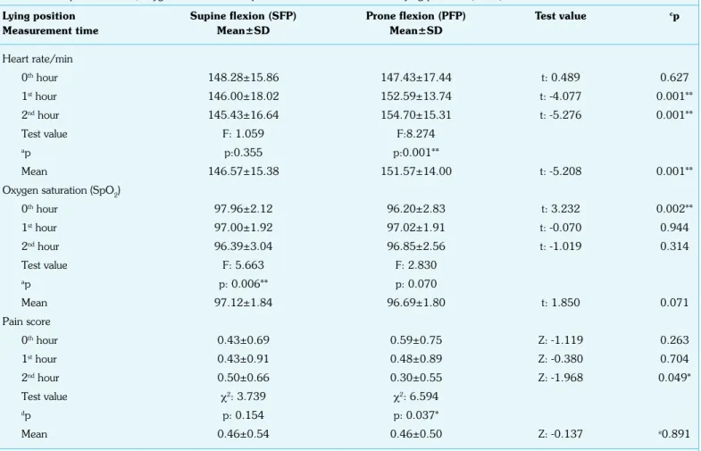
Table 1 shows all the descriptive characteristics of infants. HR de-creased gradually in infants who slept in SFP at 0, 1, and 2 h, and this decrease was not statistically significant (F=1.059; p=0.355). By contrast, HR increased gradually in infants in PFP at 0, 1, and 2 h, and this increase was statistically significant (F=8.274; p=0.001). When HR was compared in terms of positions, al-though no statistically significant difference was found in the mea-surement obtained immediately after the positioning, the HR of infants in the SFP at the 1 and 2 h was also lower at a statistically significant level compared with their HR in the PFP. This situation was also determined to be reflected on the mean HR at the end of 2 h (t=−5.208; p=0.001) (Table 2).
In the study, the SPO2 of infants in the SFP decreased imme-diately after the positioning, and this decrease was statistically significant at 1 and 2 h (F=5.663; p=0.006). By contrast, the SPO2 in the PFP did not change considerably at 0, 1, and 2 h, and no statistically significant difference was found (F=2.830; p=0.070; Table 2).
In the first assessment of the infants after positioning, the SPO2 in the SFP was significantly higher (t=3.232; p=0.002) com-pared with PFP. However, although the SPO2 observed at the first measurement between two positions was not observed at 1 and 2 h, the SPO2 was statistically similar in the first hour in both positions (Table 2).
In the study, although pain level was unchanged at 0 h immediately
Table 1. Distribution of infants in terms of descriptive characteristics (n=46)
Characteristics Min.–Max. Mean±SD
Age at birth (GW) 26–34 30.46±2.23
Age at the study (GW) 30–36 32.78±1.82
1st min Apgar score 6–9 7.61±0.88
5th min Apgar score 7–10 8.65±0.57
At birth Body weight (g) 730–2520 1500.26±383.28 Height (cm) 30–46 38.52±3.71 Head circumference (cm) 24–34 28.50±2.50 At the study Body weight (g) 950–2470 1543.98±325.11 Height (cm) 32–47 39.72±3.42 Head circumference (cm) 26–36 29.92±2.47
after SFP, pain score increased between 1 and 2 h. However, this increase was not statistically significant (χ2=3.739; p=0.154). Pain
score was 0.59±0.75 at 0 h immediately after PFP, decreased to 0.48±0.89 at 1 h, and then to 0.30±0.55 at 2 h, which were statistically significant (χ2=6.594; p=0.037) (Table 2).
DISCUSSION
Positioning of infants in their extrauterine life influences their devel-opmental period and may also affect physiological values. Position is known to affect the respiratory system, HR, and pain/discomfort feeling (18, 21–23). The literature showed that positioning impacts stress and development of preterm infants and that positioning of preterm infants should be investigated as individualized care for best practice (12, 23).
Brunherotti et al. (15) placed 16 preterm infants in the supine, prone, right lateral, and left lateral positions and evaluated their HR for 1 h. They reported that although the mean HR was 137.9/ min in the supine position, it was 140.7/min in the prone position. Thus, positions were similar in terms of presence of the difference in HR. However, the mean HR in the present study was higher than that of the study by Brunherotti et al. (15). This difference could be associated with the inclusion of infants in nasal CPAP in the sample group in their study.
Antunes et al. (14) divided 42 preterm infants into two groups: one group in the supine position (n=21) and the other group in the prone position (n=21). They assessed their HR, SPO2, ventilator values, and complications for 1 h, and they examined the effect of position on the process of disconnecting from ventilator. They reported no statistically significant difference in the HR in both positions (supine, 144/min; prone, 147/min) (14). Thus, the lack of difference in HR in terms of positions was different from our results. This difference was thought to be associated with the fact that the sample group of Antunes et al. (14) consisted of infants newly disconnected from the ventilator.
Ma et al. (24) examined cardiovascular response to short-term po-sitioned preterm infants. They reported no statistically significant difference between infants placed in changed positions in terms of HR (138±17) in a minute. In the present study, the HR was statis-tically different in terms of the infant positions. This difference may be associated with the positioning, and the infants were followed up for 2 h in each position. In the study by Ma et al. (24), cardio-vascular monitoring was followed up for 10 min in each position. Güler (25) examined its effect on spontaneous respiration in preterm infants in prone and supine positions after they were disconnected from the mechanical ventilator. They no statistical-ly significant difference between HR of infants placed in prone (153.33±14.00) and supine positions (150.50±17.06; p>0.007).
Table 2. Comparison of HR, oxygen saturations and pain score of infants in terms of lying positions (n=46)
Lying position Supine flexion (SFP) Prone flexion (PFP) Test value cp
Measurement time Mean±SD Mean±SD
Heart rate/min 0th hour 148.28±15.86 147.43±17.44 t: 0.489 0.627 1st hour 146.00±18.02 152.59±13.74 t: -4.077 0.001** 2nd hour 145.43±16.64 154.70±15.31 t: -5.276 0.001** Test value F: 1.059 F:8.274 ap p:0.355 p:0.001** Mean 146.57±15.38 151.57±14.00 t: -5.208 0.001**
Oxygen saturation (SpO2)
0th hour 97.96±2.12 96.20±2.83 t: 3.232 0.002** 1st hour 97.00±1.92 97.02±1.91 t: -0.070 0.944 2nd hour 96.39±3.04 96.85±2.56 t: -1.019 0.314 Test value F: 5.663 F: 2.830 ap p: 0.006** p: 0.070 Mean 97.12±1.84 96.69±1.80 t: 1.850 0.071 Pain score 0th hour 0.43±0.69 0.59±0.75 Z: -1.119 0.263 1st hour 0.43±0.91 0.48±0.89 Z: -0.380 0.704 2nd hour 0.50±0.66 0.30±0.55 Z: -1.968 0.049* Test value χ2: 3.739 χ2: 6.594 dp p: 0.154 p: 0.037* Mean 0.46±0.54 0.46±0.50 Z: -0.137 e0.891
Our results were similar to those of the study by Güler (25) (Table 2) in terms of HR being lower in the supine position compared with the prone position; however, they were different because the dif-ference in HR in terms of positions was not statistically significant. Additionally, the HR was close to each other in both studies. The difference between results in terms of statistical significance could be associated with the sample size and that Güler (25) compared the two different groups.
Preterm infants included in the study were placed in SFP and PFP, and the SPO2 was examined in each position for 2 h. Although a significant difference was found between the SPO2 at 0,1, and 2 h in infants in the SFP (p=0.006), no significant difference was found between the SPO2 at 0,1, and 2 h in infants in the PFP (p=0.070). Although the SPO2 in the SFP was higher than that in the PFP immediately after the infants were placed in the SFP and PFP, no difference was found between the SPO2 at 1 and 2 h. In addition, no difference was found between the mean SPO2 of infants in different lying positions (Table 2). The higher SPO2 in the SFP than in the PFP at the first evaluation might be associated with infants being placed more easily in the SFP and having lesser discomfort caused by lesser effort during the positioning.
In another study, the SPO2 of 32 preterm infants placed into the supine and prone positions for 2 h was examined. They reported that the SPO2 (98.4±2.2) in the prone position was higher than that (95±0.6) in the supine position, and the prone position was an advantageous method for oxygen need of infants on mechanical ventilator (26). The SPO2 in the prone position was higher com-pared with the supine position, which was statistically significant. In this aspect, their results were different from our study results (Ta-ble 2). This difference might be due to their infants being attached to a mechanical ventilator. Both studies were thought to be similar because the SPO2 in infants in the prone position was higher than that in the supine position.
Elder et al. (27) observed the SPO2 of 15 infants for 3 h during sleep in both supine and prone positions. They reported that the SPO2 of infants in the supine position (96.9±1.4) was statistically higher than that obtained in the prone position (95.8±2.5). Com-pared with the study by Elder et al. (27), no difference in the SPO2 was found in the present study at 1 and 2 h in terms of position. This difference might be due to the higher GW and administration of O2 support of infants in the sample group of Elder et al. (27). Additionally, another possibility is that the SPO2 might be lower than the values in the present study because infants in study group of Elder et al. (27) were placed in the same position for one more hour compared with infants w the present study.
Brunherotti et al. (15) placed 16 infants in the supine, prone, right lateral, and left lateral positions and evaluated their respiratory rate, SPO2, and HR for 1 h. They reported that the SPO2 in the prone position (97.4±1.1) was higher than that in the supine (97.1±1.13), right lateral (96.9±1.4), and left lateral (96.6±1.7) positions at a statistically significant level. In addition, oxygenation in infants was less advantageous when placed in the right and left lateral positions (15). The SPO2 in the prone and supine positions in their study and our results (Table 2) were similar. However, the SPO2 in the prone position was significantly higher than other positions because the infants were in nasal CPAP, which was different from our result.
Güler (25) placed preterm infants in the prone and supine positions after they were disconnected from the ventilator and examined its effect on spontaneous respiration. They reported that the SPO2 of infants in the prone position (96.20±2.69) was statistically higher than that in the supine position (95.36±3.01). When result of their study and results of the present study (Table 2) were compared, it was observed that although the SPO2 in the prone position in their study (25) was higher than that in the supine position, the SPO2 in the supine position was higher than that in the in prone position in the present study. The difference might be due to the infants in their study (25) were just disconnected from the mechanical ventilator. No statistically significant difference (p=0.154) was found between the pain levels of infants in the SFP at 0, 1, and 2 h, but pain score gradually increased; and their pain score in the PFP significantly decreased in terms of hour (p=0.037; Table 2).
The SPO2 of infants in the SFP was significantly higher at the first hour (Table 2). However, the HR in the PFP was higher compared with the SFP from the first minutes, and the difference at 1 and 2 h was statistically significant (Table 2). This difference might be due to infants in the SFP being more comfortable at 1 h, as reflected by the pain score after evaluating measurements immediately after positioning them. Although the pain score of the infants decreased in the PFP in the advancing hours (Table 2), no difference was found in the SPO2, and HR (Table 2) increased in the PFP with significant difference.
Thoyre and Carlson (28) and Park (29) defined normal HR as 120– 160/min. As time passed, the pain score in the SFP increased, but HR decreased. Although the pain score in the PFP decreased with advancing hour, HR increased gradually. However, no tachycardia was observed in both positions.
Cheraghi et al. (30) showed that arterial SPO2 in preterm infants had better outcomes in prone and left lateral postures than supine. In the same research, they reported that the respiratory and HRs of infants had no significantly difference when compared with the four postures (30).
CONCLUSION
Although several studies were conducted on the effect of position applied to infant in the NICU during invasive procedures on pain, a number of studies on infants’ physiological characteristics and discomfort/pain status only in terms of lying position is limited. Our study results showed that no difference in the mean pain level was found between two positions at first hour of the infants in dif-ferent lying positions. Furthermore, HR increased significantly, but pain score decreased in the PFP. Pain score was supposed to de-crease because infants felt relieved in the PFP. The inde-creased pain score of the infants in the SFP was because infants were influenced more by environmental factors in the supine position compared with the prone position.
Further studies are needed to determine the position with a mini-mum effect on physiological characteristics and comfort of infants.
Acknowledgements: The authors thank the families for participation in
the study and the nurses and neonatologists who work in NICU of İstanbul Medipol University Hospital. This study was completed as a master thesis.
Ethics Committee Approval: Ethical approval was obtained from İstan-bul Medipol University Hospital’s Ethics Committee (10840098-19), and permission was also obtained from the Medical Directorate and Nursing Services Directorate of the Hospital.
Informed Consent: Written informed consent was obtained from patients
who participated in this study.
Peer-review: Externally peer-reviewed.
Author Contributions: Concept – SÇ, DG; Design – SÇ, DG;
Supervi-sion – SÇ, DG; Resource – SÇ, DG; Materials – SÇ, DG; Data Collection and/or Processing – SÇ; Analysis and/or Interpretation – SÇ, DG; Litera-ture Search – SÇ, DG; Writing – SÇ, DG; Critical Reviews – DG.
Conflict of Interest: The authors have no conflict of interest to declare.
Financial Disclosure: The authors declared that this study has received
no financial support.
REFERENCES
1. Liu WF, Laudert S, Perkins B, Macmillan-York E, Martin S, Graven S; NIC/Q 2005 Physical Environment Exploratory Group. The develop-ment of potentially better practices to support the neurodevelopdevelop-ment of infants in the NICU. J Perinatol 2007; 27 Suppl 2: S48–74. [CrossRef]
2. Abusaad FES, El Aziz RAEAEL, Nasef NAE. The effectiveness of devel-opmentally supportive positioning on preterm ınfants’ pain response at neonatal ıntensive care units. Am J Nurs Sci 2017; 6(1): 63–71. 3. Santos AMG, Viera CS, Bertolini GRF, Osaku EF, de Macedo Costo
CRL, Giordani Grebinski ATK. Physiological and behavioural effects of preterm infant positioning in a neonatal intensive care unit. Br J Midwifery 2017; 25(19): 647–54. [CrossRef]
4. Holsti L, Grunau RE, Oberlander TF, Whitfield MF. Specific Newborn Individualized Developmental Care and Assessment Program move-ments are associated with acute pain in preterm infants in the neonatal intensive care unit. Pediatrics 2004; 114(1): 65–72. [CrossRef]
5. Spilker A, Hill C, Rosenblum R. The effectiveness of a standardised po-sitioning tool and bedside education on the developmental popo-sitioning proficiency of NICU nurses. Intensive Crit Care Nurs 2016; 35: 10–5. 6. Sonia PS. Effect of guideline on positioning of neonates in the neonatal
intensive care unit. J Biomed Sci 2019; 6(3): 8. [CrossRef]
7. Monterosso L, Kristjanson LJ, Cole J, Evans SF. Effect of postural supports on neuromotor function in very preterm infants to term equiv-alent age. J Paediatr Child Health 2003; 39(3): 197–205. [CrossRef]
8. Aydın D, Çiftçi EK. Neonatal intensive care unit nurses’ levels of infor-mation regarding therapeutic positions to be applied to preterm new-borns. J Curr Pediatr 2015; 13(1): 21–30. [CrossRef]
9. Çalışır H, Güler F. Positioning of prematüre infants receiving me-chanical ventilation in a neonatal intensive care unit: Review. Turkiye Klinikleri J Nurs Sci 2017; 9(3): 227–32. [CrossRef]
10. King C, Norton D. Does therapeutic positioning of preterm infants impact upon optimal health outcomes? A literature review. J Neonatal Nurs 2017: 23(5); 218–22. [CrossRef]
11. Ribas CG, Andreazza MG, Neves VC, Valderramas S. Effectiveness of Hammock Positioning in Reducing Pain and Improving Sleep-Wakeful-ness State in Preterm Infants. Respir Care 2019; 64(4): 384–9. 12. Graignic-Philippe R, Dayan J, Chokron S, Jacquet AY, Tordjman S.
Effects of prenatal stress on fetal and child development: a critical
liter-ature review. Neurosci Biobehav Rev 2014; 43: 137–62. [CrossRef]
13. Association of Women’s Health Obstetric and Neonatal Nurses: AW-HONN. Neonatal skin care: Evidence-based clinical practice guideline. 3rd edition. Washington DC: Johnson and Johnson Consumer Com-panies INC; 2013.
14. Antunes LC, Rugolo LM, Crocci AJ. Effect of preterm infant position on weaning from mechanical ventilation. [Article in Portuquese]. J Pe-diatr 2003; 79(3): 239–44. [CrossRef]
15. Brunherotti MA, Martinez EZ, Martinez FE. Effect of body position on preterm newborns receiving continuous positive airway pressure. Acta Paediatr 2014; 103(3): e101–5. [CrossRef]
16. Lawrence J, Alcock D, McGrath P, Kay J, MacMurray SB, Dulberg C. The development of a tool to assess neonatal pain. Neonatal Netw 1993; 12(6): 59–66.
17. Akdovan T. Assessment of pain in healthy neonates, investigation of the effects of pacifying and holding in the arms. [Thesis in Turkish]. Marmara University Institute of Health Sciences, Master’s Thesis, 1999.
18. Sarı HY, Çiğdem Z. Planning the baby’s developmental care according to gestational weeks. DEUHYO ED 2013; 6(1):40–8.
19. Ghasemi A, Zahediasl S. Normality tests for statistical analysis: A guide for non-statisticians. Int J Endocrinol Metab 2012; 10(2): 486–9. 20. Verma JP. Data Analysis in Management with SPSS Software. New
Delhi, India: Springer; 2013. [CrossRef]
21. Als H. Toward a synactive theory of development: Promise for the assessment and support of infant individuality. Infant Ment Health J 1982; 3(4): 229–43. [CrossRef]
22. Gardner S, Hagedorn MIE, Dickey LA. Pain and pain relief. In: Meren-stein BG, Gardner SL, editors. Handbook of Neonatal Intensive Care. Philadelphia (US): Mosby Elsevier; 2016.p.223–60.
23. Wiley F, Raphael R, Ghanouni P. NICU positioning strategies to reduce stress in preterm infants: a scoping review. Early Child Dev Care 2019: 1–18. [CrossRef]
24. Ma M, Noori S, Maarek JM, Holschneider DP, Rubinstein EH, Seri I. Prone positioning decreases cardiac output and increases systemic vascular resistance in neonates. J Perinatol 2015; 35(6): 424–7. 25. Güler F. Çalışır H. The effect of positioning on adapting to
sponta-neous breathing in premature infants after weaning from mechanical ventilation: A Randomized Controlled Trial. J Pediatr Res 2020; 7(2): 102–9. [CrossRef]
26. Abdeyazdan Z, Nematollahi M, Ghazavi Z, Mohhamadizadeh M. The effects of supine and prone positions on oxygenation in premature infants undergoing mechanical ventilation. Iran J Nurs Midwifery Res 2010; 15(4): 229–33.
27. Elder DE, Campbell AJ, Galletly D. Effect of position on oxygen satu-ration and requirement in convalescent preterm infants. Acta Paediatr 2011; 100(5): 661–5. [CrossRef]
28. Thoyre SM, Carlson J. Occurrence of oxygen desaturation events during preterm infant bottle feeding near discharge. Early Hum Dev 2003; 72(1): 25–36. [CrossRef]
29. Park J. Feeding outcomes in very preterm infants: Preliminary effects of positioning. Chapel Hill (US): The University of North Carolina, Doctoral dissertation, 2012.
30. Cheraghi F, Mahabadi MK, Sadeghian E, Tapak L, Basiri B. Physio-logical parameters of preterm infants in different postures: An observa-tional study. J Neonat Nur 2020; 26(4): 212–6. [CrossRef]