ABSTRACT
Introduction: Mitral valve (MV) repair is preferred over replacement for its benefits of preservation of ventricular function, lower operative mortality, superior long-term survival, and avoidance of anticoagulation. In this study, we aimed to review the repair techniques of complex MV pathologies and their outcomes. Patients and Methods: We retrospectively analyzed 56 patients (mean age 41.8 ± 16.5 years; 33 males) who underwent repair of complex MV pathologies. 44 patients had pure mitral regurgitation (MR), and 12 (21.4%) had mixed mitral disease (mitral stenosis (MS) + MR). Preoperative and operative characteristics, postoperative MR severity, operative mortality, and midterm survival were examined for each patient. Results: There was only one early death (30-day mortality: 1.8%) due to postoperative low cardiac output syndrome. The procedures were successful in all patients who underwent MV repair. Transthoracic echocardiography examinations revealed no/trivial MR in 74.6% and mild MR in 21.8% of patients at discharge. Late follow-up was obtained in 55 patients. The mean follow-up period of patients was 47.9 ± 23.1 months. Mortality developed in one (1.8%) patient with Marfan syndrome who had acute aortic dissection three years after MV surgery. During follow-up visits, mitral repair procedures were successful in 49 (90.7%) patients. Four (7.4%) patients presented with moderate MR. Only one (1.9%) patient needed reoperation because of severe MR.
Conclusion: This study showed that repair of complex MV pathologies provides excellent surgical outcomes. Repair of complex MV pathologies is safe and highly effective, but operations require considerable surgical experience.
Key Words: Mitral valve repair; mitral regurgitation; mitral stenosis
Kompleks Mitral Kapak Patolojilerin Onarımı; Uğraşmaya Değer mi? ÖZET
Giriş: Ventrikül fonksiyonun korunması, daha az cerrahi mortaliteye sahip olması, üstün uzun dönem survey ve antikoagülan kullanımının önlenmesi gibi üstünlükleri nedeniyle mitral kapak onarımı replasmana daha çok tercih edilmektedir. Bu çalışmanın amacı, kompleks mitral kapak patolojilerin onarım teknikleri ve so-nuçları sunmaktır.
Hastalar ve Yöntem: Retrospektif olarak kompleks mitral kapak patolojilerin onarımı geçiren 56 hasta incelendi (ortalama yaş 41.8 ± 16.5 yıl; 33 erkek). Kırk dört hastada saf mitral yetmezliği varken, 12 (%21.4) hastada miks mitral kapak hastalığı (mitral darlığı + mitral yetmezliği) vardı. Preoperatif ve ope-ratif özellikleri, postopeope-ratif mitral yetmezliği derecesi, cerrahi mortalite ve orta dönem sonuçları her hasta için araştırıldı.
Bulgular: Postoperatif düşük kardiyak debi sendromuna bağlı bir hastada erken mortalite (30 gün mortali-te: %1.8) görüldü. Mitral kapak onarımı ameliyatı olan bütün hastalarda mitral onarım prosedürleri başarılı olmuştur. Hastalar taburcu olduğunda yapılan ekokardiyografik değerlendirmede %74.6’sında hiç/eser yeter-sizlik ve %21.8’inde hafif yeteryeter-sizlik saptandı. 55 hastada geç dönem takibi yapıldı. Hastalarımızın ortalama takip süresi 47.9 ± 23.1 aydı. Geç mortalite mitral kapak onarımından 3 yıl sonra akut aort diseksiyonu neniyle ameliyata alınan marfan sendromlu bir hastada gözlendi. Takipler sırasında yapılan ekokardiyografik de-ğerlendirmede hastaların %90.7 (49 hasta)’sinde hiç ya da hafif yetersizlik gözlendi. Orta yetersizlik gözlenen 4 (%7.4) hastada tıbbi tedavi uygulandı. İleri yetersizlik gözlenen 1 (%1.9) hastada reoperasyon uygulandı. Sonuç: Çalışmamız kompleks mitral kapak patolojilerin onarımının sonuçları mükemmel olduğunu gösterdi. Kompleks mitral kapak patolojilerin onarım teknikleri güvenli ve sonuçları son derece etkindir, fakat ameli-yatlarda yeterli cerrahi tecrübe gereklidir.
Anahtar Kelimeler: Mitral kapak onarımı; mitral yetmezliği; mitral darlığı Salih Salihi1, H. Tarık Kızıltan2, Aşkın Ali Korkmaz1, Mustafa Güden3
1 Okan University Faculty of Medicine, Department of Cardiovascular Surgery, İstanbul, Turkey 2 Private Adana Hospital, Clinic of Cardiovascular Surgery, Adana, Turkey
3 İstanbul Medipol University Faculty of Medicine, Department of Cardiovascular Surgery, İstanbul, Turkey
Repair of Complex Mitral Valve Pathologies:
Is It Worth to Cope With?
Salih Salihi
E-mail: [email protected] Submitted: 17.05.2017 Accepted: 15.07.2017
© Copyright 2018 by Koşuyolu Heart Journal. Available on-line at
www.kosuyoluheartjournal.com Correspondence
INTrODUCTION
Mitral valve (MV) repair is preferred over replacement for its advantages of preservation of ventricular function, lower operative mortality, better long-term survival, and avoidance of anticoagulation(1-3). MV repair has been shown to have excellent
durability in patients with mitral regurgitation (MR) caused by degenerative diseaseand is indeed the method of choice in the correction of MR whenever feasible(4-7). In contrast, valve
reconstruction for rheumatic MR remains controversial as it is not only less feasible to repairbut also the repaired rheumatic valve has poorer durability when compared with a degenerative MV repair(8,9).
Most of the MV pathology involves the posterior leaflet or annulus and usually can be repaired using standard valve repair techniques. These procedures are feasible in almost 95% of patients with degenerative MR despite the presence of complex lesions(10). Difficulties may arise when trying to repair the less
common anterior leaflet prolapse or calcified mitral annulus. Although the repair for the prolapse of the posterior leaflet with valvular resection or artificial chordae is usually possible, correction of anterior or bileaflet prolapse may demand more complex repair procedures. MV repair in a complex setting such as redo repair procedure, congenital anomalies, and hypertrophic obstructive cardiomyopathy (HOCM) is often challenging because of a lack of leaflet mobility or adequate surface of coaptation. In this study, we aimed to review repair techniques of complex MV pathologies and their outcomes.
PATIENTS and METHODS
For this study, our hospital has been approved by the Scientific Ethics Committee.
Study Group and Definitions
This is a retrospective study of 56 patients who underwent repair of complex mitral pathologies using multiple procedures for MR or mitral stenosis (MS) at our hospital. Complex repair was defined as using multiple mitral valve repair techniques (three techniques or more) in the same patient. These more complex and challenging patients were selected as study group in order to assess more convincingly the efficacy of these techniques. All preoperative, intraoperative, and postoperative demographic, echocardiographic, and clinical data were collected. Additionally, all surgical notes and discharge summaries were reviewed to collect supplementary information. The data collected were focused on preoperative ejection fraction, grade of MR or MS, valve pathology, repair techniques, and intraoperative, postoperative early (< 30 days), and late (> 30 days) complications.
Surgical Techniques
Operative data were retrospectively extracted from medical records, surgery notes, and the computer-based databank from the Department of Cardiac Surgery. Surgical approach was
via a mid-sternotomy in 52 patients and a right anterolateral thoracotomy in four patients for cosmetic reasons. Aorto-bicaval cannulation was used in all. Operations were performed under cardiopulmonary bypass (CPB) at moderate hypothermia. Concomitant cardiac procedures were performed. After a right atriotomy was performed with an oblique incision, the mitral repair was completed through transseptal approach. In 14 patients, we used left atriotomy. Leaflet repair techniques were performed with principles originally reported by Carpentier et al. and Duran et al. but several modifications based on these principles were used(11,12). Our techniques of MV repair evolved over the years.
In complex mitral pathologies, chordal replacement with Gore-tex cords, leaflet resection with sliding or folding annuloplasty, or commissurotomy was performed considering the status of the mitral pathology. In rheumatic MV disease, leaflet augmentation with pericardium, commissurotomy, resection of primary or/and secondary chordae, and chordal replacement were preferred. In MR due to HOCM, we performed shortening of posterior leaflet, neochordae, and ring annuloplasty in addition to septal myectomy to prevent systolic anterior motion (SAM). The left atrial appendage was routinely ligated in patients with atrial fibrillation (AF). Upon completion of repair, MV was tested by injecting cold saline into the left ventricular cavity to observe coaptation of leaflets. Intraoperative transesophageal echocardiography (TEE) was used routinely for intraoperative assessment of MV repair after CPB. When an unsatisfactory finding was observed during TEE examination, a second cross-clamp was placed for satisfactory repair, if possible.
Follow-Up
Follow-up data were analyzed using cardiology and cardiac surgery outpatient follow-up notes, primary care and institutional computer-based databanks, and telephone interviews. All patients had a TTE before hospital discharge. Echocardiographic findings were recorded in the computer database of the hospital. The clinical parameters recorded during the follow-up period included early (< 30 days) and late mortality after surgery. All patients were anticoagulated with warfarin sodium for 3 months after surgery and permanently if they had AF or other mechanical valves.
Statistical Analysis
Data were presented as frequencies and percentages for categorical variables, and medians or means with standard deviations for continuous variables.
rESULTS
Patient Characteristics
The demographic data and preoperative characteristics for all patients are presented in Table 1. Patients’ age ranged from 5 to 77 years (mean age was 41.8 ± 16.5 years), and female sex was less frequent than male sex (23 patients; 41.1%). Twenty-five patients (44.7%) were in New York Heart Association (NYHA)
functional class III-IV. The mean preoperative LV ejection fraction was 62 ± 5%. Concomitant cardiovascular pathologies included ischemic heart disease in 4 and tricuspid regurgitation in 18 cases (Figure 1). Most patients had preoperative Grade 4 MR and underwent mitral repair according to our definition. Degenerative MV disease as the cause of MR was diagnosed in 38 patients. The distribution of MV pathologies during surgical exploration is presented in Table 2. Five patients presented with the prolapse of the posterior leaflet, whereas 25 patients had an involvement of both mitral leaflets. Commissural fusion was diagnosed in 11 patients.
Operative Data
Operative data are presented in Table 3. Most of the procedures were performed through a median sternotomy. A minimally invasive approach through a right anterior mini-thoracotomy and transthoracic aortic clamping was used in four patients for cosmetic reasons.
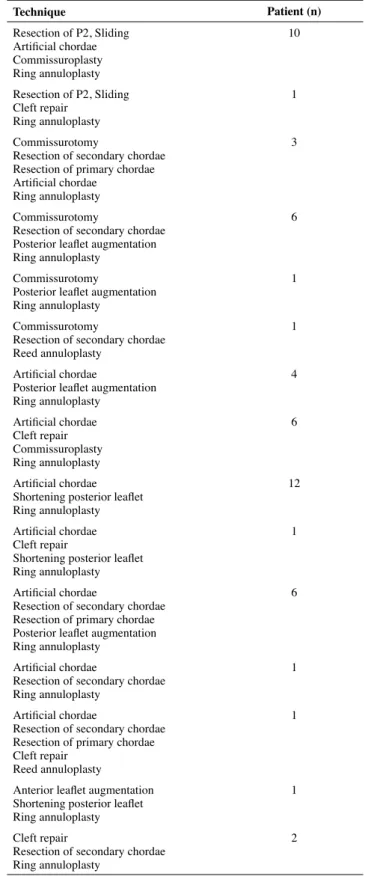
Surgical procedures involving different techniques are listed in Table 4. Ring annuloplasty was performed in 54 patients. Technically, for example, quadrangular resection of the posterior leaflet, sliding annuloplasty, ring annuloplasty, Reed annuloplasty, and chordal replacement were performed. All patients undergoing chordal replacement and posterior leaflet resection had an annuloplasty procedure.
The concomitant procedures are listed in Table 5. Four coronary artery bypass grafting, 18 tricuspid repair, left atrial radiofrequency ablation in 12 patients with preoperative AF, and left atrial appendix ligation in all patients with preoperative AF were performed. In patients presenting with MR, aortic aneurysm, and aortic regurgitation, we preferred making valve sparing aortic repair (reimplantation procedure) if patients were below 70 years of age and had a favorable physical status.
Clinical Outcomes
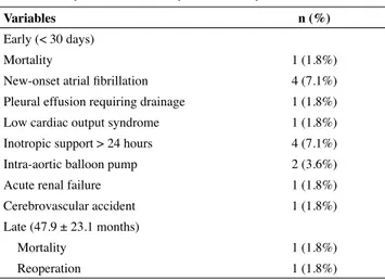
The early and late complications after mitral repair are presented in Table 6. There was only one early death (30-day
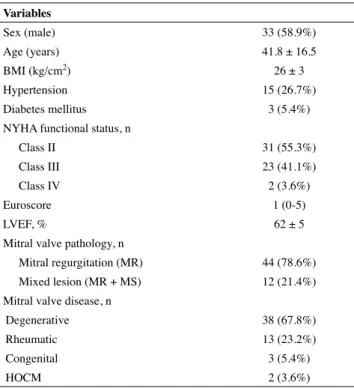
Table 1. Patient demographics and preoperative characteristics Variables Sex (male) 33 (58.9%) Age (years) 41.8 ± 16.5 BMI (kg/cm2) 26 ± 3 Hypertension 15 (26.7%) Diabetes mellitus 3 (5.4%)
NYHA functional status, n
Class II 31 (55.3%)
Class III 23 (41.1%)
Class IV 2 (3.6%)
Euroscore 1 (0-5)
LVEF, % 62 ± 5
Mitral valve pathology, n
Mitral regurgitation (MR) 44 (78.6%)
Mixed lesion (MR + MS) 12 (21.4%)
Mitral valve disease, n
Degenerative 38 (67.8%)
Rheumatic 13 (23.2%)
Congenital 3 (5.4%)
HOCM 2 (3.6%)
Data are presented as mean value ± standard deviation, median value, or number of patients. BMI: Body mass index, NYHA: New York Heart Association, LVEF: Left ventricle ejection fraction, MR: Mitral regurgitation, MS: Mitral stenosis, HOCM: Hypertrophic obstructive cardiomyopathy.
Table 2. Distribution of mitral valve pathologies
Variables N Annular dilatation 34 Leaflet prolapse Anterior leaflet 2 Posterior leaflet 5 Both leaflets 25 Commissural 9 Chordal rupture Anterior leaflet 2 Posterior leaflet 3 Mitral cleft Anterior leaflet 4 Posterior leaflet 6 Commissural fusion 11 Leaflet retraction Anterior leaflet 1 Posterior leaflet 14 Chordal retraction Primary chordae 10 Secondary chordae 20 HOCM 2
Data are presented as number of patients. HOCM: Hypertrophic obstructive cardiomyopathy.
mortality: 1.8%) due to postoperative low cardiac output syndrome in patient with significant left ventricle dysfunction. The mean intensive care unit and hospital stays of patients were 1.83 ± 0.4 and 7.12 ± 1.86 days, respectively. New-onset AF developed in four patients and medically resolved in all. Inotropic support for more than 24 hours was needed in four cases, and two of them needed an intra-aortic balloon pump.
Late follow-up was obtained in 55 patients at an average of 47.9 ± 23.1 months postoperatively. Mortality developed in one (1.8%) patient with Marfan syndrome who had acute aortic dissection three years after MV surgery. Only one (1.9%) patient needed reoperation because of severe MR. This patient was treated with mechanical valve replacement after four years of initial repair.
Echocardiographic Results
Echocardiographic data are given in Table 7. In all patients who underwent MV repair, the procedures were successful at discharge; transthoracic echocardiography examinations revealed no/trivial MR in 74.6% and mild MR in 21.8% of patients. During follow-up visits, mitral repair procedures were successful in 49 (90.7%). Only four (7.4%) patients presented with moderate MR, and they were asymptomatic under medical treatment. Unfortunately, severe MR developed in one patient. This patient was treated with mechanical valve replacement after four years of initial repair.
DISCUSSION
Current consensus guidelines on MR recommend repair over replacement whenever possible and earlier surgical intervention if there is a high likelihood of repair(13,14). Accordingly, repair
feasibility is a key factor in the decision to operate and is highly
dependent on lesion complexity and surgeon experience(15).
Repair of the MV is well known for its efficacy, durability, and
avoidance of many complications(16). As demonstrated in many
studies, MV replacement is associated with (a) gradual decline in left ventricular function, (b) hazards of anticoagulation, (c)
Table 4. Surgical repair techniques
Technique Patient (n) Resection of P2, Sliding Artificial chordae Commissuroplasty Ring annuloplasty 10 Resection of P2, Sliding Cleft repair Ring annuloplasty 1 Commissurotomy
Resection of secondary chordae Resection of primary chordae Artificial chordae
Ring annuloplasty
3
Commissurotomy
Resection of secondary chordae Posterior leaflet augmentation Ring annuloplasty
6
Commissurotomy
Posterior leaflet augmentation Ring annuloplasty
1 Commissurotomy
Resection of secondary chordae Reed annuloplasty
1 Artificial chordae
Posterior leaflet augmentation Ring annuloplasty 4 Artificial chordae Cleft repair Commissuroplasty Ring annuloplasty 6 Artificial chordae Shortening posterior leaflet Ring annuloplasty
12 Artificial chordae
Cleft repair
Shortening posterior leaflet Ring annuloplasty
1
Artificial chordae
Resection of secondary chordae Resection of primary chordae Posterior leaflet augmentation Ring annuloplasty
6
Artificial chordae
Resection of secondary chordae Ring annuloplasty
1 Artificial chordae
Resection of secondary chordae Resection of primary chordae Cleft repair
Reed annuloplasty
1
Anterior leaflet augmentation Shortening posterior leaflet Ring annuloplasty
1 Cleft repair
Resection of secondary chordae Ring annuloplasty
2 Table 3. Operative data
Variables Incision, n (%)
Sternotomy 52 (92.8%)
Right mini-thoracotomy (port access) 4 (7.2%) Surgical approach, n (%)
Left atrium 14 (25%)
Right atrium 42 (75%)
Operation duration
Cardiopulmonary bypass duration, min 144 ± 35 Aortic cross-clamp duration, min 101 ± 30
ICU stay, days 1.83 ± 0.4
Hospital stay, days 7.12 ± 1.86
thromboembolism, and (d) higher incidence of endocarditis
(3,12,17). Results from a recent seriesshow a poor survival
after valve replacement(18). Growth, marriage, and pregnancy
are important issues which are adversely affected by anticoagulation. During the last two decades, the number of MV repair procedures has increased across the world. As experience grows in this field, surgeons try to repair more
valves in complex MV disease patients. In our series consisting of 56 complex mitral valve cases that underwent MV repair, there was one early mortality after 5 days of surgery due to postoperative low cardiac output syndrome in a patient with significant left ventricle dysfunction. In the late follow-ups, there was one mortality due to acute aortic dissection after three years of surgery. This patient had Marfan syndrome, and we repaired his MV. At the time of operation, there was mild aortic regurgitation, and the diameter of the aortic root was 36 mm. Echocardiographic assessment of patients at discharge revealed no/trivial regurgitation in 74.6% and mild MR in 21.8% of all patients. Echocardiographic examination during follow-up revealed that mitral insufficiency was none or mild in 90.7% of patients. Four (7.4%) patients had moderate MR and were treated medically. Mitral insufficiency recurrence with severe regurgitation occurred in one (1.9%) patient. This patient was treated with mechanical valve replacement after four years of initial repair. We prefer surgical repair of the MV in young patients (mean age 41.8 ± 16.5), and we think that it is not a good strategy for elderly patients.
The mitral apparatus includes the leaflets, annulus, chordae tendineae, papillary muscles, and left ventricle. The goals of mitral repair are to maintain leaflet mobility, remodel the annulus, and allow normal coaptation of the anterior and posterior leaflets. In MV prolapse or Barlow’s syndrome, the leaflets and chordae become thickened and redundant, which results in leaflet prolapse beyond the plane of the annulus and MR. In our study, 38 patients had degenerative MV. Up to 2011, we repaired degenerative MVs with leaflet resection; after that, we switched to artificial chordae implantation as a routine technique. The most simple and common MV lesion, the prolapse of the posterior leaflet, can be treated with leaflet resection with excellent short-term and long-term results(19).
However, the correction of anterior, bileaflet prolapse, or even large areas of posterior prolapse is more complex(20,21).
Particularly in patients with complex degenerative MV disease, we used three or more techniques together. For example, we used a combination of artificial chordae, resection of secondary chordae, resection of primary chordae, posterior leaflet augmentation, and ring annuloplasty in six patients. Our degenerative MV repair was successful in all patients.
Table 6. Early and late morbidity and mortality
Variables n (%)
Early (< 30 days)
Mortality 1 (1.8%)
New-onset atrial fibrillation 4 (7.1%)
Pleural effusion requiring drainage 1 (1.8%)
Low cardiac output syndrome 1 (1.8%)
Inotropic support > 24 hours 4 (7.1%)
Intra-aortic balloon pump 2 (3.6%)
Acute renal failure 1 (1.8%)
Cerebrovascular accident 1 (1.8%)
Late (47.9 ± 23.1 months)
Mortality 1 (1.8%)
Reoperation 1 (1.8%)
Data are presented as number of patients (percentage).
Table 5. Concomitant surgical procedures
Concomitant surgical procedures n (%)
CABG 4 (7.1%)
TR 18 (32.1%)
Kay annuloplasty 12 (21.4%)
Ring annuloplasty 6 (10.7%)
AVR 3 (5.4%)
Aortic valve reconstruction 3 (5.4%)
Valve-sparing aortic root replacement 2 (3.6%)
Septal myectomy for HOCM 2 (3.6%)
RF ablation 12 (21.4%)
CABG: Coronary artery bypass grafting, TR: Tricuspid repair, AVR: Aortic valve replacement, HOCM: Hypertrophic obstructive cardiomyopathy, RF: Radiofrequency ablation.
Table 7. Echocardiographic follow-up data of patients
Variables Preoperative Operative TEE At discharge At follow-up
MR grade, n (%) 56 56 55 54 None/Trivial 0 46 (82.1%) 41 (74.6%) 25 (46.3%) Mild 0 10 (17.9%) 12 (21.8%) 24 (44.4%) Moderate 4 (7.1%) 0 2 (3.6%) 4 (7.4%) Severe 52 (92.9%) 0 0 1 (1.9%) 25 (43%) 89 (63%) 0.024
Echocardiographic examination during follow-up revealed that mitral insufficiency was none or mild in 37 patients. One patient had moderate MR and was treated medically.
MV repair has been shown to have excellent durability in patients with MR caused by degenerative disease(4,5). In contrast,
valve reconstruction for rheumatic MR remains controversial as it not only suffers from an inferior feasibility of repair, but also the repaired rheumatic valve is less stable, with inferior
durability when compared with a degenerative MV repair(8,9).
The utilization of leaflet mobilization and extension with the pericardium to increase the leaflet area and the surface of
coaptation may provide satisfactory results(22-24). Chauvaud
et al. on the other hand, had demonstrated good long-term results in repairing diseased rheumatic MVs using Carpentier’s reconstruction techniques(22,23). Dillon and colleagues reported
that, after leaflet extension in rheumatic MV reconstruction, MR grade was none/trivial in 64.5% of patients, mild in 22.6%, moderate in 6.5%, moderately severe in 4.8%, and severe in 1.6%. Two patients had redo mitral surgery. At 5 years postoperatively, the estimated rates of freedom from reoperation was 96.8%(25).
13 of our patients had diseased rheumatic MV. We repaired their valves using commissurotomy, resection of primary or/ and secondary chordae, artificial chordae, ring annuloplasty, or posterior leaflet augmentation. Echocardiographic examination during follow-up revealed that mitral insufficiency was none or mild in 10 patients. Two patients had moderate MR and were treated medically. One patient had redo mitral surgery after four years of surgery. In the follow-up, regurgitation was seen once often in rheumatic valves. Retraction of the pericardial patch and the on-going process of rheumatic disease were considered to be the undergoing pathologies in these cases.
In contrast, in children with congenital MR, conventional repair of the valve is not always successful. In part, this reflects the complicated abnormalities of the valvular structures and the associated cardiac malformations. When planning the optimal surgical repair of the MV, attention must be directed at the annular attachment, the valvar leaflets, and the tension apparatus of the valve. In patients with congenital MR, the annular attachment is commonly dilated, and the papillary muscles, as well as their attachments to the ventricular wall, are frequently abnormal(26,27). In some patients with prolapse of the leaflets of
the MV, use of artificial chords has been suggested to provide efficient short-term results(28).
In our study, three patients underwent mitral reconstructive operations for congenital mitral diseases. The pathologic findings of the first patient’s MV were short and thickened chordae and annular dilatation. Her MV was repaired using artificial chordae, resection of secondary chordae, resection of primary chordae, posterior leaflet augmentation, and ring annuloplasty. The second’s echocardiography showed severe mitral stenosis related to a hammock MV, and his valve was repaired using commissurotomy, resection of secondary chordae, and Reed
annuloplasty. The third’s MV was repaired using artificial chordae, resection of secondary chordae, resection of primary chordae, cleft repair, and Reed annuloplasty.
Kawahira et al. used artificial cords in 11 children with congenital MR, and they reported that, in two patients, regurgitation recurred within 1 year of the operation(29). Early
and late results of reconstructive operation for congenital MR in 66 pediatric age group patients were reported by Okita et al. Valvuloplasty failed in 19 of the long-term survivors, and one of these patients underwent MV replacement 11 years after initial operation(30). During follow-up, there was no reoperation, and
one of the patients had moderate MR during echocardiographic examination. Valve repair was particularly preferred in this patient because he had mental retardation and warfarin use and regular INR follow-up were not feasible. The recurrence of MR in this patient may be explained by the fact that mitral ring was not used in the repair surgery to avoid development of functional stenosis in the following years.
The MV in HOCM usually has an increased length of the anterior and posterior mitral leaflets. The MV, specifically the SAM of the MV leaflets, is an important component of the obstruction(31). In HOCM, abnormal anatomy and valve
displacement induce drag forces that cause SAM. This condition can be corrected by an autologous pericardial patch in the anterior mitral leaflet(32).
We routinely excise sufficient septal muscles to leave a residual septal thickness within the normal range. Patients with more severe forms of hypertrophic obstructive cardiomyopathy with MV involvement may require a more complex reconstructive operation. The anterior leaflet is reconstructed using an ovoid patch of glutaraldehyde-treated autologous pericardium sutured to the edges of the leaflet incision. Whenever the posterior leaflet was higher than 20 mm, we reduced it to less than 20 mm by an ovoid resection. Finally, in severe forms with an excessively small annulus and a hyperkinetic ventricle, a rigid annuloplasty ring is implanted. There were two HOCM patients at this study. In the first case, we repaired the MV using artificial chordae, shortening posterior leaflet, commissuroplasty, and ring annuloplasty. The other’s MV was repaired using shortening posterior leaflet, anterior leaflet augmentation, and ring annuloplasty. There is no MR in echocardiographic examination during follow-up.
CONCLUSION
MV repair for complex pathologies is a feasible and safe procedure with excellent surgical outcomes in experienced hands. We demonstrated that MV repair can be performed for mixed MV disease patients with results similar to those in pure MR patients. Autologous pericardium is a useful leaflet substitute that facilitates MV repair. Combining multiple techniques of MV repair may extend valve repair into a wider spectrum of complex valve pathologies.
Limitations of the Study
The major limitations of this study are the retrospective design, the small number of patients, and the short follow-up period in some patients.
CONFLICT of INTEREST
The authors reported no conflict of interest related to this article. AUTHOrSHIP CONTrIBUTIONS Concept/Design: SS, TK, AK Analysis/Interpretation: SS, AK Data Acquisition: SS, MG Writting: SS, TK Critical Revision: SS, MG Final Approval: All of authors
REFERENCES
1. Ren JF, Askut S, Lightly GW Jr, Vigilante GJ, Sink JD, Segal BL, et al. Mitral valve repair is superior to valve replacement for early preservation of cardiac function: relation of ventricular geometry to function. Am Heart J 1996;131:974-81.
2. Suri RM, Schaff HV, Dearani JA, Sundt TM 3rd, Daly RC, Mullany CJ, et
al. Survival advantage and improved durability of mitral repair for leaflet prolapse subsets in the current era. Ann Thorac Surg 2006;82:819-26. 3. Enriquez-Sarano M, Schaff HV, Orszulak TA, Tajik AJ, Bailey KR, Frye
RL. Valve repair improves the outcome of surgery for mitral regurgitation. A multivariate analysis. Circulation 1995;91:1022-8.
4. David TE, Armstrong S, Sun Z, Daniel L. Late results of mitral valve repair for mitral regurgitation due to degenerative disease. Ann Thorac Surg 1993;56:7-12; discussion 13-4.
5. Gillinov AM, Cosgrove DM, Blackstone EH, Diaz R, Arnold JH, Lytle BW, et al. Durability of mitral valve repair for degenerative disease. J Thorac Cardiovasc Surg 1998;116:734-43.
6. Korkmaz AA, Onan B, Demir AS, Tarakçı S, Gündoğdu R, Akdemir I, et al. Clinical outcomes of mitral valve repair in mitral regurgitation: a prospective analysis of 100 consecutive patients. Anadolu Kardiol Derg 2011;11:542-50.
7. Onan B, Erkanlı K, Onan IS, Ersoy B, Aktürk IF, Bakır I. Clinical outcomes of mitral valve repair, a single center experience in 100 patients. Türk Kalp Damar Cerrahisi Dergisi 2014;22:19-28.
8. Cosgrove DM, Steward WJ. Mitral valvuloplasty. Current Probl Cardiol 1989;14:359-415.
9. Yau TM, Ei-Ghoneimi YA, Armstrong S, Ivanov J, David TE. Mitral valve repair and replacement in rheumatic disease. J Thorac Cardiovasc Surg 2000;119:53-60.
10. David TE, Ivanov J, Armstrong S, Christie D, Rakowski H. A comparison of outcomes of mitral valve repair for degenerative disease with posterior, anterior, and bileaflet prolapse. J Thorac Cardiovasc Surg 2005;130:1242-9. 11. Carpentier A. Cardiac valve surgery-the “French correction”. J Thorac
Cardiovasc Surg 1983;86:323-37.
12. Duran CG, Revuelta JM, Gaite L, Alonso C, Fleitas MG. Stability of mitral reconstruction surgery at 10-12 years for predominantly rheumatic valvular disease. Circulation 1988;78:91-6.
13. Vahanian A, Alfieri O, Andreotti F, Antunes MJ, Barón-Esquivias G, Baumgartner H, et al. Guidelines on the management of valvular heart disease. The Joint Task Force on the Management of Valvular Heart
Disease of the European Society of Cardiology (ESC) and the European Association for Cardio-Thoracic Surgery (EACTS). Eur J Cardiothorac Surg 2012;42:S1-44.
14. Bonow RO, Carabello BA, Chaterjee K, de Leon AC, Faxon DP, Freed MD, et al. Focused update incorporated into the ACC/AHA 2006 guidelines for the management of patients with valvular heart disease: a report of the American College of Cardiology/American Heart Association Task Force on Practice Guidelines. Circulation 2008;118:e523-661.
15. Adams DH, Anyanwu AC, Rahmanian PB, Filsoufi F. Current concepts in mitral valve repair for degenerative disease. Heart Fail Rev 2006;11: 241-57. 16. Kumar AS, Rao PN, Saxena A. Results of mitral valve reconstruction in
children with rheumatic heart disease. Ann Thorac Surg 1995;60:1044-7. 17. Erez E, Kanter KR, Isom E, Williams WH, Tam VK. Mitral valve
replacement in children. J Heart Valve Dis 2003;12:25-9.
18. Gao G, Wu Y, Grunkemeier GL, Furnary AP, Starr A. Forty-years survival with the Starr-Edwards heart valve prosthesis. J Heart Valve Dis 2004;13:91-6.
19. Braunberger E, Deloche A, Berrebi A, Abdallah F, Celestin JA, Meimoun P, et al. Very long-term results (more than 20 years) of valve repair with Carpentier’s techniques in nonrheumatic mitral valve insufficiency. Circulation 2001;104:8-11.
20. Gillinov AM, Blackstone EH, Alaulaqi A, Sabik JF, Mihaljevic T, Svensson LG, et al. Outcomes after repair of the anterior mitral leaflet for degenerative disease. Ann Thorac Surg 2008;86:708-17.
21. Kasegawa H, Shimokawa T, Shibazaki I, Koyanagi T, Ida T. Mitral valve repair for anterior leaflet prolapse with expanded polytetrafluoroethylene sutures. Ann Thorac Surg 2006;81:1625-31.
22. Chauvaud S, Jebara V, Chachques JC, El Asmar B, Mihaileanu S, Perier P, et al. Valve extension with glutaraldehyde-preserved autologous pericardium: results inmitral valve repair. J Thorac Cardiovasc Surg 1991;102:171-8.
23. Chauvaud S, Fuzellier JF, Berrebi A, Deloche A, Fabiani JN, Carpentier A. Long term (29 years) results of reconstructive surgery in rheumatic mitral valve insufficiency. Circulation 2001;104(Suppl I):I-12-5.
24. Zakkar M, Amirak E, John Chan KM, Punjabi PP. Rheumatic mitral valve disease: current surgical status. Prog Cardiovasc Dis 2009;51:478-81. 25. Dillon J, Yakub MA, Nordin MN, Pau KK, Krishna Moorthy PS. Leaflet
extension in rheumatic mitral valve reconstruction. Eur J Cardiothorac Surg 2013;44:682-9.
26. Kadoba K, Jonas RA, Mayer JE, Castaneda AR. Mitral valve replacement in the first year of life. J Thorac Cardiovasc Surg 1990;100:762-8. 27. Carpentier A, Branchini B, Cour JC, Asfaou E, Villani M, Deloche A, et al.
Congenital malformations of the mitral valve in children. Pathology and surgical treatment. J Thorac Cardiovasc Surg 1976;72:854-8.
28. Murakami T, Yagihara T, Yamamoto F, Uemura H, Yamashita K, Ishizaka T. Artificial chordae for mitral valve reconstruction in children. Ann Thorac Surg 1998;65:1377-80.
29. Kawahira Y, Yagihara T, Uemura H, Ishizaka H, Yoshizumi K, Kitamura S.Use of expanded polytetrafluoroethylene sutures as artificial tendinous cords in children with congenital mitral regurgitation. European Journal of Cardio-thoracic Surgery 1999:15:289-293.
30. Okita Y, Miki S, Kusuhara K, Ueda Y, Tahata T, Tsukamoto Y, et al. Early and late results of reconstructive operation for congenital mitral regurgitation in pediatric age group. The Journal of Thoracic and Cardiovascular Surgery 1988;96:294-8.
31. Grigg LE, Wigle ED, Williams WG, Daniel LB, Rakowski H. Transesophageal Doppler echocardiography in obstructive hypertrophic cardiomyopathy: clarification of pathophysiology and importance in intraoperative decision making. J Am Coll Cardiol 1992;20:42-52. 32. Van der Lee C, Kofflard MJM, van Herwerden LA, Vletter WB, ten Cate
FJ. et al. Sustained improvement after combined anterior mitral leaflet extension and myectomy in hypertrophic obstructive cardiomyopathy. Circulation 2003;108:2088-92.