CASE REPORT
Nonextraction Treatment of a Class III Malocclusion
Case Using Mini-Screw-Assisted Lower Molar
Distalization
ABSTRACT
Mini-screw assisted lower molar distalization was planned for a present mild Class III malocclusion case. Two mini-screws were in-serted into the available inter-root area: one on the left, and the other on the right side in the posterior region in the mandible. Dis-talization of lower molars, premolars and canines were achieved. Orthodontic treatment lasted approximately 2.5 years with 1 year of molar distalization. Minimal relapse was seen in the postretention period. Dentoalveolar changes with mini-screw assisted lower molar distalization are reported in the present case.
Keywords: Mini-screw, lower molar distalization, Class III malocclusion
INTRODUCTION
Camouflage treatment of mild Class III malocclusion may include distalization of mandibular dentition besides a number of other treatment modalities. Mostly, intermaxillary elastics with fixed appliances have been used for this purpose (1). However, Class III elastic wear causes unwanted side effects, such as maxillary incisor proclina-tion, maxillary molar and mandibular incisor elongation and it also tends to widen maxillary molars, roll their crowns lingually besides requiring patient compliance (2).
To prevent these undesirable effects, absolute anchorage systems have been applied for either en-masse distaliza-tion of mandibular dentidistaliza-tion or molar distalizadistaliza-tion (3-10). In the present case report, we introduce a nonextracdistaliza-tion and nonsurgical treatment of Class III malocclusion using mini-screw-assisted mandibular molar distalization. CASE PRESENTATION
The patient was a 18-year-old Turkish man who had a sightly concave profile, symmetric face and retrusive lips with an acute nasolabial angle. Intraoral examination revealed Angle Class III molar relationship, anterior cross-bite and moderate crowding in both arches. Overjet was -2mm and overcross-bite was 0.5mm (Figure 1).
Lateral cephalometric analysis indicated mild skeletal Class III relationship with maxillary retrusion, optimum mandibular plane angle and normal upper and lower incisor positions (Table 1).
Treatment Plan and Procedure
In the present case, the extraction of mandibular third molars and mini-screw supported lower molar distaliza-tion was planned to provide Angle Class I molar reladistaliza-tionship and solve crowding. Bone anchorage was provided
Belma Işık Aslan1 , Ebru Küçükkaraca2
1Department of Orthodontics, Gazi University School of Dentistry, Ankara, Turkey 2Department of Orthodontics, Dr. Rıdvan Ege Training and Research Hospital, Ankara, Turkey
Address for Correspondence: Ebru Küçükkaraca, Department of Orthodontics, Dr. Rıdvan Ege Training and Research Hospital, Ufuk University School of Dentistry, Ankara, University
E-mail: [email protected]
©Copyright 2018 by Turkish Orthodontic Society - Available online at www.turkjorthod.org
Received: 3 April 2018 Accepted: 2 July 2018 Available Online Date: 8 January 2019 Cite this article as: Işık Aslan B, Küçükkaraca E. Nonextraction Treatment of a Class III Malocclusion Case Using Mini-Screw-Assisted Lower Molar
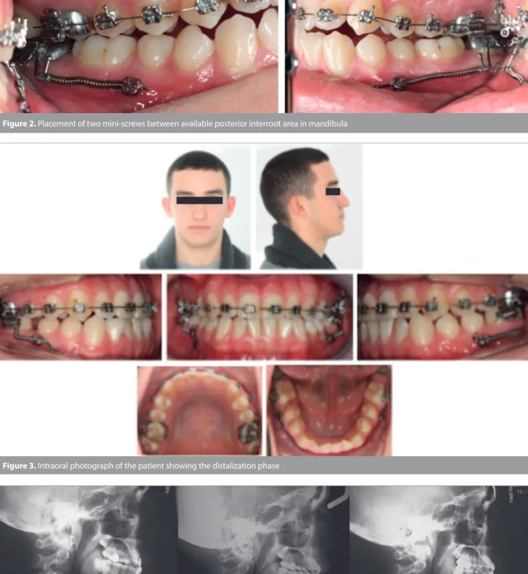
by two mini-screws (1.6×8mm Metin mini-screws (MTN), Medi-farm, Ankara, Turkey] placed into an available inter-root area. On the right side, one of the mini-screws was inserted between the first molar and second premolar, whereas on the left side it was inserted between the premolars (Figure 2).
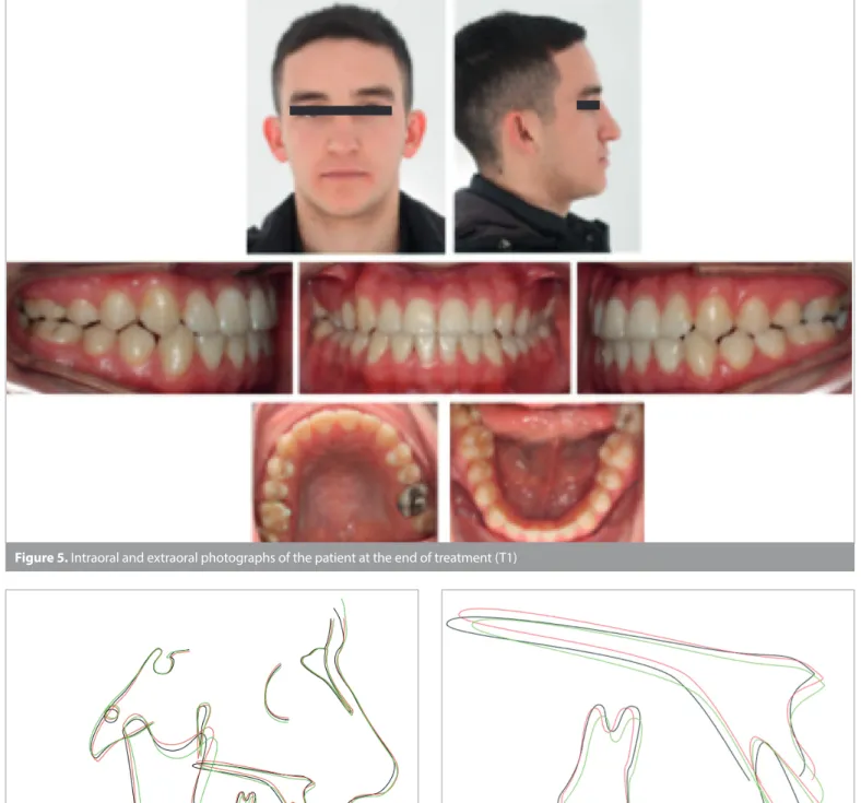
A segmented archwire bent from 0.017×0.025” stainless steel archwire was inserted between the slot of the mini-screw and an auxiliary tube of the second molar. Force (200g) was applied via a compressed open coil for second molar distalization. After the second molar distalization, the first molars were distalized using mini-screws as second molars, then premolars were distalized on the continuous archwire with closed coils while the first molars were kept in place using mini-screws (Figure 3).
In the maxillary arch, protrusion of incisors was planned to align the anterior teeth and correct cross-bite. Lateral cephalograms of the patient were obtained prior to (T0) and at the end of full-fixed orthodontic treatment (T1), 1.8 years after full-fixed orthodon-tic treatment (T2; Figure 4).
Treatment Results
Orthodontic treatment lasted approximately 2.5 years, with 1 year of molar distalization. At the end of the full-fixed treatment,
Table 1. Skeletal, dental, and soft-tissue measurements prior to
treatment (T0), at the end of treatment (T1), and after a postreten-tion period (T2) T0 T1 T2 SNA (°) 78 78 78 SNB (°) 79 79 79 ANB (°) -1 -1 -1 S-Go (mm) 91 91 91 ANS-Me (mm) 75 75 75 SNGoGn (°) 31 32 32 U1-NA (mm) 5 8 7 U1/PP (°) 115 131 128 L1-NB (mm) 5 6 6.5 IMPA (°) 89 91 93 Overjet (mm) -2 2 0.5 Overbite (mm) 0.5 1 1 Upper lip-SL (mm) -4 -2 -2 Lower lip-SL (mm) 0 -1 -1 Nasolabial (°) 117 109 110
Figure 1. Intraoral and extraoral photographs of the patient prior to treatment (T0)
120
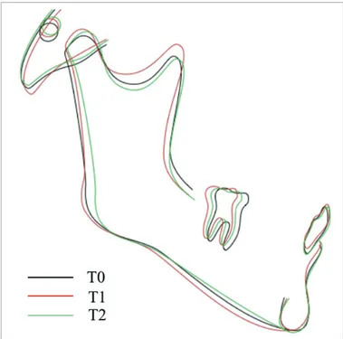
crowding was eliminated and Class I canine and super Class I mo-lar relationship with 2mm of overjet and 1mm of overbite was
obtained (Figure 5). Slight advancement in profile was achieved owing to the protrusion of upper lip position (Figure 6). Local
su-Figure 2. Placement of two mini-screws between available posterior interroot area in mandibula
Figure 3. Intraoral photograph of the patient showing the distalization phase
Figure 4. Lateral cephalograms of patient were taken at prior to treatment (T0), at the end of treatment (T1), after postretention period (T2)
perimpositions revealed prominent protrusion of upper incisors, slight protrusion of lower incisors, and distalization of lower mo-lars (Figure 7, 8; Table 1). Minimal relapse was seen in the postre-tention period (Figure 9).
DISCUSSION
The severity of the skeletal problem, growth pattern, facial pro-file and patient requirements are important in managing skeletal Class III malocclusions (7). In this mild skeletal Class III case, we preferred camouflage treatment. After treatment, his facial pro-file slightly improved owing to the protrusion of the upper lip. Another treatment option, in this case, was to extract four pre-molars; however, this would lead to more retrusive lips, which
Figure 5. Intraoral and extraoral photographs of the patient at the end of treatment (T1)
Figure 7. Local maxillary superimpositions
Figure 6. Total cephalometric superimpositions
122
could have worsened the profile. Another camouflage treatment option was to extract one mandibular incisor. However, the
pa-tient rejected either extractions. Therefore, mini-screw assisted mandibular molar distalization was preferred to correct the Class III malocclusion without teeth extractions, and positive overjet was achieved with the protrusion of maxillary incisors.
Class III elastic, which is one of the most widely used mecha-nisms for Class III correction, has disadvantages, such as the need for patient cooperation, tipping movement, anchorage loss and extrusion of maxillary molars (2). Here, extrusion and mesial-ization of maxillary molars would have increased the arch dis-crepancy and caused an open-bite tendency. However, with this system, direction of distalization force was passing through the center of resistance of molars, which avoided extrusion. Thus, mini-screw-assisted distal movement of the mandibular poste-rior teeth eliminated these undesirable effects.
In the previously reported mechanotheraphy, mini-screws or mini-implants were inserted into different areas for mandibular molar distalization (3-10). Some authors placed mini-plates or mini-screws into the anterior border of mandibular ramus and performed either en-masse distalization of mandibular denti-tion or tooth distalizadenti-tion (3-6). The posterior alveolar bone is an alternative site for posterior anchorage. Chung et al. inserted a C-shaped mini-implant into the maxillary molar area for Class III elastic usage through this implant (8). Later, Chung et al. in-serted C-implants between the mandibular first molar and
sec-Figure 8. Local mandibular superimpositions. 1: LMcs. Sagital linear
change of molar mesiobuccal cusp tip
Figure 9. Intraoral and extraoral photographs of the patient after postretention period (T2)
ond premolar, like in our system, as close as possible to the first molar root. In this system, second molars were distalized using a sliding jig connected to the main archwire that transferred the elastic forces to second molars applied from the mini-screws (9). Jing et al. (10) vertically implanted the mini-screws into external oblique ridge areas of the bilateral mandibular ramus between the first mandibular and second molar for en-masse distaliza-tion. This area reportedly offers more simple and stable force systems (11). Here, a mini-screw was inserted into the available mandibular posterior inter-root area. The implant site was based on cortical bone thickness, bone hardness, anatomic structures, and soft-tissue functional movements. The quantity and quality of the cortical bone greatly influenced the failure force of mini-screw implants (12, 13). Different from in the other studies, the present system of posterior inter-root area can be used for mini-screw insertion. Also, there is no need for full-fixed systems or to wait for leveling at the beginning of the treatment. Distalization can be immediately started. Further, this system differs in that the lower second molar is distalized by the frictionless system; it distalizes with the arch and does not slide on the archwire. In the present case, mandibular molars distalized 3 mm of each side of the arch. In the literature, molar distalization amounts with the as-sistance of mini-implants or mini-plates vary between 2-6 mm. Suga-wara et al. (3) achieved mandibular molar distalization of 3.5 mm at the crown level and 1.8 mm at the root level, and the average amount of relapse was 0.3 mm at both the crown and root apex levels. Poletti et al. (4) reported 4mm of molar distalization with a tipping of 10°. A case report stated that a mandibular dentition was distalized 5 and 2 mm on the left and right sides, respectively. Jing et al. (10) reported 4 mm of distalization without undesirable tipping.
The relapse amount in distalized mandibular molars during the postretention period, in this case, was 1mm. There are different re-ports about correlations between tipping and relapse. Chung et al. (9) stated that the larger the amount of tooth movement and the more the teeth are tipped, the greater is the relapse. However, Suga-wara et al. (3) found no significant correlations between the amount of relapse and tipping ratio and the amount of tooth movement. CONCLUSION
Thus, mini-screw supported mandibular molar distalization can be proposed as an effective treatment alternative for avoiding routine teeth extractions in borderline Class III cases.
Informed Consent: Written informed consent was obtained from the patient who participated in this study.
Peer-review: Externally peer-reviewed.
Author Contributions: Concept - B.I.A., E.K.; Design - B.I.A., E.K.; Supervi-sion - B.I.A., E.K.; Data Collection and/or Processing - B.I.A., E.K.; Analysis and/or Interpretation - B.I.A., E.K.; Writing Manuscript - B.I.A., E.K.; Critical Review - B.I.A., E.K.
Conflict of Interest: The authors have no conflict of interest to declare. Financial Disclosure: The authors declared that this study has received no financial support.
REFERENCES
1. Baek SH, Shin SJ, Ahn SJ, Chang YI. Initial effect of multiloop edge-wise archwire on the mandibular dentition in Class III malocclusion subjects, A three-dimensional finite element study. Eur J Orthod 2008; 30: 10-5. [CrossRef]
2. Proffit WR. Interarch elastics: their place in modern orthodontics. Hosl E, Baldauf A, editors. “Mechanical and biological basics in or-thodontic therapy.” Huthig, Germany: 1991; pp. 173-8.
3. Sugawara J, Daimaruya T, Umemori M, Nagasaka H, Takahashi I, Kawamura H, et al. Distal movement of mandibular molars in adult patients with the skeletal anchorage system. Am J Orthod Dentofa-cial Orthop 2004; 125: 130-8. [CrossRef]
4. Poletti L, Silvera AA, Ghislanzoni LTH.Dentoalveolar class III treat-ment using retromolar miniscrew anchorage. Prog Orthod 2013; 14: 7. [CrossRef]
5. Agarwal S, Shah N, Yadav S, Nanda RS. Mandibular arch retraction with retromolar skeletal anchorage in a Class III open-bite patient. J Clin Orthod 2014; 48: 775-82.
6. Anhoury PS. Retromolar miniscrew implants for Class III camouflage treatment. J Clin Orthod 2013; 47: 706-15.
7. Kerr WJS. Changes in soft tissue profile during the treatment of Class III malocclusion. Br J Orthod 1987; 14: 243-9. [CrossRef]
8. Chung K, Kim S, Kook YC. Orthodontic microimplant for distaliza-tion of mandibular dentidistaliza-tion in Class III correcdistaliza-tion. Angle Orthod 2005; 75: 119-28.
9. Chung K, Kim S, Choo H, Kook Y, Cope JB. Distalization of the man-dibular dentition withmini-implants to correct a Class III maloc-clusion with a midline deviation. Am J Orthod Dentofacial Orthop 2010; 137: 135-46. [CrossRef]
10. Jing Y, Han X, Guo Y, Li J, Bai D. Nonsurgical correction of a Class III malocclusion in an adult by miniscrew-assisted mandibular denti-tion distalizadenti-tion. Am J Orthod Dentofacial Orthop 2013; 143: 877-87. [CrossRef]
11. Chaimanee P, Suzuki B, Suzuki EY. “Safe zones” for miniscrew im-plant placement in different dentoskeletal patterns. Angle Orthod 2011; 813: 397-403. [CrossRef]
12. Lijima M, Takano M, Yasuda Y, Muguruma T, Nakagaki S, Sakakura Y, et al. Effect of the quantity and quality of cortical bone on the failure force of a miniscrew implant. Eur J Orthod 2013; 35: 583-9.
[CrossRef]
13. Motoyoshi M, Hirabayashi M, Uemura M, Shimizu N. Recommended placement torque when tightening an orthodontic mini-implant. Clin Oral Implants Res. 2006; 17: 109-14.[CrossRef]