CASE REPORT
Bilateral vertebral artery stenosis present
with vertigo
Dilcan Kotan,
1Saadet Sayan,
2Bilgehan Atilgan Acar,
2Pinar Polat
31Department of Neurology,
Sakarya University Medical Faculty, Sakarya, Turkey
2Department of Neurology, SB
Sakarya University Education and Research Hospital, Sakarya, Turkey
3Department of Radiology,
Kosuyolu Medipol Hospital, Istanbul, Turkey
Correspondence to Dr Dilcan Kotan, dilcankotan@ yahoo.com
To cite: Kotan D, Sayan S, Acar BA, et al. BMJ Case Rep Published online: [ please include Day Month Year] doi:10.1136/bcr-2012-007544
SUMMARY
Of ischaemic stroke patients, about 25% rise from the posterior or vertebrobasilar system. The ischaemia of vertebral arteries may emerge for different vascular pathological reasons, at different localisations and with different clinicalfindings. Despite its low morbidity and mortality risk, early diagnosis and treatment is of importance. Vertebrobasilar ischaemia symptoms can be observed clinically such as vertigo, tinnitus, double vision, headache, hypokinesis and hearing disorders, etc. In this article, a 42-year-old stroke patient case is presented, who applied to the emergency service with vertigo and then, was diagnosed with bilateral vertebral artery stenosis by means of cranial MR angiography.
BACKGROUND
Vertebral arteries appear immediately before the thyr-ocervical trunk on both sides of the neck as two branches of both subclavian arteries; they combine to form the basilar artery on the skull base upon extend-ing through a neural cavity between cervical 1 and 7 vertebras.1 Posterior or vertebrobasilar system
occlusions are responsible for one-fourth of ischae-mic stroke. Atherosclerosis of vertebral arteries is the main cause of posterior circulation infarcts. Vertebrobasilar ischaemia may occur in combination with symptoms such as vertigo, tinnitus, double vision, headache, drowsiness and hearing disorders.2
Vertebral artery stenosis is one of the recoverable causes of strokes in the posterior system. Although
the risk of morbidity and mortality is low, early diag-nosis and treatment is important.3 In recent years,
the number of diagnosed and treated patients has been increasing in accordance with advancements in medical-imaging methods. In this study, the case of a man with vertigo and young stroke is presented, who was diagnosed with bilateral vertebral artery stenosis.
CASE PRESENTATION
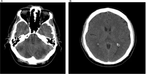
A 42-year-old hypertensive man consulted emer-gency service with headache and vertigo com-plaints. When he was admitted, his neurological examination was normal, except for left-beating nystagmus with horizontally rotating quick phase and minimal central facial palsy on the right side. He was admitted to hospital as an inpatient upon seeing hypodense area concordant with infarct in distinct bilateral cerebellar on the right and in thal-amus on the left side (figure 1A,B) in CT scan. Heparin with low molecular weight and 300 mg/ day acetylsalicylic acid two times a day was given to the patient during the period of hospitalisation. Ramipril 10 mg/day was included in the treatment with reasons of high arterial tension. On the second day of hospitalisation, right central facial palsy, slight dysarthria, left distinct dysmetria, left dysdiadochokinesia and left truncal ataxia were present in examination of the patient with advanced neurologicalfindings. Pervasive, diffusive infarct areas were seen on cranial MRI bilateral
Figure 1 (A and B) Hypodense area concordant with infarct in bilateral cerebellar area distinct on the left side (A) and in thalamus on the left side in CT scan.
Kotan D, et al. BMJ Case Rep 2013. doi:10.1136/bcr-2012-007544 1
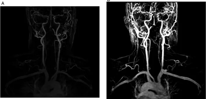
cerebellar hemisphere, left occipital, right mesencephalon and left thalamic area (figure 2A–C). Diffusion restriction was seen in these areas in diffusion MRI (figure 3A,B). It was impossible to see right vertebral flow in Doppler ultrasonography (USG) examination of carotis vertebral arteries and 70% stenosis was detected in left vertebral artery. As a result, in the intended carotis vertebral MR angiography, distal occlusion was observed in right vertebral artery’s intermediate and distal sections and artery distal of left vertebral (figure 4A,B). Balloon angioplasty was applied to the patient diagnosed with bilateral vertebral artery stenosis.
DISCUSSION
Vertebral artery roots from proximal subclavian. Vertebral artery reaches up to cervical vertebra after going through transverse foramens of the fifth and sixth vertebral after leaving the sub-clavian artery. The segment out of foramen ‘V1’, cervical segment within transverse foramens ‘V2’, segment up to the point where it enters subarachnoid space after going through dura mater‘V3’ form ‘V4’ segment after it enters into the sub-arachnoid space, and forms the basilar artery by combining with ‘V4’ in the frontal part of the bulb upon heading towards frontal-upper axis.4 Ischaemia of vertebral arteries may occur due to different vascular pathological causes, in different loca-tions and upon different clinicalfindings. Vertebral artery sten-osis is accompanied with a wide range of posterior system findings from a simple vertigo to occipital cortex dysfunction (vertigo, loss of balance, ataxia, tinnitus and drop attacks).5
When our patient was admitted to the hospital, there was only one symptom which was vertigo, and 2 days later, ataxia and other cerebellarfindings were involved. In the images of emer-gency tomography, he was monitored closely and anticoagulant treatment was initiated due to the presence of suspected infarct.
As the vertebral artery system has a structure that may be con-sidered complex, the evaluation of stenosis with ultrasonog-raphy (USG) with colour Doppler may be difficult. It is known that a good image quality, high level of technical knowledge and experience are all required in stenosis evaluation with ultrason-ography with colour Doppler.6Vertebral MR angiography has been introduced as a routine practice in recent years and as a supplementary method for detailed insights of vascular path-ology.7In our case, it was impossible to see right vertebralflow
with USG with colour Doppler and flow rate of left vertebral decreased remarkably. Vertebral artery angiography revealed that intermediate and distal sections of right and left vertebral distal were occluded. In our case, USG with colour Doppler and verte-bral artery MR angiography suggested the same vascular pathology.
Antithrombotic and anticoagulant medicines were used in order to reduce risk of stroke in vertebral artery stenosis.8 9 In
our patient, anticoagulant treatment was initiated starting from the early stages. Interventional treatment was considered, since the clinical findings were progressive despite the treatment. Surgical treatment is avoided in vertebral artery stenosis due to frequent occurrence of complications such as Horner syndrome (15–28%), lymphocele (4%), recurrent laryngeal nerve injury (2%).10In cases where symptoms are persistent following a suit-able medical treatment, there is an indication for treatment with balloon angioplasty and stenting.11However, there is no other study to compare the success rates of two methods.12 Our case
was admitted to the neuroradiology department with consider-ation that endovascular intervention would be suitable and a balloon angioplasty was applied.
Figur e 2 (A –C) Diffuse infar ct ar eas in bila ter al cer ebellar hemispher es and right mesencephalon (A), left occipital (B), left thalamic ar ea (C) in cr anial MRI.
2 Kotan D, et al. BMJ Case Rep 2013. doi:10.1136/bcr-2012-007544
Learning point
As a result, vertebral artery pathologies should be taken into consideration in individuals with central system-related vertigo regardless of the presence of vascular risk factors.
Competing interests None. Patient consent Obtained.
Provenance and peer review Not commissioned; externally peer reviewed.
REFERENCES
1 Savitz SI, Caplan LR. Vertebrobasilar disease. N Engl J Med 2005;352:2618–26. 2 Caplan LR, Wityk RJ, Glass TA, et al. New England medical center posterior
circulation registry. Ann Neurol 2004;56:389–98.
3 Flossmann E, Rothwell PM. Prognosis of vertebrobasilar transient ischaemic attack and minor stroke. Brain 2003;126:1940–54.
4 Bahar S Zarko, Bakac G. Anatomy of brain vascular circulation. In: Emre O, ed. Istanbul Medicine Faculty Neurology Book. 3rd Edn. Istanbul: Istanbul Medicine Faculty, 2012.
Figure 3 (A and B) Diffusion MRIfindings concordant with acute phase infarct in B1000 (A), and apparent diffusion coefficient map.
Figure 4 (A and B) Occlusion in right vertebral artery’s intermediate and distal sections and artery distal of left vertebral in MR angiography.
Kotan D, et al. BMJ Case Rep 2013. doi:10.1136/bcr-2012-007544 3
5 Khan S, Cloud G, Kerry S, et al. Imaging of vertebral artery stenosis: a systematic review. J Neurol Neurosurg Psychiatry 2007;78:1218–25.
6 Salonen JT, Salonen R. Ultrasound B-mode imaging in observational studies of atherosclerotic progression. Circulation 1993;87(3 Suppl):II56–65.
7 Kim SH, Lee JS, Kwon OK, et al. Prevalence study of proximal vertebral artery stenosis using high-resolution contrast-enhanced magnetic resonanceangiography. Acta Radiol 2005;3:314–21.
8 Prognosis of patients with symptomatic vertebral or basilar artery stenosis. The Warfarin-Aspirin Symptomatic Intracranial Disease (WASID) Study Group. Stroke 1998;29:1389–92.
9 Schonewille WJ, Algra A, Serena J, et al. Outcome in patients with basilar artery occlusion treated conventionally. J Neurol Neurosurg Psychiatry
2005;76:1238–41.
10 Jenkins JS, White CJ, Ramee SR, et al. Vertebral artery stenting. Catheter Cardiovasc Interv 2001;54:1–5.
11 Chastain HD, Campbell MS, Iyer S, et al. Extracranial vertebral artery stent placement: in-hospital and follow-up results. J Neurosurg 1999;91:547–52. 12 Cloud GC, Markus HS. Diagnosis and management of vertebral artery stenosis. QJM
2003;96:27–34.
Copyright 2013 BMJ Publishing Group. All rights reserved. For permission to reuse any of this content visit http://group.bmj.com/group/rights-licensing/permissions.
BMJ Case Report Fellows may re-use this article for personal use and teaching without any further permission. Become a Fellow of BMJ Case Reports today and you can:
▸ Submit as many cases as you like
▸ Enjoy fast sympathetic peer review and rapid publication of accepted articles ▸ Access all the published articles
▸ Re-use any of the published material for personal use and teaching without further permission For information on Institutional Fellowships contact [email protected]
Visit casereports.bmj.com for more articles like this and to become a Fellow
4 Kotan D, et al. BMJ Case Rep 2013. doi:10.1136/bcr-2012-007544