117
Common mistakes in the Dual-Energy X-ray Absorptiometry (DXA) in Turkey.
A Retrospective Descriptive Multicenter Study
Ali Yavuz Karahan1,*, Bugra Kaya2, Banu Kuran3, Ozlem Altındag4, Pelin Yildirim5, Sevil Ceyhan Dogan6, Aynur Basaran1, Ender Salbas7, Turgay Altınbilek8, Tuba Guler5, Sena Tolu9, Zekiye Hasbek10, Banu Ordahan1,
Ercan Kaydok11, Ufuk Yucel11, Selcuk Yesilyurt12, Almula Demir Polat13, Murat Cubukcu14, Omer Nas15, Umit Sarp15, Ozan Yasar16, Seher Kucuksarac1, Gozde Turkoglu1, Ahmet Karadag17, Sinan Bagcaci18,
Kemal Erol19, Emel Guler20, Serpil Tuna21, Ahmet Yildirim22, Savas Karpuz1 1 Department of Physical Medicine and Rehabilitation, Beyhekim State Hospital of Konya Konya/Turkey 2 Department of Nuclear Medicine of Necmettin Erbakan University, Meram Faculty of Medicine Konya/Turkey 3 Department of Physical Medicine and Rehabilitation, Sisli Etfal Training and Research Hospital Istanbul/Turkey 4 Department of Physical Medicine and Rehabilitation, Gaziantep University Sahinbey Research and Training Hospital, Gaziantep/Turkey
5 Department of Physical Medicine and Rehabilitation, Derince Training and Research Hospital Kocaeli/Turkey 6 Department of Physical Medicine and Rehabilitation, Cumhuriyet University, Faculty of Medicine Sivas/Turkey 7 Department of Physical Medicine and Rehabilitation, State Hospital of Agri/Turkey
8 Department of Physical Medicine and Rehabilitation, Physical therapy High school of Health Sciences of University of Halic, Istanbul/Turkey
9 Department of Physical Medicine and Rehabilitation, Medipol University, Faculty of Medicine Istanbul/Turkey 10 Department of Nuclear Medicine, Cumhuriyet University, Faculty of Medicine Sivas/Turkey
11 Department of Physical Medicine and Rehabilitation, State Hospital of Nevsehir/Turkey
12. Department of Physical Medicine and Rehabilitation, Physical Medicine and Rehabilitation Hospital of Yoncali, Kutahya/ Turkey
13 Department of Physical Medicine and Rehabilitation, State Hospital of Afyon/Turkey 14 Department of Physical Medicine and Rehabilitation, State Hospital of Denizli/Turkey 15 Department of Physical Medicine and Rehabilitation, State Hospital of Yozgat/Turkey
16 Department of Physical Medicine and Rehabilitation, Amasya University Sabuncuoglu Serefeddin Research and Training Hospital, Amasya/Turkey
17 Department of Physical Medicine and Rehabilitation, State Hospital of Sivas/Turkey 18 Department of Physical Medicine and Rehabilitation, State Hospital of Hakkari/Turkey 19 Department of Physical Medicine and Rehabilitation, State Hospital of Nigde/Turkey
20 Department of Physical Medicine and Rehabilitation, Kayseri Training and Research Hospital Kayseri/Turkey 21 Department of Physical Medicine and Rehabilitation, Akdeniz University, Faculty of Medicine Antalya/Turkey 22 Department of Orthopedics and Traumatology, Beyhekim State Hospital of Konya Konya/Turkey
* Corresponding author: Department of Physical Medicine and Rehabilitation, State Hospital of Konya/Turkey, Yunusem-remhnuranşsk no1 meramkonya; e-mail: [email protected]
Summary: Background: Osteoporosis is a widespread metabolic bone disease representing a global public health problem currently affecting more than two hundred million people worldwide. The World Health Organization states that dual-energy X-ray absorptiometry (DXA) is the best densitometric technique for assessing bone mineral density (BMD). DXA provides an accurate diagnosis of osteoporosis, a good estimation of fracture risk, and is a useful tool for monitoring patients undergo-ing treatment. Common mistakes in BMD testundergo-ing can be divided into four principal categories: 1) indication errors, 2) lack of quality control and calibration, 3) analysis and interpretation errors, and 4) inappropriate acquisition techniques. The aim of this retrospective multicenter descriptive study is to identify the common errors in the application of the DXA technique in Turkey. Methods: All DXA scans performed during the observation period were included in the study if the measure-ments of both, the lumbar spine and proximal femur were recorded. Forearm measurement, total body measuremeasure-ments, and measurements performed on children were excluded. Each examination was surveyed by 30 consultants from 20 different centers each informed and trained in the principles of and the standards for DXA scanning before the study. Results: A total of 3,212 DXA scan results from 20 different centers in 15 different Turkish cities were collected. The percentage of the discovered erroneous measurements varied from 10.5% to 65.5% in the lumbar spine and from 21.3% to 74.2% in the prox-imal femur. The overall error rate was found to be 31.8% (n = 1021) for the lumbar spine and 49.0% (n = 1576) for the
ORIGINAL ARTICLE
ACTA MEDICA (Hradec Králové) 2016; 59(4):117–123 https://doi.org/10.14712/18059694.2017.38
Introduction
Bone mineral density (BMD) is used in clinical practice as an indirect indicator of osteoporosis and fracture risk (1). Bone densitometry has quickly become the internationally accepted in-vivo bone mass measurement (1–3). The modal-ities of bone densitometry instruments include dual-energy X-ray absorptiometry (DXA), quantitative ultrasound, and quantitative computed tomography. DXA is realized as the reference technique to measure BMD in the lumbar spine, proximal femur, forearm, and whole body (2–4).
The World Health Organization (WHO) considers DXA to be the best densitometric technique for assessing BMD. DXA allows accurate diagnosis of osteoporosis, helps to de-termine the estimate fracture risk and monitoring of patients undergoing treatment (1–5). The primary target of DXA is to quantify BMD accurately and reproducibly and to compare that measurement with a reference population of asympto-matic individuals. Low measurement values on DXA predict the risk of fractures of the spine and hip, analogous to the relationship between high serum cholesterol and the risk of heart disease, or between high blood pressure and the risk of stroke. DXA is also useful in evaluating the effectiveness of FDA-approved therapies for osteoporosis, such as alen-dronate, risealen-dronate, and raloxifene (1, 2, 4–6).
Positive features of DXA include the safety of its per-formance, the short investigation time and the ease of use. A DXA measurement may take several minutes to complete with minimal radiation exposure. The densitometer produces ionizing radiation in the form of X-rays and uses laser ra-diation to position scans. However, the rara-diation exposure is so low that no shielding of rooms or health profession-als is required. The radiation from a DXA scan is less than one would receive during a round trip cross-country air-plane flight or during a day of normal background radiation (4, 7, 8).
In measuring BMD, DXA provides a high degree of ac-curacy, although precision of DXA varies across operators and equipment. Many factors related to the equipment, the operator and the patient determine DXA precision. Opera-tor-related factors are known to contribute to both long- and short-term precision errors. Differences in patient position-ing and definposition-ing the regions of interest (ROI), both heavily operator dependent, contribute to these variations (3, 7–9). It is estimated that more than five thousand DXA instruments are in regular use worldwide and it is well known that the operators have had widely varied instruction and
experi-ence. Operator training in different centers and countries is not standardized, and a lack of knowledge and skill can introduce errors in acquisition, analysis and interpretation of the scans (6, 7, 10).
Common errors in BMD testing can be separated into four categories: 1) indication errors, 2) lack of quality con-trol and calibration, 3) analysis and interpretation errors, and 4) acquisition errors (8–15). Acquisition errors have been well defined in previous studies including improper patient positioning, inappropriate scan mode, invalid skeletal site, persistent artifacts from the scanned area and incorrect de-mographic information (8–15). To our knowledge, there is no multicenter study that identifies and quantifies acquisition errors in DXA scanning in Turkey.
The aim of this retrospective descriptive multicenter sur-vey is to identify common errors in the acquisition of DXA scanning in Turkey.
Materials and Methods
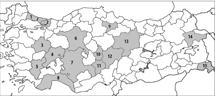
This descriptive study was conducted retrospectively over a period of 6 months from January 2014 to June 2014, in widely dispersed regions of Turkey. Data analysis and the study were approved by both the local scientific and ethical committees. We analyzed DXA scan results of 20 differ-ent cdiffer-enters in 15 differdiffer-ent cities in Turkey (Figure 1). The project was approved by the Necmettin University Human Research Ethics Committee (NEUHREC) (Approval num-ber: 2014/662) Individual ethics approval was also obtained from the (NEUHREC) responsible for each of the centers that participated in the project.
Consecutively performed DXA scan results were includ-ed the study if each containinclud-ed a measurement of the lumbar spine and the proximal femur. All available scans performed over the observation period were considered. Forearm meas-urements, total body measurements or measurements carried out on children were excluded.
Studies were surveyed by 30 consultants from 22 differ-ent cdiffer-enters that were informed and trained on DXA scanning principles and standards before the study. Screening was performed according to the following guidelines:
For the lumbar spine (Figure 2a):
– The spine must be centered and straight (centered spinous processes).
– The scan must accurately show L1 through L5, as well as the ribs attached to T12 at the top of the view. proximal femur. Conclusion: In Turkey, DXA measurements of BMD have been in use for over 20 years, and examination processes continue to improve. There is no educational standard for operator training, and a lack of knowledge can lead to significant errors in the acquisition, analysis, and interpretation.
Fig. 1: Locations of DXA sites that contributed to the study: 1. Istanbul, 2. Kocaeli, 3. Kütahya, 4. Afyon, 5. Denizli, 6. Ankara, 7. Konya,
8. Antalya, 9. Amasya, 10. Nevşehir, 11. Niğde, 12. Kayseri, 13. Sivas, 14. Ağrı, 15. Hakkari.
– A small amount of the iliac crest must be visible in the lower corners of the view.
– Similar amounts of soft tissue must appear on each side of the entire spine.
– The region of interest (ROI) must be correctly oriented. ROI areas must include an adequate amount of soft tissue but must not include excessive ribs or iliac crests. – Marking of vertebrae located in the ROI must be in the
correct order.
– ROI must not contain artifacts or foreign bodies like met-als, surgical clips, contrast medium, plastic materimet-als, jewelry, body piercings, zippers or buttons.
– The scan must be free of distorting anatomies like lami-nectomy or spina bifida occulta.
– There must be no conditions that can affect the region being examined or invalidate the measurements like Pa-get’s disease, ankylosing spondylitis, aortic calcification, severe scoliosis or degenerative changes.
For the proximal femur (Figure 2b):
– The image must include the entire femoral head, neck, and approximately 7.5 cm (3 inches) of the femoral shaft. – The femoral shaft must be aligned parallel to the long
dimension of the measurement rectangle.
– The correct amount of soft tissue must be visible lateral to the greater trochanter.
– Internal rotation of the hip must be confirmed by little or no visible lesser trochanter.
– The preferred position for the femoral neck ROI differs depending on equipment manufacturers. The selection of the ROI (greater trochanter, proximal femoral neck
or femoral head) must be correct. Fig. 2: Correct positioning in lumbar and proximal femur scanning.
– There must be no conditions that can affect the region being scanned or invalidate the measurements like frac-tures, callus formation or bladder stone.
Also, the specifics of the equipment and the department responsible for the DXA scanning for each institution were recorded.
Statistical analysis of data was performed using the computerized software program SPSS version 13 (SPSS, Inc., Chicago, IL, USA). Variables designed as some cat-egorical (true or false) and derived variables (absolute and percent change) for the statistical analysis plan. Descriptive data were presented as mean ± standard deviation. Demo-graphic and clinical characteristics were compared using the chi-square test. Independent samples t test was used for the comparison of the two groups. A “p” value less than 0.05 was considered as statistically significant.
Results
A total of 3,212 DXA scan results were examined, ac-quired from 22 different centers in 15 different Turkish cities. Ten of the 22 centers were state hospitals, 8 were university hospitals, and 4 were training and research hospitals. The Radiology department was responsible for DXA scanning in 12 centers, and the Department of Nuclear Medicine was in charge in 10 centers.
According to our criteria, the percentage of unaccept-able results ranged from 10.5% to 65.5% (average 31.7%, n = 1021) for measurement of the lumbar spine and from
21.3% to 74.2% (average 49.0%, n = 1576) in the proximal Fig. 3: Error rate in lumbar and proximal femur measurements in each of the 20 centers.
Fig. 4: Common sources of error for lumbar spine DXA measurements.
a/b: Incorrect placing of the ROI (lines should not coincide with the vertebral bodies). c/d: Region of interest must be marked in correct
order. e–h: Artifacts and foreign bodies (e. Button and zipper; f. Vertebral internal fixator; g. Calcification of the omentum and zipper;
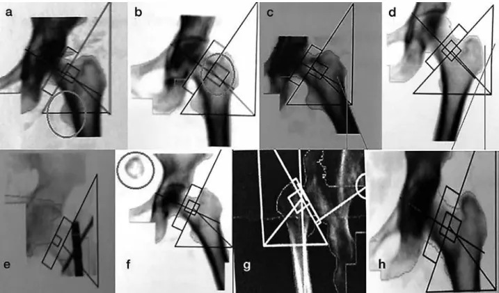
Fig. 5: Common sources of error for proximal femur measurements.
a: Inadequate internal rotation of hip (too much of the lesser trochanter is showing [circle]). b: Incorrect femoral neck region of interest
placement (region of interest must be set on proximal femur neck). c: Exaggerated abduction of the hip. d: Exaggerated adduction of the hip. e–f: Artifacts or foreign bodies (e. Internal fixator in the proximal femur; f: Bladder calculus [circle]). g: Congenital hip dislocation.
h: Inappropriately restricted and narrow scan area.
femur (Figure 3). Errors in defining the ROI was the most common error in lumbar spine results (16.2%, n = 522). Other disqualifying problems included the presence of ar-tifacts and foreign bodies, lateral misalignment of the ROI, laminectomy defects, Paget’s disease, severe scoliosis and degenerative changes (Figure 4).
Inadequate internal rotation of hip was the most common disqualifying error in proximal femur measurements. Other errors included the faulty definition of the ROI, exaggerated adduction or abduction of the hip, artifacts or foreign bodies in the scanned area, congenital hip dislocation, and restricted or narrow scan area (Figure 5).
There were no statistically significant differences in er-rors between the state hospitals, the university hospitals or the training and research hospitals (p > 0.05). There also were no statistically significant differences between the ra-diology and the nuclear medicine departments responsible for the measurements (p > 0.05).
Discussion
The results of this study show seriously high error rates in DXA assessment of the lumbar spine (31.7%) and the proximal femur (49.0%) in 22 centers in Turkey. Different
types of acquisition errors detected in both the lumbar and proximal femur DXA scans were responsible for the mis-takes. Data were collected from three classes of hospitals, state, university, and training and research hospitals between which there were no significant differences.
DXA is a clinically proven technique of measuring BMD in the lumbar spine, proximal femur, forearm, and whole body. With DXA, it is possible to measure BMD accurate-ly and reproducibaccurate-ly and to compare that measurement to a reference population of asymptomatic individuals (1–3). Therefore, in the diagnosis and management of osteopo-rosis and in determining future fracture risk, DXA is the most frequently used method. Some of the challenges in performing reliable DXA measurements include differences in equipment, acquisition techniques, reference databases, reporting methods, and descriptive terminology (7, 8, 10). Much of the responsibility of DXA fall to the operator such as reviewing the patient’s skeletal health history, entering demographic data into the computer, performing the image acquisition, and analyzing the scan (8, 10, 13). The operator must evaluate and, if necessary, adjust the computer-selected bone edges and markers of bone ROI. Also, hip bone edges and ROI must be correct, with particular attention paid to the location of the femoral neck box (7–11, 13, 16). Therefore,
DXA clearly is an operator-dependent method. This situa-tion marks the crucial need for educasitua-tion and experience to perform high-quality DXA.
The International Society for Clinical Densitometry (ISCD) is a society with a mission to 1) enhance knowledge, skill, and quality of densitometry among healthcare profes-sionals, 2) educate and certify clinicians and technologists, 3) increase patient awareness, 4) improve access to densi-tometry, 5) support clinical and scientific advances in the field, and 6) foster the exchange of scientific information. To address these issues, the ISCD periodically holds Position Development Conferences (1, 5, 9). Many guidelines and studies have drawn attention to frequent errors in the clini-cal use of DXA. Also, there are two available Turkish DXA guidelines (17, 18). However, to our knowledge, no detailed quality assessment and error flagging study of DXA bone mineral density measurement has been carried out across this country. The present study is the first one.
The selection of the ROI can be a major source of error even for experienced operators, especially in roto-scoliosis of the lumbar spine (7, 16, 19). In a typical DXA evaluation, there is usually a gradual increase in vertebral area progress-ing from L2 to L4. If this is not evident in the results, it is necessary to check the selection of ROI levels (7, 16, 19). In our study, ROI definition errors were the most common problem in lumbar spine measurements (Figure 4a–d).
Kendler et al.(14) used undissolved calcium (Ca) as an artifact to detect the effect on DXA results. They placed the Ca tablets in the soft tissue field, overlying bone and over-lapping both bone and soft tissue. An overlying Ca tablet had a considerable effect on BMD, resulting in 12.6% increase in the density of a single vertebral body (14). The Ca tablet ar-tifact showcases the importance of other spine DXA arar-tifacts on imprecision (14). Lumbar spine osteophytes explained 22% of the variation in spine BMD in men and 17% in wom-en. Artifacts such as mal-positioning, osteophytes, and aortic calcification, which could affect BMD, could be seen in the Ca tablet trials (1, 17, 20). In our study, we encountered different types of artifacts such as buttons, zippers, gall-stones or nonspecific omental calcification (Figure 4e–h). Abnormal densities are obtained in Paget’s disease because of the larger and denser vertebrae. An overlying calcified aorta may raise apparent vertebral density. In addition, ab-sent bony structures, as in laminectomy or spina bifida, or displaced bone as in vertebral rotation due to idiopathic sco-liosis could decrease the BMD estimation.
The BMD response following anti-resorptive or anabolic therapy is greater in the lumbar spine than in the proximal femur. However, this progress could be obscured in the pres-ence of degenerative skeletal conditions or by the internal fixation of the lumbar spine. Correct positioning of the pa-tient during the measurement of proximal femur BMD is also vital to obtaining precise values (15, 21). According to cadaveric studies, the minimum BMD in the femoral neck was recorded when the femoral neck was parallel to the ex-amination surface, and gradual increase in BMD occurred
when the femur was rotated either internally or externally (22, 23). Therefore, accurate estimation of BMD requires that the axis of the femoral neck be maintained parallel to the inspection surface and perpendicular to the X-ray beam. DXA equipment manufacturers provide different types of positioning aids to achieve this objective. Girard et al. (22) found a significant change in femoral neck BMD when the leg was rotated 10–15° from its neutral position. But Leka-mwasam S. et al. (21) examined the effect of leg rotation by 10 degrees either internally or externally on hip BMD in living subjects and concluded that their results show the effect of mal-positioning of the hip during DXA scanning were more significant in longitudinal than in cross-sectional BMD analysis (21). According to our results, inadequate internal rotation of hip was the most common error in prox-imal femur results. Definition errors of the ROI, excessive adduction or abduction of the hip, artifacts, foreign bodies, congenital hip dislocation, and inappropriate scan area were the other sources of error (Figure 5).
DXA estimates of BMD have been in use for over 20 years in Turkey, and examination protocols continue to improve results (17). There is no educational standard for operator training, and a significant lack of knowledge can lead to flaws in an acquisition, analysis, and interpretation of the scan. Clinical and technical awareness of potential sourc-es of error and artifact are pivotal to maximize the value of DXA measurements of BMD.
Limitations of study
The existing data was gleaned from records of 22 center from 15 provinces of Turkey. Turkey has 81 provinces, and our results that obtained from this study could not generalize to the generalize to the whole country. Although precision errors for DXA scanning evaluated for this study, we don’t know the full effects of these errors to the results and how these incorrect measurements effects on the clinical deci-sions. But this retrospective study helps to focus the question of “frequencies and definitions of common mistakes in the DXA for Turkey” and determines an appropriate sample size. A particularly useful application of this retrospective study is a pilot study that should be completed in anticipation of a prospective trial.
Acknowledgements
Only personal funds were used for this study. No ex-ternal funds were obtained. Financial disclosure statements have been achieved, and no conflicts of interest have been reported by the authors.
References
1. Sindel D, Gula G. Assessment of Bone Mineral Density in Osteoporosis. Türk Osteoporoz Dergisi 2015; 21: 23–9.
2. Tunç G, Doğan SC, Hizmetli S, Hayta E. In urban areas of Sivas city the determi-nation of bone mineral density reference values of healthy women who admitted to
Cumhuriyet University Hospital Physical Medicine and Rehabilitation outpatient clinic. Türk Osteoporoz Dergisi 2014; 20: 104–9.
3. Sindel D. Osteoporozda görüntüleme yöntemlerinde gelişmeler. Türkiye Klinikleri Journal of Physical Medicine Rehabilitation Special Topics 2009; 2(1): 50–61. 4. Aslan A, Uysal E, Karakoyun Ö. Bone Mineral Density Value in Kastamonu and
Area of Turkish Society Women J Clin Anal Med 2013; 4(3): 209–12. 5. Guglielmi G, Ferrari F, Bazzocchi A. Bone Mineral Density and Quantitative
Ima-ging. In: W. C. G. Peh ed. Pitfalls in Diagnostic Radiology. Springer, 2015: 109–32. 6. Allin S, Munce S, Carlin L, Butt D, Tu K, Hawker G, et al. Fracture risk assessment after BMD examination: whose job is it, anyway? Osteoporosis International 2014; 25(5): 1445–53.
7. Lewiecki EM, Lane NE. Common mistakes in the clinical use of bone mineral density testing. Nature clinical practice Rheumatology 2008; 4(12): 667–74. 8. El Maghraoui A, Roux C. DXA scanning in clinical practice. QJM: monthly
jour-nal of the Association of Physicians 2008; 101(8): 605–17.
9. Guglielmi G, Diano D, Ponti F, Bazzocchi A. Quality assurance in bone densito-metry. Current Radiology Reports 2014; 2(2): 1–6.
10. Khan AA, Colquhoun A, Hanley DA, Jankowski LG, Josse RG, Kendler DL, et al. Standards and guidelines for technologists performing central dual-energy X-ray absorptiometry. Journal of clinical densitometry: the official journal of the International Society for Clinical Densitometry 2007; 10(2): 189–95.
11. Messina C, Bandirali M, Sconfienza LM, DʼAlonzo NK, Di Leo G, Papini GD, et al. Prevalence and type of errors in dual-energy x-ray absorptiometry. European radiology 2015; 25(5): 1504–11.
12. Ott SM, Ichikawa LE, LaCroix AZ, Scholes D. Navel jewelry artifacts and intra-vertebral variation in spine bone densitometry in adolescents and young women. Journal of clinical densitometry: the official journal of the International Society for Clinical Densitometry 2009; 12(1): 84–8.
13. Lewiecki EM, Binkley N, Petak SM. DXA quality matters. Journal of clinical den-sitometry: the official journal of the International Society for Clinical Densitometry 2006; 9(4): 388–92.
14. Kendler DL, Kiebzak GM, Ambrose CG, Dinu C, Robertson S, Schmeer P, et al. Effect of calcium tablets on interpretation of lumbar spine DXA scans. Journal of
clinical densitometry: the official journal of the International Society for Clinical Densitometry 2006; 9(1): 97–104.
15. Wong JCH, Ong B. Evaluation of Femur Angle Abduction/Adduction and Bone Mineral Density Values. Journal of Clinical Densitometry 2005; 8(4): 472–5. 16. Vasić J, Gojković F, Zvekić-Svorcan J, Ćulafić-Vojinović V, Elez J, Filipović K.
The most common mistakes in bone mineral density testing with DXA method. MD-Medical data 2013; 5(3): 271–8.
17. Manisali M, Ozaksoy D, Dogan S. Osteoporozda Radyolojik Görüntüleme. Tur-kiye Klinikleri Journal of Orthopaedics and Traumatology Special Topics 2010; 3(2): 29–38.
18. Erselcan T, Ozen A, Yuksel D, Altun D, Ozturk E, Balci TA, et al. Kemik mi-neral yoğunluğu ölçümü uygulama kılavuzu. Turk J Nucl Med 2009; 18(1): 31–40.
19. Krueger D, Vallarta-Ast N, Libber J, Checovich M, Gangnon R, Binkley N. Po-sitioner and clothing artifact can affect one-third radius bone mineral density measurement. Journal of clinical densitometry: the official journal of the Interna-tional Society for Clinical Densitometry 2013; 16(2): 154–9.
20. Richmond B. DXAscanning to diagnose osteoporosis: Do you know what the results mean? Cleveland Clinic Journal of Medicine 2003; 20(4): 353–60. 21. Lekamwasam S, Lenora RS. Effect of leg rotation on hip bone mineral density
measurements. Journal of clinical densitometry: the official journal of the Inter-national Society for Clinical Densitometry 2003; 6(4): 331–6.
22. Girard MS, Sartoris DJ, Moscona AA, Ramos E. Measured femoral density by dual-energy X-ray absorptiometry as a function of rotation. Orthopaedic review 1994; 23(1): 38–40.
23. Cheng XG, Nicholson PH, Boonen S, Brys P, Lowet G, Nijs J, et al. Effects of anteversion on femoral bone mineral density and geometry measured by dual energy X-ray absorptiometry: a cadaver study. Bone 1997; 21(1): 113–7.
Received: 23/05/2016 Accepted: 29/08/2016