Address for Correspondence / Yazışma Adresi: Mustafa Hasbahceci. MD, Bezmialem Vakif University Faculty of Medicine, Dept of General Surgery, Vatan Str, 4th Floor,
34093, Fatih, Istanbul, Turkey. E-mail: [email protected]
©Telif Hakkı 2015 Gazi Üniversitesi Tıp Fakültesi - Makale metnine http://medicaljournal.gazi.edu.tr/ web adresinden ulaşılabilir. ©Copyright 2015 by Gazi University Medical Faculty - Available on-line at web site http://medicaljournal.gazi.edu.tr/
doi:http://dx.doi.org/10.12996/gmj.2015.50
Usefulness of Computed Tomography for Differentiation of Primary Inguinal Hernia
Primer İnguinal Herni Ayrımında Bilgisayarlı Tomografinin Yararlılığı
1Bezmialem Vakif University Faculty of Medicine, Dept of General Surgery, Vatan Str, 4th Floor, 34093, Fatih, Istanbul, Turkey 2Istanbul Medipol University, Faculty of Medicine, Dept of Radiology, TEM Road, 34214, Bagcilar, Istanbul, Turkey
ABSTRACT
Objective: The perception of inguinal anatomy during hernia formation may be an important issue for the evaluation of hernias. This study was conducted to describe detailed anatomy of inguinal region in patients with primary inguinal hernia by using computed tomography.
Methods: Study group included 18 inguinal hernia patients with computed tomography images. Images with multi-planar reformatting were evaluated with regard to anatomical landmarks found in inguinal region.
Results: Visualization of inguinal ligament and inferior epigastric artery was possible in all patients. Type of inguinal hernia was defined correctly in all patients by using inferior epigastric artery as an anatomic landmark. Pubic tubercle and lateral crescent sign in differentiation between direct and indirect inguinal hernias had variable sensitivity and specificity (0.69 and 0.60 for pubic tubercle, and 0.69 and 0.80 for lateral crescent sign, respectively). Conclusion: Multi-planar reformatting helps physicians to differentiate types of inguinal hernia by using the inferior epigastric artery as an anatomic landmark.
Key Words: Hernia, inguinal, diagnosis, imaging, computed tomography.
Received: 02.10.2015 Accepted: 08.04.2015
ÖZET
Amaç: Herni oluşumu sürecinde inguinal bölge anatominin algılanması, hernilerin değerlendirilmesi için önemli bir sorun olabilir. Bu çalışma, bilgisayarlı tomografi tardımı ile primer inguinal herni hastalarındaki inguinal bölge anatomisinin detaylı bir şekilde tanımlanması için düzenlenmiştir. Yöntemler: Çalışma grubu bilgisayarlı tomografi görüntüleri olan 18 inguinal herni hastasından oluşmakta idi. Çok kesitli yeniden biçimlendirme ile elde edilen görüntüler, inguinal bölgede bulunan anatomik belirleyiciler gözönüne alınarak değerlendirildi.
Bulgular: İnguinal ligament ve inferior epigastrik arterin görüntülenmesi hastaların tamamında gerçekleşti. Hastaların tamamında, inferior epigastrik arterin anatomik belirleyici olarak kullanılması ile herni türü doğru bir şekilde belirlendi. Direkt ve indirekt herni ayrımında pubik tüberkül ve lateral hilal bulgularının özgüllük ve duyarlılığı değişkenlik göstermekte idi (pubik tüberkül bulgusu için 0.69 ve 0.60; lateral hilal bulgusu için 0.69 ve 0.80).
Sonuç: Çok kesitli yeniden biçimlendirme, anatomik belirleyici olarak inferior epigastrik arterin kullanılması ile inguinal herni tiplerinin belirlenmesinde hekimlere yardımcı olmaktadır.
Anahtar Sözcükler: İnguinal herni, tanı, görüntüleme, bilgisayarlı tomografi. Geliş Tarihi: 10.02.2015 Kabul Tarihi: 04.08.2015
Mustafa Hasbahceci
1, Cengiz Erol
2INTRODUCTION
Inguinal hernia is a frequent condition, and inguinal hernia repair is one of the most commonly performed operations in practice of general surgery throughout the world (1-3). The perception of inguinal anatomy with regard to the pathologic changes occurred during hernia formation may be an important issue for preoperative diagnosis of direct and indirect inguinal hernias (4).
Although clinical diagnosis of these two types of inguinal hernias is possible by physical examination, computed tomography (CT) has dramatically improved preoperative diagnosis (1-6). Fine detailed anatomy of the inguinal region can also be well visualized with CT to improve the diagnosis and the differentiation between the hernia types (7-9).
The purpose of this study was to describe the spectrum of CT findings of inguinal region in patients with primary inguinal hernia, and to evaluate the ability of CT to differentiate direct and indirect inguinal hernia during preoperative period.
METHODS
The study was approved by the institutional review board. An approval by an ethics committee and informed patient consent were not required because of its retrospective design and not including patients’ data, respectively.
Patients
The study group was selected among 452 patients who underwent 513 Lichtenstein inguinal hernia repair between February 2003 and November 2010 and were taken for unrelated reasons except inguinal hernia by using cross-reference with Hospital and Radiology Information Systems. Preoperative CT scan was regarded as the inclusion criteria. The subjects included 13 male patients (mean age 62.6 years, age range 45-78 years) with primary inguinal hernia on preoperative CT images. There were 18 hernias that were right sided in 10 and direct in 13 of them. There was no femoral hernia diagnosed pre- and intra-operatively in the study group. The mean time interval between preoperative CT and inguinal hernia repair was 72 ± 93 days (median 21 days).
All the stored images of the identified patients in the picture archiving and communication system (PACS; GE Healthcare, Milwaukee, Wisconsin, USA) were reviewed by one radiologist, who was blinded to the final diagnosis, to determine whether the CT findings were or were not compatible with the surgical report.
Evaluation and Imaging Techniques
All abdominal CT examinations were performed with a 64-detector CT scanner (Lightspeed VCT; GE Healthcare, Milwaukee, Wisconsin, USA). Images were obtained in the cranio-caudal direction with detector collimation 64 × 0.625 mm; voltage 80-120 kVp; and tube current 150-250 mAs.
CT examinations were performed with oral and intravenous contrast medium. Water soluble oral contrast agents between 750-1500 ml (2%) were consumed 1-2 hour before the examinations. An upper extremity 18-20 gauge IV cannula was used for venous access. Between 50-100 mL of non-ionic contrast medium with 300 mg/ml iodine concentration was injected at a flow rate of 2-3 mL/s, followed by a 50 mL saline chaser with the same flow rate. The scan was started after 60-70 sec delay. All images were reconstructed as 2.5 mm axial sections and sent to the PACS. Stored image data sets in the PACS were analyzed again for the study group.
Axial images were reformatted to sagittal, coronal and oblique planes, and each patient’s scans were reviewed with regard to specific anatomic landmarks including the inguinal ligament, the inferior epigastric artery and the radiological femoral triangle. In addition, the inferior epigastric vein, the pubic tubercle and the spermatic cord were sought. Lateral compression of and stretching of the inguinal canal fat and contents by the hernia sac was accepted as the lateral crescent sign of direct inguinal hernia.
For each patient, the parameters listed below were noted: the anatomical structure contained within the hernia; medial or lateral situation of the hernia to the inferior epigastric artery; after schematic construction of orthogonal lines focused on the pubic tubercle, location of the hernia, ventral or dorsal to the X-axis and strictly lateral or crossing medially the Y-axis, and the presence of femoral vein compression. All inguinal hernias were defined as hernias located to ventral or superior to the X-axis. Direct and indirect inguinal hernias were defined when the hernias were located strictly lateral to the Y-axis and crossing medially the Y-axis among the hernias located ventral to the X-axis, respectively.
Intra-operative diagnosis of type of inguinal hernia was accepted as the standard reference for exact diagnosis. Any protrusion of viscus or peritoneal sac out of the peritoneal cavity was defined as a hernia. During the operation, a hernia via the internal ring lateral to the inferior epigastric artery and a hernia through the external ring medial to the inferior epigastric artery were defined as indirect and direct inguinal hernia, respectively. A femoral hernia was defined as the one posterior to the inguinal ligament through the femoral ring. A structured retrospective chart review side of the hernia, intra-operative diagnosis of the hernia with regard to its type and its content from surgical reports and CT findings was completed.
Statistical Analysis
Continuous variables were recorded as stated in medical records; other variables were marked as either absent or present. If the variable was missing for a specific finding, it was coded as such and not included in the final analysis. Sensitivity and specificity of all parameters used for the differentiation of the inguinal hernias from the femoral hernias and direct inguinal hernias from indirect ones were calculated.
RESULTS
In all hernias, it was possible to evaluate the inguinal ligament and the radiological femoral triangle (Table 1).
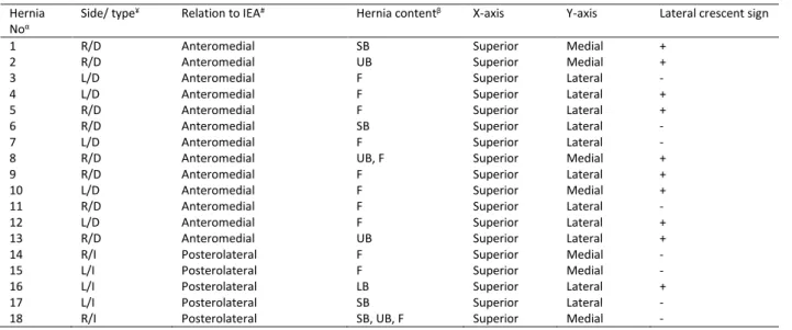
Table 1: Computed tomography findings. Hernia
Noα
Side/ type¥ Relation to IEA# Hernia contentβ X-axis Y-axis Lateral crescent sign
1 R/D Anteromedial SB Superior Medial +
2 R/D Anteromedial UB Superior Medial +
3 L/D Anteromedial F Superior Lateral -
4 L/D Anteromedial F Superior Lateral +
5 R/D Anteromedial F Superior Lateral +
6 R/D Anteromedial SB Superior Lateral -
7 L/D Anteromedial F Superior Lateral -
8 R/D Anteromedial UB, F Superior Medial +
9 R/D Anteromedial F Superior Lateral +
10 L/D Anteromedial F Superior Medial +
11 R/D Anteromedial F Superior Lateral -
12 L/D Anteromedial F Superior Lateral +
13 R/D Anteromedial UB Superior Lateral +
14 R/I Posterolateral F Superior Medial -
15 L/I Posterolateral F Superior Medial -
16 L/I Posterolateral LB Superior Lateral +
17 L/I Posterolateral SB Superior Lateral -
18 R/I Posterolateral SB, UB, F Superior Medial -
α: 18 hernias in 13 patients.
¥: R for right side, L for left side, D for direct hernia, I or indirect hernia. #: The inferior epigastric artery.
β: SB for the small bowel, UB for the urinary bladder, F for pre-peritoneal fatty tissue, LB for the large bowel.
GMJ
2015; 26: 170-173
Hasbahceci and Erol
All hernias were found to be located superior to the inguinal ligament without the presence of femoral vein compression indicating inguinal hernia. Type of the inguinal hernia as direct or indirect was defined correctly in all patients by using the inferior epigastric artery as an anatomic landmark (Figure 1). Pre-peritoneal fatty tissue was the most commonly encountered tissue in hernia sacs as seen in 12 out of 18. The small bowel and the urinary bladder were detected more than the large bowel in these hernia sacs. Evaluation of the hernias with regard to the relation to pubic tubercle revealed that all inguinal hernias were located superior to the X-axis (Figure 2). However, direct and indirect differentiation based on the relation to Y axis showed significant discordant results (Table 2). Only 12 out of the 18 hernias were classified in accordance with intra-operative definitions. The lateral crescent sign of direct inguinal hernia were detected only in nine out of 13 direct hernias (Figure 2).
Figure 1. Contrast enhanced coronal CT image of direct and indirect inguinal hernia. Right-sided direct inguinal hernia which is located medial to the inferior epigastric vessels (arrow) contains urinary bladder (asterix) and mesenteric fat, and left sided indirect inguinal hernia that is located lateral to the inferior epigastric vessels (arrow) contains colon and its mesentery (target).
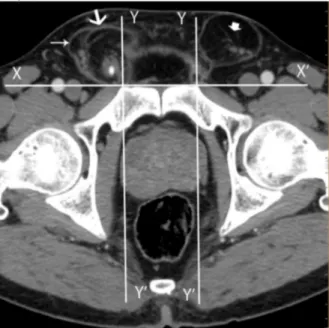
Figure 2. Axial CT image of the same patient at the level of pubic tubercle shows that actually right sided hernia is direct (thick arrow at the right side), however crossing medial to the Y-Y’ axis, and with a lateral crescent sign (thin arrow). The hernia on the left is indirect (arrowhead), however it is located lateral to the Y-Y’ axis.
Table 2. Sensitivity and specificity of the imaging parameters used for differentiation of inguinal hernia types.
Parameter Differentiation Sensitivity (%)
Specificity (%) Inferior epigastric artery Direct vs. indirect 100 100 Pubic tubercle (y axis) Direct vs. indirect 69.0 60.0 Lateral crescent sign Direct vs. indirect 69.0 80.0
DISCUSSION
Although the classification of inguinal hernia is usually based on the findings detected during preoperative physical examination and the surgical repair, new technology gives detailed anatomic criteria to diagnose and classify inguinal hernia. It is also difficult to distinguish between direct and indirect inguinal hernia only by physical examination (1, 2, 10). A patient presenting with inguinal pain alone has also some diagnostic controversy which may necessitate surgical intervention. Therefore, accurate imaging is vital to ensure diagnosis of inguinal hernia in patients with such presentations (10, 11). Scanner technology with multi-planar reformatting makes it possible to produce high resolution sagittal, coronal and oblique images in any plane from raw axial images that allows visualization of relevant anatomic structures (4, 8, 12, 13). It is reported that the inferior epigastric artery has been visible in 90% of the cases, especially on unenhanced computed tomography examinations (1). However, efficacy of such technology to visualize important anatomic structures found in the inguinal region was reported by several authors previously (12). In this study, it was possible to visualize the inferior epigastric artery and the vein, the inguinal ligament, the spermatic cord, the radiologic femoral triangle and the pubic tubercle in all cases. We believe that it can be accomplished with the use of multi-planar reformatting technique. However, the radiation exposure and higher cost are important issues for a case with an inguinal hernia. Therefore, CT scans were performed only for unrelated causes except inguinal hernia. It has been also speculated that the inguinal ligament could not be demonstrated on CT examinations in all cases (8). Therefore, the pubic tubercle has been proposed to be a valuable CT reference point in distinguishing direct inguinal, indirect inguinal and femoral hernias (1, 2, 8). Besides the study by Delabrousse (1), we could get discordant results by using the pubic tubercle as a reference point in distinguishing the direct and indirect inguinal hernias. The presence of direct inguinal hernia at the right side and indirect inguinal hernia at the left side were demonstrated in one of our patients by using the inferior epigastric artery as the anatomic landmark, in accordance with the intraoperative findings. However, differentiation based on the Y-axis erroneously resulted in reverse results. Parallel to our experience, the differentiation of the larger inguinal hernias based on the pubic tubercle have been thought to be difficult, because these hernias might be confined to more than one quadrant in relation to the pubic tubercle (2). In the present study, it was shown that it is possible to visualize the inguinal ligament in all cases. Both the visualization of the inferior epigastric artery in all cases and the highest sensitivity and specificity by using this artery as a reference point support that the inferior epigastric artery should be more reliable landmark than the pubic tubercle in distinguishing the types of inguinal hernia during CT.
In cases of direct inguinal hernia, it has been thought that contents of the inguinal canal including testicular vessels and the vas deferens or the round ligament is compressed and laterally stretched by the hernia causing a semicircular fatty tissue found in the inguinal canal. It has been reported that this lateral crescent of fat is a useful diagnostic sign for direct hernias (2). However, sensitivity and specificity of this sign was calculated as 0.69 and 0.80, respectively, questioning the effectiveness in distinguishing direct hernias from indirect ones.
Use of CT for the diagnosis of inguinal hernia hosts some inherent flaws that make it a less than ideal tool (2, 5, 12, 13). First, acquisition with the patients in the supine position causes the reduction of reducible hernias in most of the cases (4). Although there may be some improvement by using the Valsalva maneuver during CT, it is not possible to perform this maneuver in all cases (2, 14, 15).
There were some limitations belonging to our study. Retrospective design and small number of the cases were the major ones. Lack of femoral hernia was another important factor to prevent differentiation between inguinal and femoral hernias.
GMJ
2015; 26: 170-173
Hasbahceci and Erol
Usefulness of CT
CONCLUSION
Although radiation exposure and higher cost should be regarded as the factors to avoid for taking CT in all cases, multi-planar reformatting helps physicians to understand the inguinal anatomy in patients with inguinal hernia during pre-operative period in selected patients. The inferior epigastric artery should be the major reference point for differentiation of the inguinal hernia types.
Conflict of Interest
No conflict of interest was declared by the authors.
REFERENCES
1. Delabrousse E, Denue PO, Aubry S, Sarliève P, Mantion GA, Kastler BA. The pubic tubercle: a CT landmark in groin hernia. Abdom Imaging 2007; 32: 803-6.
2. Yeung VH, Pearl JM, Coakley FV, Joe BN, Westphalen AC, Yeh BM. Computed tomographic appearance of Prolene Hernia System and polypropylene mesh plug inguinal hernia repair. J Comput Assist Tomogr 2008; 32: 529-32.
3. Holzheimer RG. Inguinal hernia repair: what to do with the evidence? World J Surg 2009 33:2056-7.
4. Cherian PT, Parnell AP. The diagnosis and classification of inguinal and femoral hernia on multisection spiral CT. Clin Radiol 2008; 63: 184-92.
5. Suzuki S, Furui S, Okinaga K, Sakamoto T, Murata J, Furukawa A, et al. Differentiation of femoral versus inguinal hernia: CT findings. AJR Am J Roentgenol 2007; 189: W78-83.
6. Garvey JF. Computed tomography scan diagnosis of occult groin hernia (2012) Hernia 16:307-14.
7. Burkhardt JH, Arshanskiy Y, Munson JL, Scholz FJ. Diagnosis of inguinal region hernias with axial CT: the lateral crescent sign and other key findings. Radiographics 2011; 31: E1-12.
8. Kitami M, Takase K, Tsuboi M, Rikimaru Y, Hakamatsuka T, Yamada T, et al. Differentiation of femoral and inguinal hernias on the basis of anteroposterior relationship to the inguinal ligament on multidimensional computed tomography. J Comput Assist Tomogr 2009; 33:678-81.
9. Hoshiai S, Mori K, Shiigai M, et al (2014) Reduction en masse of inguinal hernia: MDCT findings of two cases. Hernia 2014 Jan 12. doi 10.1007/s10029-014-1217-9
10. Robinson A, Light D, Kasim A, Nice C. A systematic review and meta-analysis of the role of radiology in the diagnosis of occult inguinal hernia. Surg Endosc 2012; 27: 11-8.
11. Fataar S. CT of inguinal canal lipomas and fat-containing inguinal hernias. J Med Imaging Radiat Oncol 2011; 55: 485-92.
12. Cherian PT, Parnell AP. Radiologic anatomy of the inguinofemoral region: insights from CT. AJR Am J Roentgenol 2007; 189: W177-83. 13. Shizukuishi T, Abe K, Takahashi M, Sakaguchi M, Aizawa T, Narata M, et
al. Inguinal bladder hernia: multi-planar reformation and 3-D reconstruction computed tomography images useful for diagnosis. Nephrology (Carlton) 2009; 14: 263.
14. Højer AM, Rygaard H, Jess P. CT in the diagnosis of abdominal wall hernias: a preliminary study. Eur Radiol 1997; 7: 1416-8.
15. Emby DJ, Aoun G. CT technique for suspected anterior abdominal wall hernia. AJR Am J Roentgenol. 2003; 181: 431-3.