91
Diseases of the Esophagus (2004) 17, 91–94
© 2004 ISDE
Blackwell Publishing, Ltd.
Original article
Esophageal perforation: the importance of early diagnosis and primary repair
Atilla Eroglu,1 Ibrahim Can Kürkçüoglu,1 Nurettin Karaoglanoglu,1 Celal Tekinbaß,1 Ömer Yımaz,2
Mahmut Baßoglu3
Departments of 1Thoracic Surgery, 2Gastroenterology, and 3General Surgery, Faculty of Medicine, Atatürk
University, Erzurum, Turkey
SUMMARY. Esophageal perforation is associated with high morbidity and mortality rates, particularly if not
diagnosed and treated promptly. Despite the many advances in thoracic surgery, the management of patients with esophageal perforation remains controversial. We performed a retrospective clinical review of 36 patients, 15 women (41.7%) and 21 men (58.3%), treated at our hospital for esophageal perforation between 1989 and 2002. The mean age was 54.3 years (range 7–76 years). Iatrogenic causes were found in 63.9% of perforations, foreign body perforation in 16.7%, traumatic perforation in 13.9% and spontaneous rupture in 5.5%. Perfora-tion occurred in the cervical esophagus in 12 cases, thoracic esophagus in 13 and abdominal esophagus in 11. Pain was the most common presenting symptom, occurring in 24 patients (66.7%). Dyspnea was noted in 14 patients (38.9%), fever in 12 (33.3%) and subcutaneous emphysema in 25 (69.4%). Management of esophageal perforation included primary closure in 19 (52.8%), resection in seven (19.4%) and non-surgical therapy in 10 (27.8%). The 30-day mortality was found to be 13.9%, and mean hospital stay was 24.4 days. In the surgically treated group the mortality rate was three of 26 patients (11.5%), and two of 10 patients (20%) in the conserv-atively managed group. Survival was significantly influenced by a delay of more than 24 h in the initiation of treatment. Primary closure within 24 h resulted in the most favorable outcome. Esophageal perforation is a life threatening condition, and any delay in diagnosis and therapy remains a major contributor to the attendant mortality.
KEY WORDS: diagnosis, esophageal perforation, treatment.
INTRODUCTION
Despite advances in the treatment of esophageal
perforations, the mortality remains high.1,2
The rate of perforation of the esophagus by medical apparatus increases in parallel with the recent and rapid devel-opment in upper gastrointestinal system endoscopic techniques.3,4
However, the incidence of perforation also shows increases during the endoscopic extrac-tion of foreign bodies.4
Morbidity resulting from perforation of the esophagus depends on the corrosive nature of the gastrointestinal fluid and the spread of the intaken foods and bacteria to the paraesophageal spaces. Perforation may or may not extent to the pleura. If pleura is intact, the contents of gastro-intestinal systems initially cause chemical and then
severe bacterial mediastinitis under the pleura. The severity of the lesion and the clinical symptoms depend on the site, extent of the perforation and any delay in diagnosis. The purpose of this report is to review the diagnostic examinations, therapy and outcome in 36 patients with esophageal perforation treated at our clinic.
METHODS
From 1989 to 2002, 36 patients with esophageal perforation were diagnosed and treated at our clinic. There were 21 men and 15 women with a mean age of 54.3 years (range 7–76 years). The causes of esoph-ageal perforations included endoscopic instru-mentation (63.9%), foreign bodies (16.7%), external trauma (13.9%) and spontaneous rupture (5.5%) (Table 1). Specifically excluded from analysis were the patients with postoperative anastomotic leakage or perforations secondary to neoplasm. This analysis focused on etiology, location of perforation, signs
Address correspondence to: Dr Atilla Eroglu, Department of Thoracic Surgery, Faculty of Medicine, Atatürk University, 25240 Erzurum, Turkey. Tel: +90 442 3166333; Fax: +90 442 3166340;
Email: [email protected]
92 Diseases of the Esophagus
and symptoms, diagnostic methods, time interval at presentation, specific treatment and morbidity and mortality.
RESULTS
The etiologies of perforation varied widely with instrumental causes predominating over other causes. In 23 (63.9%) of the 36 patients, the perforation was caused by instrumentation. Instrumental perforation occurred during or within 6 h of the procedure. Benign underlying esophageal disease was documented in four patients and malignant disease in seven (30.5%). The perforation was located in the upper third of the esophagus in 12 patients, middle third in 13 patients, and lower third in 11 patients.
Pain, the most common presenting symptom, was noted in 24 of the 36 cases (66.7%). Other symp-toms included dyspnea (38.9%), fever (33.3%) and dysphagia (5.5%). Subcutaneous emphysema was the most common sign and was recorded in nearly two-third of the patients. The time delay from the onset of symptoms to the admission to our clinic varied from 0 to 96 h (mean 13.4 h).
Chest radiographs were performed in all patients and revealed mediastinal air in 18 (50%), mediastinal widening in 11 (30.5%), hydropneumothorax in nine (25%) and pleural effusion in eight (22.2%). Diag-nosis was confirmed by contrast radiography in 17 patients (47.2%), and by endoscopy in all patients. The interval between rupture and initial treatment was less than 24 h in 27 patients (75%), and longer than 72 h in seven (19.4%). Perforations secondary to endoscopy were identified earlier, but identification of perforations resulting from foreign bodies was delayed. Primary closure was performed in 19 patients, followed in frequency by non-surgical treatment (10 patients), and resection (seven patients). Ten perfora-tions in the cervical esophagus were treated with primary closure and two were treated non-operatively. No deaths were observed in this group. Eight perfora-tions in the thoracic esophagus were treated oper-atively and five were treated non-operoper-atively. In the
operative group, there were four primary closures with wide mediastinal drainage, and four esoph-agogastrostomies were performed. There were four deaths (30.7%) in this group. Eight abdominal esophageal perforations were treated with primary closure and three were treated with esophagogas-trostomy. One patient in this group died. All patients received antibiotic therapy and fluid resuscitation. The mainstay of non-operative treatment was broad-spectrum antibiotics, hyperalimentation and naso-gastric suction.
Complications occurred in 11 patients (30.5%) and were as follows: anastomotic leakage in two patients, sepsis in three, pleural effusion in five, wound infection in two, respiratory failure in three and renal failure in two. This constituted 30.7% in the operative group and 30% in the non-operative group.
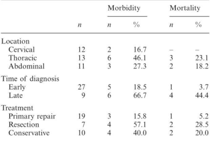
The overall mortality was five of 36 patients (13.9%), with four of five patients dying from causes related to their esophageal perforation. Three deaths occurred in surgically-treated patients (11.5%) and two occurred in medically-treated patients (20%). Mortality among patients treated within 24 h of sustaining the injury was substantially less than among those for whom diagnosis and treatment were delayed. Cervical esophageal perforations resulted in less mortality than thoracic and abdominal per-forations (Table 2).
The mean hospital stay was 24.4 days (range, 7–76 days). At discharge all patients were on a normal diet without dysphagia. Follow-up informa-tion was available in 18 of the 31 survivors (58%). The mean follow up period was 37 months (2–121 months). No patients required re-operation on the esophagus. Of the 18 patients, 14 (77.7%) have no complaints and can swallow freely.
DISCUSSION
The incidence of esophageal perforation from a particular cause varies depending on the patient
Table 1 Etiology and mortality of the esophageal perforations
Etiology No Results Survived Died Esophagoscopy 11 10 1 Foreign body 6 5 1 Trauma 5 3 1 Prostheses placement 4 3 1 Dilatation 3 3 0 ERCP 3 2 1
Endotracheal tup placement 2 2 0
Boerhaave 2 2 0
Total 36 31 5
Table 2 Features of the esophageal perforations
n Morbidity Mortality n % n % Location Cervical 12 2 16.7 – – Thoracic 13 6 46.1 3 23.1 Abdominal 11 3 27.3 2 18.2 Time of diagnosis Early 27 5 18.5 1 3.7 Late 9 6 66.7 4 44.4 Treatment Primary repair 19 3 15.8 1 5.2 Resection 7 4 57.1 2 28.5 Conservative 10 4 40.0 2 20.0
Esophageal perforation: early diagnosis and repair 93
population. The most common cause of esophageal
perforation is instrumentation.4,5 The incidence of
rupture increasing with the increasing rate of endo-scopic procedures. The reported incidence of perfora-tion for rigid esophagoscopy is 0.11%, and fiber
endoscopy varies from 0.018 to 0.03%.3 Therapeutic
endoscopy is associated with a much higher
fre-quency of perforation (1–10%).6,7 Spontaneous
perforation, foreign body penetration, traumatic intubation, paraesophageal operation, penetrating trauma, placement of intraesophageal prostheses
and pneumatic dilatation have also been implicated.4,5
In our study most perforations were caused by instruments.
Perforation can occur at any level, but it is most common at the cervical and the distal end of the esophagus. According to recent studies the third
area of narrowing is seldom involved.8 But in our
series all areas were equally involved with ruptures. A high incidence of underlying esophageal disease has been reported in recent series.4,5,8 In our series
underlying esophageal disease was present in 11 of 36 patients (30.5%).
Diagnosis of esophageal perforation can be diffi-cult, as the presentation is often non-specific and is easily confused with other disorders such as spon-taneous pneumothorax, myocardial infarction, aortic aneurysm, peptic ulcer, pancreatitis and pneumo-nia.5,9,10 The signs and symptoms of the perforation
depend on the location, the causes of the perfora-tion and the time of the rupture. Pain is the most
common complaint of esophageal perforation.8 It
can occur anywhere in the chest or epigastrium. Less often, dysphagia, dyspnea, cyanosis are other common signs. Physical examination may reveal subcutaneous emphysema and signs related to the development of hydropneumothorax. When pain and subcutaneous emphysema develops after instru-mentation, perforation should be suspected. Radio-graphic examination may reveal in varying degrees, pneumomediastinum, pleural effusion, hydropneu-mothorax, subcutaneous emphysema and subdia-phragmatic air. Han et al. noted normal plain film
findings in 12% of patients.11 Radiographic findings
were noted in about 75% of the patients in our series. Pneumomediastinum and subcutaneous emphysema were frequently observed. Diagnosis can be confirmed with the use of contrast radiographs, CT scans or endoscopy. Moghissi and Pender recommend the
use of flexible esophagoscopy.12 We applied
pre-operative flexible or rigid esophagoscopy. This will reveal the site and extent of the perforation and will be of assistance in designing a therapeutic approach for the patient.
Discussions about the treatment of esophageal ruptures have been continuing.5,13,14 Various factors
have important impacts on the treatment approach. These are as follows: the cause and location of the
perforation, the presence of underlying esophageal disease, the time interval between the perforation and diagnosis and the age and general status of the patient. Treatment options include medical or sur-gical interventions. Medical modalities can include antibiotic administration, nasogastric suctioning, administration of H2 receptor blockers, pleural drainage, restricted oral intake and a feeding entero-stomy or total parental nutrition. Non-operative therapy that can be applied in selected cases
resulted in a 22% mortality rate in a review.5 This
rate was 20% in our series.
Surgical interventions may include an esopha-geal resection or exclusion, or chest drainage with or without esophageal repair. The primary repair of perforation of the esophagus within 24 h, in the absence of pre-existing esophageal disease, remains the gold standard of therapy and it is the approach
most commonly advocated in the literature.5,14
Prim-ary repair with or without reinforcement was performed in 52.7% of patients in our series, with a 94.7% survival rate. The layers were closed primarily and separately after muscular and mucosal deprit-mant. Reinforcement of the primary repair has been
advocated by many surgeons.15–17 We favor use of a
pleural flap for middle third injuries and the omen-tal flap for lower third ruptures. Primary repair is not advisable in some situations, such as: underly-ing malignant disease, scleroderma, grade IV reflux esophagitis and stage III achalasia. These situations are best treated by esophageal resection.12,18,19
The outcome for perforation depends on its location, causes, promptness of treatment, the pres-ence of underlying esophageal disease and type of treatment. The mortality rate was 8.7% in our series due to instrumental injuries but it was 23% due to other causes. The morbidity and mortality rates are found to be significantly low in perfora-tions of the cervical esophagus. In the cases that were diagnosed in the first 24 h with no underlying disease Jones and Ginsberg (1992) reported a mor-tality of 22% in their collective review.5 The overall
mortality was 13.9% in our series. We can explain this by the early diagnosis and the cause being instrumentation in most of our cases.
In summary, perforation of the esophagus remains a potentially fatal condition and requires early diagnosis and accurate treatment to prevent mor-bidity and mortality. Our experience suggest that primary repair should be attempted whenever pos-sible for all patients with esophageal perforation.
References
1 Larsen K, Skov Jensen B, Axelsen F. Perforation and rup-ture of the esophagus. Scand J Thorac Cardiovasc Surg 1983; 17: 311–16.
2 Nesbitt J C, Sawyers J L. Surgical management of esoph-ageal perforation. Am Surg 1987; 53: 183 –91.
94 Diseases of the Esophagus
3 Wesdorp I C, Bartelsman J F, Huibregtse K, den Hartog Jager F C, Tytgat G N. Treatment of instrumental oesoph-ageal perforation. Gut 1984; 25: 398 – 404.
4 Michel L, Grillo H C, Malt R A. Operative and nonoperat-ive management of esophageal perforations. Ann Surg 1981; 194: 57–63.
5 Jones W G, Ginsberg R J. Esophageal perforation: a contin-uing challenge. Ann Thorac Surg 1992; 53: 534 – 43. 6 Okike N, Payne W S, Neufeld D M, Bernatz P E, Pairolero P C,
Sanderson D R. Esophagomyotomy versus forceful dilation for achalasia of the esophagus: results in 899 patients. Ann Thorac Surg 1979; 28: 119–25.
7 Miller R E, Bossart P W, Tiszenkel H I. Surgical manage-ment of complications of upper gastrointestinal endoscopy and esophageal dilation including laser therapy. Am Surg 1987; 53: 667–7.
8 Bladergroen M R, Lowe J E, Postlethwait R W. Diagnosis and recommended management of esophageal perforation and rupture. Ann Thorac Surg 1986; 42: 235–9.
9 Finley R J, Pearson F G, Weisel R D, Todd T R J, Ilves R, Cooper J. The management of nonmalignant intrathoracic esophageal perforations. Ann Thorac Surg 1980; 30: 575 – 83.
10 Han S Y, McElvein R B, Aldrete J S, Tishler J M. Perfora-tion of the esophagus: correlaPerfora-tion of site and cause with plain film findings. AJR 1985; 145: 537– 40.
11 Moghissi K, Pender D. Instrumental perforations of the oesoph-agus and their management. Thorax 1988 August; 43: 642–6. 12 Ökten I, Cangir A K, Özdemir N, Kavukçu S, Akay H, Yavuzer S. Management of esophageal perforation. Surg Today 2001; 31: 36–9.
13 Bufkin B L, Miller J I, Mansour K A. Esophageal perfora-tion: Emphasis on management. Ann Thorac Surg 1996; 61: 1447–52.
14 Grillo H C, Wilkins E W Jr. Esophageal repair following late diagnosis of intrathoracic perforation. Ann Thorac Surg 1975; 20: 387–99.
15 Bryant L R, Eiseman B. Experimental evaluation of inter-costal pedicle grafts in esophageal repair. J Thorac Cardio-vasc Surg 1965; 50: 626–9.
16 Millard A H. ‘Spontaneous’ perforation of the oesophagus treated by utilization of a pericardial flap. Br J Surg 1971; 58: 70–2.
17 Wright C D, Mathisen D J, Wain J C, Moncure A C, Hilgenberg A D, Grillo H C. Reinforced primary repair of thoracic esophageal perforation. Ann Thorac Surg 1995; 60: 245–9.
18 Orringer M B, Stirling M C. Esophagectomy for esophageal disruption. Ann Thorac Surg 1990; 49: 35 – 43.
19 Attar S, Hankins J R, Suter C M, Coughlin T R, Sequeira A, McLaughlin J S. Esophageal perforation: a therapeutic challenge. Ann Thorac Surg 1990; 50: 45 –51.