Ankara Univ Vet Fak Derg, 66, 407-416, 2019 DOI: 10.33988/auvfd.571632
Determination of antibiotic susceptibility, ESBL genes and
pulsed-field gel electrophoresis profiles of extended-spectrum
β-lactamase-containing Escherichia coli isolates
Hidayet TUTUN
1,a,, Alper KARAGÖZ
2,b, Levent ALTINTAŞ
3,c,
Nadir KOÇAK
4,d1Burdur Mehmet Akif Ersoy University, Faculty of Veterinary Medicine, Department of Pharmacology and Toxicology, Burdur; 2Uşak University, Department of Molecular Biology and Genetics, Uşak; 3Ankara University, Faculty of Veterinary Medicine,
Department of Pharmacology and Toxicology, Ankara; 4Selçuk University, Faculty of Medicine, Medical Genetics, Konya, Turkey. aORCID: 0000-0001-9512-8637; bORCID: 0000-0002-8178-223X; cORCID: 0000-0002-5148-723X;
dORCID: 0000-0002-1727-1582.
Corresponding author: [email protected]
Received date: 29.05.2019- Accepted date: 05.08.2019
Abstract: The purpose of this study was to determine the phenotypic antibiotic susceptibility patterns, extended-spectrum β-lactamase (ESBL) genes and genotypic profiles of ESBL-positive Escherichia coli strains isolated from urine samples obtained from outpatients with urinary tract infection in Turkey. A total of 120 E. coli strains during 2017, 2018, and 2019 (40 patients per year) were examined for antibiotic susceptibility patterns by disc diffusion method, for ESBL genes using PCR and sequencing and for molecular typing by pulsed-field gel electrophoresis (PFGE) method. The isolates were evaluated for their sensitivity to 21 different antibiotics. Four different antimicrobial resistance patterns were determined according to antibiotic susceptibility status of the isolates. The β-lactamase genes detected in the isolates were CTX-M-15 + OXA-1 (n= 14), CTX-M-15 (n= 24), 1 + CTX-M-15 (n= 52), TEM-1 + SHV-TEM-12 (n=6), SHV-TEM-12 TEM-1 (n= 6), TEM-TEM-1 + CTX-M-TEM-1 (n= 6), TEM-TEM-1 + CTX-M-TEM-16 (n= 6) and TEM-TEM-1 + CTX-M-9 (n= 6). The CTX-M-15 was the most prevalent ESBL enzyme in the isolates. As a result of PFGE analysis performed by XbaI enzyme restriction process, one major PFGE profile and three main groups (Group I-II-III) were observed. While antibiotic resistance profiles of the strains showed four groups (RI-RII-RIII-RIV), PFGE band profiles showed a major group (90% similarity ratio). High ESBL production and decreased susceptibility to broad-spectrum cephalosporins were observed in E. coli strains. In addition, PFGE analysis showed high clonal similarity among E. coli isolates.
Keywords: Antimicrobial drug resistance, ESBL genes, Escherichia coli, molecular subtyping, pulsed-field gel electrophoresis.
Genişlemiş spektrumlu β-laktamaz üreten Escherichia coli izolatlarının antibiyotik duyarlılıklarının,
GSBL genlerinin ve pulsed-field jel elektroforez yöntemiyle genotipik profillerinin belirlenmesi
Özet: Bu çalışmada Türkiye’de idrar yolu enfeksiyonu bulunan hastalardan alınan idrar örneklerinden izole edilmiş olan genişlemiş spektrumlu β-laktamaz (GSBL) pozitif E. coli suşlarının antibiyotik duyarlılık paternlerinin (fenotipik), GSBL genlerinin ve genotipik profillerinin belirlenmesi amaçlandı. Çalışmada 2017, 2018 ve 2019 yıllarında ve her yıl 40 hastadan olmak üzere, toplam 120 adet E. coli suşunun disk difüzyon yöntemi ile antibiyotik duyarlılıkları, PCR ve sekanslama ile GSBL genleri, pulsed-field jel elektroforez (PFGE) yöntemi ile moleküler tipleri belirlendi. Numunelerden elde edilen izolatların 21 farklı antibiyotiğe karşı duyarlılığı değerlendirildi. İzolatların antibiyotik duyarlılık durumlarına göre, değerlendirmede dört farklı antimikrobiyal direnç paterni tespit edildi. İzolatlarda β-laktamaz genleri olarak CTX-M-15 + OXA-1 (n= 14), CTX-M-15 (n= 24), TEM-1 + CTX-M-15 (n= 52), TEM-1 + SHV-12 (n= 6), SHV-12 1 (n= 6), TEM-1 + CTX-M-1 (n= 6), TEM-1 + CTX-M-16 (n= 6) ve TEM-1 + CTX-M-9 (n= 6) tespit edildi. CTX-M-15 izolatlarda en yaygın görülen GSBL enzim tipi olarak belirlendi. PFGE analizi sonucunda, bir majör PFGE profili ve üç ana grup (Grup I-II-III) gözlendi. Suşların antibiyotik direnç profilleri, dört grupta (RI-RII-RIII-RIV) bulunurken, PFGE bant profilleri ise bir majör grup içinde bulunduğu (% 90 benzerlik oranı) belirlendi. Numunelerde E. coli yüksek GSBL üretimi ve geniş spektrumlu sefalosporinlere karşı azalan bir duyarlılık gözlendi. Ayrıca, PFGE analizi ile bu izolatların yüksek klonal benzerliğe sahip olduğu da gösterildi.
Anahtar sözcükler: Antimikrobiyal direnç, GSBL genleri, Escherichia coli, moleküler alt tiplendirme, pulsed-field jel elektroforez.
Introduction
Escherichia coli is a commensal bacteria of the
digestive tract microflora of humans and animals, some of them cause intestinal and extraintestinal pathologies (25). Animals are defined as important zoonotic reservoirs for human intestinal pathogenic E. coli and extraintestinal pathogenic E. coli (ExPEC) causing diseases in farm and pet animals (4, 53). Extraintestinal pathogenic E. coli is an important cause of diverse infections, which includes urinary tract infections (UTI) in human and animals (26, 32, 53). UTI is a significant bacterial infection that causes serious complications including emphysematous cystitis and pyelonephritis when infection is inadequately managed. Although most of the patients face a single or rare episode of the disease, many patients experience recurrent UTIs (19, 50). Approximately 80 % of patients who suffer from UTI is caused by E. coli and
Staphylococcus saprophyticus (15).
Antibiotic resistance leads to failure in the treatment of both community- and hospital-acquired infections and appears to be a growing problem worldwide. The application of antibacterial drugs in clinical therapy results in the emergence of microorganisms that are resistant to these drugs. One of the most common bacterial-resistance mechanism against antimicrobial drugs is the inactivation of the drug by the enzymes they synthesize (8). β-lactamases are enzymes that are produced by bacteria providing multiple resistance to β-lactam compounds by hydrolyzing the β-lactam ring in these antibiotic groups. The prolonged exposure of bacterial strains to a large number of β-lactam antibiotics has increased their activities by inducing mutation of the β-lactamases, which are known as extended-spectrum β-lactamases (ESBLs), against the third-generation cephalosporins (42). Types of ESBL generated by mutations in genes coding the narrow-spectrum β-lactamases (TEM-1, TEM-2, or SHV-1) are TEM, SHV, CTX-M, OXA and Amp C. The most frequently identified ESBL genes produced by E. coli and
Klebsiella spp. are TEM and SHV (34). The emergence of
ESBL-producing bacteria has been commonly reported in veterinary medicine since β-lactam antibiotics have been used mostly for therapeutic and prophylactic reasons in livestock (35, 47). Several studies showed that similar ESBL isolates were found in human and livestock, suggesting a zoonotic transfer (11, 22, 28).
The emergence of antibiotic resistance is accelerated by the overuse and misuse of antibiotics and the lack of development of new antimicrobial drugs (48). Antimicrobial agents are widely used for therapeutic or nontherapeutic purposes in animal husbandry. Use of these drugs results in selection for antimicrobial resistant
E. coli in the microflora of these animals. Subsequently,
antimicrobial-resistant E. coli can be transferred from animals to humans through cross-contamination or
consumption of raw or insufficiently cooked meat contaminated with antimicrobial resistant bacteria (4, 38). Pulsed-field gel electrophoresis is a molecular fingerprinting method considered the “gold standard” among molecular typing methods to classify bacteria. The method is based on the determination and interpretation of the profiles formed by the appropriate restriction endonuclease enzyme of genomic DNA isolated from the bacterial cell embedded in low melting agarose without deterioration of the structural integrity. PFGE technique has been used safely in the typing of many bacteria such as Salmonella typhimurium, Neisseria gonorrhoeae, methicillin-resistant Staphylococcus aureus, Acinetobacter baumannii, and E. coli (12, 14, 16, 20, 49).
The purpose of the present study was to determine the antibiotic susceptibility patterns and genotypic profiles of ESBL positive E. coli strains isolated from urine samples collected between 2017 and 2019 and also determine the prevalence of ESBL genes among the isolates. The most effective antibiotic selection was provided for the empirical treatment of E. coli-induced UTI by forming an antibiotic susceptibility pattern of E.
coli. In addition, by determining the possible clonal
relationship between isolates via PFGE analysis, the similarity of antibiotic susceptibilities of strains with common band profiles was investigated.
Material and Methods
Sample collection: A total of 120 E. coli isolates obtained from UTI patient’s urine samples collected from outpatients (n= 120) in Public hospital in Konya province of Turkey between 2017-2019 were evaluated in the present study. Patient data anonymized in this study. These samples were collected with collection containers and transported to the microbiology laboratory.
Isolation and identification of Escherichia coli: E. coli isolates were incubated in Eosin Methylene-blue
(EMB) and Nutrient agar (NA) media overnight at 37ºC by a single colony incubation technique. Colonies which appeared metallic sheen were again fished out into nutrient broths and subcultures were maintained on nutrient agar and used for further identification and antimicrobial sensitivity testing. A single colony picked up and identified as E. coli using IMVIC test (citrate, methyl red, Voges-Proskauer, citrate, ornithine, urea, indole, kligler iron agar media). The strains were confirmed as E. coli using with gram-negative crystal identification kit (BBL Crystal ID System, Becton Dickinson, Cockeysville). ESBL production was confirmed if the presence of a β-lactamase inhibitor enlarged, the zone size of inhibition by ≥5 mm for all 120 isolates (10).
Antimicrobial susceptibility testing: Disc diffusion method according to Clinical and Laboratory Standards Institute (10) was used for antibiotic susceptibility test.
Tested antibiotics (BBL, Becton Dickinson) were cefoxitin (FOX, 30 µg), cefotaxime (CTX, 30 µg), cefepime (FEP, 30 µg), ceftazidime (CAZ, 30 µg), cefazolin (CFZ, 30 µg), cephalothin (CEF, 30 µg), cefuroxime (CXM, 5 µg), ampicillin (AMP, 10 µg), ampicillin-sulbactam (SAM, 10 µg), amoxicillin-clavulanate (AMC, 30 µg), imipenem (IPM, 10 µg), piperacillin (PIP, 100 µg), trimethoprim-sulfamethoxazole (SXT, 23.75 µg/1.25 µg), ofloxacin (OFX, 5 µg), amikacin (AMK, 30 µg), gentamicin (GEN, 10 µg), sulfisoxazole (SXZ, 0.25 µg), nitrofurantoin (NIT, 100 µg), piperacillin-tazobactam (TZP, 110 µg), ticarcillin-clavulanate (TIM, 85 µg), carbenicillin (CAR, 100 µg). Quality control was performed with E. coli ATCC 25922 strain. Isolates were grouped as resistant (R), intermediate-resistant (I), or susceptible (S) according to the CLSI (10).
Characterization of ESBL genes: The presence of genes encoding TEM, SHV, OXA and CTX-M type β-lactamases was examined in this study. PCR screening for the presence of different β-lactamase genes, blaTEM-type, blaCTX-M-blaTEM-type, blaSHV-type and blaOXA-1-blaTEM-type, were performed as described previously (6, 7, 24). Amplicons obtained from PCR were sequenced on both strands and sequences were compared to those reported in the Database of the GenBank and on the Lahey Clinic beta-lactamase website (http://www.lahey.org/Studies/) to identify the β-lactamase genes.
PFGE analysis: The isolates, which were identified as E. coli by biochemical and molecular methods and confirmed as ESBL-producing E. coli by antibiotic susceptibility test, were subjected to PFGE to analyze the genetic diversity of them in order to investigate genetic similarities. A single colony of each isolate was suspended with CSB buffer (cell suspension buffer, 10 mM Tris-HCl, 50 mM EDTA, 20 mM NaCl, pH 7.2). 2 % low-melting agarose (LMA, Sigma-Aldrich) was prepared in the CSB buffer supplemented with Sodium dodecyl sulfate (a final concentration of 1 %) (SDS, Sigma-Aldrich). The agarose-buffer mixture was melted by heating in magnetic stirrer to 45-50°C. Bacteria suspension was added to the
agarose tubes by means of a pipette, and the pipette was used for mixing the suspension. This mixture was transferred to plug molds (10mm x 5mm x 1.5mm, Bio-Rad) and after solidification of agarose for the preparation of high-quality DNA, the plugs were incubated overnight at 55°C in lysis buffer (50 mmol Tris-HCI, pH 8.0; 50 mmol EDTA, pH 8.0; 1 % sarcosine; 1 mg of proteinase K/ml). The agarose was washed three times with sterile distilled water, followed by three washes with TE buffer (10 mmol Tris, pH 8.0; 1 mmol EDTA, pH 8.0) Agarose-embedded DNA was transferred to the mixture containing XbaI restriction enzyme (Thermo Scientific), and incubated to digest at 37°C for 2 hours. PFGE was carried out with the CHEF-DR® II system (Bio-Rad Laboratories, Nazareth, Belgium) using a 1 % of pulsed-field certified agarose prepared in standard 0.5xTris-boric acid-EDTA (TBE buffer). The electrophoresis condition was set as follows: Initial switch time, 2 s; final switch time, 35 s, run time, 20 h; gradient, 6V/cm; angle 120°; temperature, 14°C. After electrophoresis, the gel was stained by putting into 400 ml ultrapure water solution containing 5 µg/ml ethidium bromide for 20 minutes and the fingerprinting profile was photographed under ultraviolet light using a Gel Logic 220 imaging system (Kodak Company, USA). The band profiles were analyzed using the Gel Compar II software system (version 3.0, Applied Maths, Sint-Martens-Latem). First of all, three external standard strains (1, 7, 15, carried out in wells, E. coli ATCC 25922 strain) in each gel were used to normalize the images. Dendrograms and clustering analysis of PFGE profiles were performed using “the unweighted-pair group method with mathematical averaging” (UPGMA), the Dice coefficient with a 1-1.5 % band position tolerance and optimization. The interpretation of PFGE patterns was categorized as follows: indistinguishable, closely related, possibly related, or different according to the criteria of Tenover et al. (44).
Results
All isolates were tested for antibiotic resistance using Disc diffusion method. Table 1 shows the resistant
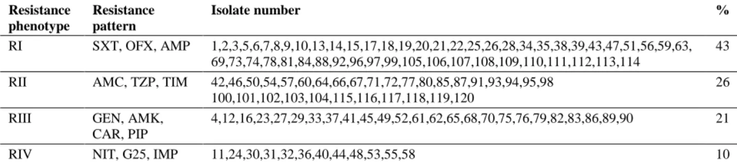
Table 1. Antibiotic resistance profiles for E. coli isolates. Resistance phenotype Resistance pattern Isolate number % RI SXT, OFX, AMP 1,2,3,5,6,7,8,9,10,13,14,15,17,18,19,20,21,22,25,26,28,34,35,38,39,43,47,51,56,59,63, 69,73,74,78,81,84,88,92,96,97,99,105,106,107,108,109,110,111,112,113,114 43
RII AMC, TZP, TIM 42,46,50,54,57,60,64,66,67,71,72,77,80,85,87,91,93,94,95,98 100,101,102,103,104,115,116,117,118,119,120
26
RIII GEN, AMK,
CAR, PIP
4,12,16,23,27,29,33,37,41,45,49,52,61,62,65,68,70,75,76,79,82,83,86,89,90 21
RIV NIT, G25, IMP 11,24,30,31,32,36,40,44,48,53,55,58 10
SXT: Trimethoprim-sulfamethoxazole; OFX: Ofloxacin; AMP: Ampicilin; AMC: Amoxicillin-clavulanate; TZP: Piperacillin-tazobactam; TIM: Ticarcillin-clavulanate; GEN: Gentamicin; AMK: Amicasin; CAR: Carbenicillin; PIP: Piperacillin; NIT: Nitrofurantoin; G25: Gentamicin high resistance; IMP: Imipenem.
phenotypes of E. coli isolates. Among the isolates, the predominant resistance profile was RI (SXT-OFX-AMP). Approximately 43 % of all isolates were RI phenotype, whereas isolates with RII phenotype (Combinations of β-lactam-β-lactam inhibitors) accounted for 26 %. 21 % of all isolates were resistant to 4 antibiotics belonging to RIII phenotype (GEN, AMK, CAR, and PIP). Aminoglycoside antibiotics (GEN, AMK) were considered as the most important ones in this group. Isolates (10 %) which are resistant to other antibiotics were classified as RIV phenotype (NIT, G25, and IMP).
In the PFGE study for genotypic typing, after the E.
coli DNAs were cut with FastDigest XbaI enzyme with
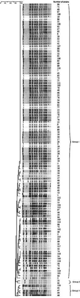
restriction endonuclease activity, PFGE gel images in which various band patterns were formed were determined. Dendrogram analysis was performed in the next stage of gel images of E. coli strains in which PFGE band profiles were observed. After the band profile analysis using Gel-Compare-II, PFGE profile dendrograms were formed and the relationships between the strains were determined according to the Dice similarity coefficient. When the dendrogram of 120 E. coli strains of 2017, 2018 and 2019 were examined; based on Tenover criteria (44), 120 strains were found to be related to each other according to 85% and higher similarity rates. Although there is only one major clone, the strains are divided into three pulsotypes. Group I (90-3/113 strains), Group II (51-81/2 strains), Group III (120-61-67-64-66/5 strains) (Figure 1).
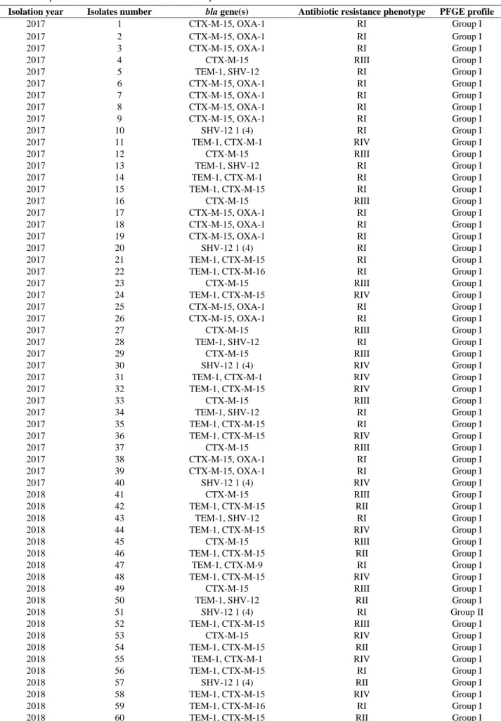
After all the studies, epidemiological data were obtained from the clinical files of the patients in order to correlate all the data with significant results. In the present study that examined patients admitted to the hospital at different times, a data (Table 2) with information about epidemiological information, antimicrobial susceptibility patterns and PFGE types of 120 isolates collected from 2017 to 2019 were obtained.
Group I (113 strains) and Group III (5 strains) were the most common PFGE profile groups in the genotypic investigation of E. coli strains. Among the 113 strains in Group I, 50 had RI resistance and had SXT, OFX and AMP resistance. In addition, 27, 24, and 12 strains had RII, RIII and RIV resistances, respectively. The two identical strains in Group II showed the same antibiotic resistance pattern (RI). The 4 strains in Group III show the same antibiotic resistance pattern (RII). Furthermore, there is also a strain with RIII resistance (Table 2).
All the phenotypic ESBL-producing E. coli isolates were confirmed by PCR and sequencing for detection of genes encoding TEM, SHV, OXA and CTX-M type β-lactamases. The β-lactamase genes detected in the isolates were CTX-M-15 + OXA-1 (n= 14), CTX-M-15 (n= 24), TEM-1 + CTX-M-15 (n= 52) , TEM-1 + SHV-12 (n= 6),
Figure 1. PGFE XbaI digestion patterns and clonal analysis of 120 ESBL-producing E. coli isolates (2017-2019) (1-120). The dendrogram using the Dice coefficient and UPGMA clustering methods showing the relationships between E. coli strains.
Table 2. Comparison of PFGE and antibiotic resistance profiles of E. coli strains.
Isolation year Isolates number bla gene(s) Antibiotic resistance phenotype PFGE profile
2017 1 CTX-M-15, OXA-1 RI Group I 2017 2 CTX-M-15, OXA-1 RI Group I 2017 3 CTX-M-15, OXA-1 RI Group I 2017 4 CTX-M-15 RIII Group I 2017 5 TEM-1, SHV-12 RI Group I 2017 6 CTX-M-15, OXA-1 RI Group I 2017 7 CTX-M-15, OXA-1 RI Group I 2017 8 CTX-M-15, OXA-1 RI Group I 2017 9 CTX-M-15, OXA-1 RI Group I 2017 10 SHV-12 1 (4) RI Group I
2017 11 TEM-1, CTX-M-1 RIV Group I
2017 12 CTX-M-15 RIII Group I 2017 13 TEM-1, SHV-12 RI Group I 2017 14 TEM-1, CTX-M-1 RI Group I 2017 15 TEM-1, CTX-M-15 RI Group I 2017 16 CTX-M-15 RIII Group I 2017 17 CTX-M-15, OXA-1 RI Group I 2017 18 CTX-M-15, OXA-1 RI Group I 2017 19 CTX-M-15, OXA-1 RI Group I 2017 20 SHV-12 1 (4) RI Group I 2017 21 TEM-1, CTX-M-15 RI Group I 2017 22 TEM-1, CTX-M-16 RI Group I 2017 23 CTX-M-15 RIII Group I
2017 24 TEM-1, CTX-M-15 RIV Group I
2017 25 CTX-M-15, OXA-1 RI Group I 2017 26 CTX-M-15, OXA-1 RI Group I 2017 27 CTX-M-15 RIII Group I 2017 28 TEM-1, SHV-12 RI Group I 2017 29 CTX-M-15 RIII Group I 2017 30 SHV-12 1 (4) RIV Group I
2017 31 TEM-1, CTX-M-1 RIV Group I
2017 32 TEM-1, CTX-M-15 RIV Group I
2017 33 CTX-M-15 RIII Group I
2017 34 TEM-1, SHV-12 RI Group I
2017 35 TEM-1, CTX-M-15 RI Group I
2017 36 TEM-1, CTX-M-15 RIV Group I
2017 37 CTX-M-15 RIII Group I
2017 38 CTX-M-15, OXA-1 RI Group I
2017 39 CTX-M-15, OXA-1 RI Group I
2017 40 SHV-12 1 (4) RIV Group I
2018 41 CTX-M-15 RIII Group I
2018 42 TEM-1, CTX-M-15 RII Group I
2018 43 TEM-1, SHV-12 RI Group I
2018 44 TEM-1, CTX-M-15 RIV Group I
2018 45 CTX-M-15 RIII Group I
2018 46 TEM-1, CTX-M-15 RII Group I
2018 47 TEM-1, CTX-M-9 RI Group I
2018 48 TEM-1, CTX-M-15 RIV Group I
2018 49 CTX-M-15 RIII Group I
2018 50 TEM-1, SHV-12 RII Group I
2018 51 SHV-12 1 (4) RI Group II
2018 52 TEM-1, CTX-M-15 RIII Group I
2018 53 CTX-M-15 RIV Group I
2018 54 TEM-1, CTX-M-15 RII Group I
2018 55 TEM-1, CTX-M-1 RIV Group I
2018 56 TEM-1, CTX-M-15 RI Group I
2018 57 SHV-12 1 (4) RII Group I
2018 58 TEM-1, CTX-M-15 RIV Group I
2018 59 TEM-1, CTX-M-16 RI Group I
2018 61 CTX-M-15 RIII Group III
2018 62 CTX-M-15 RIII Group I
2018 63 TEM-1, CTX-M-15 RI Group I
2018 64 TEM-1, CTX-M-15 RII Group III
2018 65 CTX-M-15 RIII Group I
2018 66 TEM-1, CTX-M-9 RII Group III
2018 67 TEM-1, CTX-M-15 RII Group III
2018 68 TEM-1, CTX-M-15 RIII Group I
2018 69 TEM-1, CTX-M-15 RI Group I
2018 70 CTX-M-15 RIII Group I
2018 71 TEM-1, CTX-M-15 RII Group I
2018 72 TEM-1, CTX-M-16 RII Group I
2018 73 TEM-1, CTX-M-15 RI Group I
2018 74 TEM-1, CTX-M-15 RI Group I
2018 75 CTX-M-15 RIII Group I
2018 76 CTX-M-15 RIII Group I
2018 77 TEM-1, CTX-M-15 RII Group I
2018 78 TEM-1, CTX-M-15 RI Group I
2018 79 TEM-1, CTX-M-1 RIII Group I
2018 80 TEM-1, CTX-M-15 RII Group I
2019 81 TEM-1, CTX-M-9 RI Group II
2019 82 TEM-1, CTX-M-15 RIII Group I
2019 83 CTX-M-15 RIII Group I
2019 84 TEM-1, CTX-M-15 RI Group I
2019 85 TEM-1, CTX-M-16 RII Group I
2019 86 TEM-1, CTX-M-15 RIII Group I
2019 87 TEM-1, CTX-M-15 RII Group I
2019 88 CTX-M-15 RI Group I
2019 89 TEM-1, CTX-M-15 RIII Group I
2019 90 TEM-1, CTX-M-15 RIII Group I
2019 91 TEM-1, CTX-M-15 RII Group I
2019 92 TEM-1, CTX-M-16 RI Group I
2019 93 TEM-1, CTX-M-15 RII Group I
2019 94 TEM-1, CTX-M-9 RII Group I
2019 95 TEM-1, CTX-M-15 RII Group I
2019 96 CTX-M-15 RI Group I
2019 97 TEM-1, CTX-M-15 RI Group I
2019 98 TEM-1, CTX-M-15 RII Group I
2019 99 TEM-1, CTX-M-15 RI Group I
2019 100 TEM-1, CTX-M-15 RII Group I
2019 101 TEM-1, CTX-M-1 RII Group I
2019 102 TEM-1, CTX-M-15 RII Group I
2019 103 TEM-1, CTX-M-15 RII Group I
2019 104 TEM-1, CTX-M-15 RII Group I
2019 105 CTX-M-15 RI Group I 2019 106 TEM-1, CTX-M-15 RI Group I 2019 107 TEM-1, CTX-M-9 RI Group I 2019 108 TEM-1, CTX-M-15 RI Group I 2019 109 TEM-1, CTX-M-15 RI Group I 2019 110 TEM-1, CTX-M-9 RI Group I 2019 111 TEM-1, CTX-M-15 RI Group I 2019 112 CTX-M-15 RI Group I 2019 113 TEM-1, CTX-M-15 RI Group I 2019 114 TEM-1, CTX-M-15 RI Group I
2019 115 TEM-1, CTX-M-16 RII Group I
2019 116 TEM-1, CTX-M-15 RII Group I
2019 117 TEM-1, CTX-M-15 RII Group I
2019 118 TEM-1, CTX-M-15 RII Group I
2019 119 TEM-1, CTX-M-15 RII Group I
SHV-12 1 (4) (n= 6), TEM-1 + CTX-M-1 (n= 6), TEM-1 + CTX-M-16 (n= 6) and TEM-1 + CTX-M-9 (n= 6) (Table 3). One hundred and eight out of 120 isolates were found to harbor a blaCTX-M gene, with the blaCTX-M-15 group being the most common type. Most of the blaCTX-M-15–containing E. coli isolates also harbored different β-lactamase genes, including especially blaTEM-1 and
blaOXA-1. The blaTEM-1 was found in 76 isolates, alone
and in combination with other genes. Sixteen out of all isolates harbored the blaCXT-M-9 gene, 14 isolates harbored a blaOXA-1 gene and other genes found in the isolates were blaCXT-M-1 (n= 6), blaCXT-M-9 (n= 6),
blaSHV-12 (n= 6) and blaSHV-12 1 (4) (n= 6).
Table 3. Detected ESBL genes of E. coli isolates.
bla gene(s) Number of E. coli isolates (%)
CTX-M-15, OXA-1 14 (12) CTX-M-15 24 (20) TEM-1, CTX-M-15 52 (43) TEM-1, SHV-12 6 (5) SHV-12 1 (4) 6 (5) TEM-1, CTX-M-1 6 (5) TEM-1, CTX-M-16 6 (5) TEM-1, CTX-M-9 6 (5)
Discussion and Conclusion
In Turkey, as well as in the world, especially outbreaks of infections with ESBL-producing
Enterobacteriaceae has an increasing frequency. Although ESBLs have been described in almost all enteric bacteria, they are frequently found in E. coli and K.
pneumoniae. The high prevalence of ESBL-positive E. coli isolates reported for farm animals, especially poultry,
due to misuse and overuse of antimicrobial agents is a zoonotic risk factor for human (5, 35, 47). A study on examining the ESBL prevalence of E. coli isolated from urine samples of patients. The results of the study demonstrated that the prevalence of ESBL producers was a significant increase among isolates from inpatients (12.5 % to 44.7 %) and from outpatients (9.6 % to 22.8 %) (41). In 2012, 66 (37.1 %) of a total of 178 patients were ESBL positive-E. coli isolated from urine samples (n= 322) collected from Ankara Training and Research Hospital in Ankara province of Turkey (27). In a study conducted in Turkey, Akçam et al. (1) reported that the production of ESBL was found in 7.2 % of E. coli and 35 % of Klebsiella spp. In another study, although ESBL positivity was observed in 52.2 % of 52 E. coli strains and in 58.2 % of 12 K. pneumoniae strains, it was not observed in 6 Proteus spp. (2). In a similar study, Sahin et al. (40) reported that positivity rates of ESBL for Enterobacteriaceae were
detected 19.4 % for E. coli (n= 108), 15.9 % for Klebsiella spp. (n= 44) and 13.6 % for Proteus spp. (n= 22). In studies conducted in other countries, the frequency of ESBLs were reported to be 11.0 % to 63.6 % in E. coli and 13.0 % to 86.6 % in Klebsiella spp. (21, 23, 46).
The differences in ESBL production rates in both Turkey and other countries are related to the fact that the production in bacteria changes with certain conditions. It is known that the increase in the production is closely related to the use of broad-spectrum β-lactam antibiotics and in parallel with the increase in β-lactam resistance. Ozkan et al. (33) found that E. coli and K. pneumoniae strains were sensitive to 80-85 % and 60-63 % of third-generation cephalosporins including cefinaxone, ceftazidime, and cefotaxime, respectively. The third-generation cephalosporin resistance of E. coli strains in the present study is also proportional to the ESBL production in these bacteria.
In Turkey, SHV-2, SHV-5, SHV-12, OXA-1, CTX-M-2, CTX-M-15, CTX-M-16, and TEM-1 type ESBLs were reported in E. coli isolates (17, 18, 47). Sequencing of β-lactamase genes revealed that blaCTX-M-15 was the most prevalent (90/120) in the ESBL-producing E. coli isolates, followed by blaTEM-1(76/120), blaCTX-M-9 (16/120), blaOXA-1 (14/120), blaCXT-M-1 (6/120),
blaCXT-M-9 (6/120), blaSHV-12 (6/120) and blaSHV-12
1 (4) (6/120) in this study. A study conducted in Izmir province of Turkey between 2004 and 2005 showed that
E. coli isolated from patients with UTI produced an ESBL,
of which CTX-M-15 was predominant (53 %) (51). Similarly, CTX-M-15 group has been reported to be found in 86.8 % of E. coli isolated from inpatients and outpatients at the hospital of İstanbul Faculty of Medicine between 2002 and 2004 (17). Among ESBLs, CTX-M-15 was found as the most prevalent type enzymes as reported in different studies from Turkey and several other countries (9, 17, 18, 29, 30, 31, 51). In several studies conducted in different province of Turkey, it has been reported that ESBL-positive E. coli isolates harbored
blaTEM gene (3, 7). TEM-1 was found to be harbored in
63 % of ESBL-positive E. coli isolates in combination with other genes in this study. In animal studies performed in Turkey, it has been reported that CTX-M-15 was the most frequent ESBL enzyme type in ESBL-positive E.
coli isolates obtained from both healthy broilers (47) and
laying hens (35). The results of our study are similar to the results of these studies. CTX-M-15 is the predominant ESBL in both human and poultry. This may be caused by the transmission of ESBL-producing bacteria between humans and animals.
In the present study, the standard criteria of Tenover (13, 43-45) were used for the analysis of E. coli strains using PFGE system. If the restriction patterns of isolates according to the Tenover criteria have the same number of
bands and the reciprocal bands are the same size, these isolates are the same. In the present study, 120 strains which constitute the main cluster and evaluated as 3 groups in this cluster may be epidemiologically identical strains. When these strains were examined epidemiologically, it was determined that patients admitted to the hospital from the same region. However, four different groups were observed when resistance profiles were compared. According to XbaI digestion profiles, 120 strains were found to be related to each other epidemiologically.
Studies in different countries show that resistance to β-lactam antibiotics is increasing and this is an emerging threat in today’s world. In the last decade, plasmid-encoded ESBL-producing organisms have increased rapidly (13, 36, 37). Currently, most of these strains can be treated with combinations of β-lactam and β-lactamase inhibitor. However, the number of strains that are not treated with these combinations is also increasing (52). In addition, ESBL-producing strains are becoming more and more resistant to other antimicrobial drugs such as aminoglycosides, sulphonamides, and tetracyclines through various mechanisms (39), and it is foreseen that our antibiotic options will be severely restricted if no precautions are taken.
CTX-M-15-positive E. coli is more widespread in the isolates and the presence of different types of enzymes in each isolates shows that the epidemiology of ESBLs in the hospital is complex. High ESBL production and decreased susceptibility to broad-spectrum cephalosporins are present in E. coli strains in our hospital. If empirically treated with these broad-spectrum antimicrobial agents, treatment of ESBL-producing E. coli-associated
infections with these agents may result in failure. The resistance problem is still observed for infections with E.
coli. However, it is known that resistance properties can
be transferred to different types of bacteria. In the coming years, resistance can be encountered in different bacteria in our hospital.
Consequently, in order to help slow the increasing antibiotic resistance, it is necessary to develop activities and policies to promote more rational use of antibiotics. The local prevalence and antibiotic susceptibility of the bacterial organism should be considered during the selection of empirical antibiotic therapy. High levels of ofloxacin, trimethoprim-sulfamethoxazole, and ampicillin resistance were found in the E. coli strains isolated from the patients who admitted to our hospital. Overuse of these antibiotics may have led to the development of resistance to bacteria.
Conflict of Interest
The authors declared that there is no conflict of interest.
References
1. Akçam FZ, Gonen I, Kaya O, et al (2004): Hastane
enfeksiyonu etkeni cesitli Gram-negatif bakterilerde genislemis spektrumlu beta-laktamaz yapiminin iki yontemle arastirilmasi. Klimik, 17, 47-49.
2. Aktas AE, Sahin AU, Yigit N, et al (2001): Gram negatif
bakterilerde genislemis spektrumlu beta-laktamazların cift disk sinerji ve E-test yontemleri ile arastirilmasi. Infek
Derg, 15, 325-328.
3. Alpay-Karaoglu S, Ozgumus OB, Sevim E, et al (2007):
Investigation of antibiotic resistance profile and TEM-type β-lactamase gene carriage of ampicillin-resistant Escherichia coli strains isolated from drinking water.
Annals of Microbiology, 57, 281.
4. Bélanger L, Garenaux A, Harel J, et al (2011):
Escherichia coli from animal reservoirs as a potential source of human extraintestinal pathogenic E. coli. FEMS
Immunol Med Microbiol, 62, 1-10.
5. Börjesson S, Ny S, Egervärn M, et al (2016): Limited
dissemination of extended-spectrum β-lactamase–and plasmid-encoded AmpC–producing Escherichia coli from food and farm animals, Sweden. Emerg Infects Dis, 22, 634.
6. Briñas L, Moreno MA, Zarazaga M, et al (2003):
Detection of CMY-2, CTX-M-14, and SHV-12 β-lactamases in Escherichia coli fecal-sample isolates from healthy chickens. Antimicrob Agents Chemother, 47, 2056-2058.
7. Burcu BE, Açik L, Sultan N (2010): Phenotypic and
molecular characterization of SHV, TEM, CTX-M and extended-spectrum–lactamase produced by Escherichia coli, Acinobacter baumannii and Klebsiella isolates in a Turkish hospital. Afr J Microbiol Res, 4, 650-654.
8. Bush K, Jacoby GA, Medeiros AA (1995): A functional
classification scheme for B-lactamases and its correlation with molecular structure. Antimicrob Agents Chemother,
39, 1211-1233.
9. Celik AD, Yulugkural Z, Kuloglu F, et al (2010): CTX-M
type extended spectrum β-lactamases in Escherichia coli isolates from community acquired upper urinary tract infections at a university in the european part of Turkey. J
Microbiol Immunol Infect, 43, 163-167.
10. CLSI (2012). M100-S25: Performance Standards for Antimicrobial Susceptibility Testing. Twenty-Fifth Informational Supplement.
http://file.qums.ac.ir/repository/mmrc/CLSI2015.pdf. (18 July 2018).
11. Dahms C, Hübner NO, Kossow A, et al (2015):
Occurrence of ESBL-producing Escherichia coli in livestock and farm workers in Mecklenburg-Western Pomerania, Germany. PloS one, 10, e0143326.
12. Durmaz R, Otlu B, Koksal F, et al (2009): The
optimisation of a rapid pulsed field gel electrophoresis protocol for the typing of Acinetobacter baumannii, Escherischia coli and Klebsiella spp. Jpn J Infect Dis, 62,
372-377.
13. Esther-Maria A, Lothar HW, Christa E (2009): Adhesive
threads of extraintestinal pathogenic Escherichia coli. Gut
Pathogens, 1, 1-22.
14. Gardiner K (1991): Pulsed-Field Gel Electrophoresis. Anal Chem, 63, 658-665.
15. Glover M, Moreira CG, Sperandio V, et al (2014):
Recurrent urinary tract infections in healthy and nonpregnant women. Urol Sci, 25, 1-8.
16. Goering RV (2010): Pulsed field gel electrophoresis: a
review of application and interpretation in the molecular epidemiology of infectious disease. Infect Genet Evol, 10,
866-875.
17. Gonullu N, Aktas Z, Kayacan CB, et al (2008):
Dissemination of CTX-M-15 b-lactamase genes carried on Inc FI and II plasmids among clinical isolates of Escherichia coli in a University Hospital in İstanbul, Turkey. J Clin Microbiol, 46, 1110-1112.
18. Gulamber C, Altindis M, Kalayci R, et al (2012):
Molecular characterization of nosocomial CTX-M type ß-Lactamase producing Enterobacteriaceae from University Hospital in Turkey. Afr J Microbiol Res, 6, 5552-5557.
19. Gupta K, Hooton TM (2004): Duration of therapy for
urinary tract infection: the long and the short of it. Clin
Infect Dis, 39, 665-666.
20. Guven GT, Kalayci Y, Yaman A, et al (2018): Molecular
characterization of methicillin-resistant Staphylococcus aureus strains by spa typing and pulsed field gel electrophoresis methods. BMC Microbiol, 18, 155-162.
21. Ho PL, Tsang DN, Que TL, et al (2000): Comparison of
screening methods for detection extended spectrum betalactamases and their prevalance among Escherichia coli and Klebsiella species in Hong Kong. APMIS, 108,
237-240.
22. Huijbers PM, Graat EA, Haenen AP, et al (2014):
Extended-spectrum and AmpC beta-lactamase-producing Escherichia coli in broilers and people living and/or working on broiler farms: prevalence, risk factors and molecular characteristics. J Antimicrob Chemother, 69,
2669-2675.
23. Jain A, Roy I, Gupta MK, et al (2003): Prevalance of
extended spectrum beta-lactamase producing Gram negative bacteria in septiceamic neonates in tertiary care hospital. J Med Microbiol, 52, 421-425.
24. Jouini A, Vinue L, Slama KB, et al (2007):
Characterization of CTX-M and SHV extended-spectrum β-lactamases and associated resistance genes in Escherichia coli strains of food samples in Tunisia. J Antimicrob
Chemother, 60, 1137-1141.
25. Kaper JB, Nataro JP, Mobley HL (2004): Pathogenic
Escherichia coli. Nat Rev Microbiol, 2, 123–140.
26. Katouli M (2010): Population structure of gut Escherichia
coli and its role in development of extra-intestinal infections. Iran J Microbiol, 2, 59.
27. Köksal E, Tulek N, Sonmezer MC, et al (2019):
Investigation of risk factors for communityacquired urinary tract infections caused by extended-spectrum beta-lactamase Escherichia coli and Klebsiella species. Investig
Clin Urol, 60, 46-53.
28. Leverstein-van Hall MA, Dierikx CM, Cohen Stuart J, et al (2011): Dutch patients, retail chicken meat and poultry
share the same ESBL genes, plasmids and strains. Clin
Microbiol Infect, 17, 873-80.
29. Livermore DM, Canton R, Gniadkowski M, et al (2007):
CTX-M: changing the face of ESBLs in Europe. Journal of
Antimicrobial Chemotherapy, 59, 165-174.
30. Maciuca IE, Williams NJ, Tuchilus C, et al (2015): High
prevalence of Escherichia coli-producing CTX-M-15 extended-spectrum beta-lactamases in poultry and human clinical isolates in Romania. Microb Drug Resist, 21,
651-662.
31. Mendonça N, Leitão J, Manageiro V, et al (2007): Spread
of extended-spectrum β-lactamase CTX-M-producing Escherichia coli clinical isolates in community and nosocomial environments in Portugal. Antimicrob Agents
Chemother, 51, 1946-1955.
32. Olin SJ, Bartges JW (2015): Urinary tract infections:
treatment/comparative therapeutics. Vet Clin North Am
Small Anim Pract, 45, 721-746.
33. Ozkan C, Oldacay M, Erdem G (2002): Hastane
infeksiyonu etkeni olarak izole edilen Eschrerichia coli ve Klebsiella pneumoniae suslarinda genislemis spektrumlu beta-laktamaz sikligi. ANKEM Derg, 16, 65-68.
34. Paterson DL, Bonomo RA (2005): Extended-spectrum
β-lactamases: a clinical update. Clinical Microbiology
Reviews, 18, 657-686.
35. Pehlivanoğlu F, Türütoğlu H, Öztürk D, et al (2017):
Characterization of extended-spectrum beta-lactamase-producing fecal Escherichia coli isolates in laying hens.
Ankara Univ Vet Fak Derg, 64, 301-306.
36. Perez F, Endimiani A, Hujer KM, et al (2007): The
continuing challenge of ESBLS. Curr Opin Pharmacol, 7,
459-469.
37. Ramphal R, Ambrose PG (2006): Extended spektrum
beta-lactamases and clinical outcomes: Current data. Clin
Inf Dis, 42, 164-172.
38. Rasheed MU, Thajuddin N, Ahamed P (2014):
Antimicrobial drug resistance in strains of Escherichia coli isolated from food sources. Rev Inst Med Trop Sao Paulo,
56, 341-346.
39. Rawat D, Nair D (2010): Extended-spectrum β-lactamases
in Gram-negative bacteria. J Glob Infect Dis, 2, 263.
40. Sahin I, Kaya D, Oksuz C et al (2003): Klinik orneklerden
izole edilen Gram-negatif comaklarda genislemis
spektrumlu beta-laktamaz sikligi ve antibiyotik duyarliligi.
Infek Derg, 17, 45-48.
41. Senbayrak S, Boz ES, Cevan S, et al (2017): Antibiotic
resistance trends and the ESBL prevalence of Escherichia coli and Klebsiella spp. urinary isolates in in-and outpatients in a tertiary care hospital in İstanbul, 2004-2012. Jundishapur J Microbiol, 10, e13098.
42. Shaikh S, Fatima J, Shakil S, et al (2015): Antibiotic
resistance and extended spectrum beta-lactamases: types, epidemiology and treatment. Saudi J Biol Sci, 22, 90-101.
43. Tenover FC, Arbeit RD, Goering RV, et al (1995):
Interpreting chromosomal DNA restriction patterns produced by pulsed- field gel electrophoresis: criteria for bacterial strain typing. J Clin Microbiol, 33, 2233-2239.
44. Tenover FC, Arbeit RD, Goering RV (1997): How to
select and interpret molecular strain typing methods for epidemiological studies of bacterial infections: a review for healthcare epidemiologists. Infect Control Hosp Epidemiol,
18, 426-439.
45. Tenover FC, Mohammed JM, Gorton T, et al (1999):
Detection and reporting of organisms producing extended-spectrum β-lactamases: survey of laboratories in connecticut. J Clin Microbiol, 37, 4065-4070.
46. Tzelepi E, Magana CH, Platsouka E, et al (2003):
Extended spectrum beta-lactamase types in Klebsiella pneumoniae and Escherichia coli in two Greek hospitals.
47. Ünal N, Karagöz A, Aşkar Ş, et al (2017):
Extended-spectrum β-lactamases among cloacal Escherichia coli isolates in healthy broilers in Turkey. Turk J Vet Anim Sci,
41, 72-76.
48. Ventola CL (2015): The antibiotic resistance crisis: part 1:
causes and threats. Pharm Ther, 40, 277.
49. Viau RA, Kiedrowski LM, Kreiswirth BN, et al (2017):
A comparison of molecular typing methods applied to Enterobacter cloacae complex: hsp60 sequencing, Rep-PCR, and MLST. Pathog Immun, 2, 23-33.
50. Wagenlehner FME, Naber KG (2006): Treatment of
bacterial urinary tract infections: presence and future. Eur
Urology, 49, 235-244.
51. Yumuk Z, Afacan G, Nicolas-Chanoine MH, et al (2008):
Turkey: a further country concerned by community-acquired Escherichia coli clone O25-ST131 producing
CTX-M-15. J Antimicrob Chemother, 62, 284-288.
52. Zaoutis ET, Goyal M, Chu JH, et al (2005): Risk factors
for outcomes of bloodstream infection caused by extended spectrum beta-lactamase-producing E. coli and Klebsiella species in children. Pediatrics, 115, 942-949.
53. Zogg AL, Zurfluh K, Schmitt S, et al (2018):
Antimicrobial resistance, multilocus sequence types and virulence profiles of ESBL producing and non-ESBL producing uropathogenic Escherichia coli isolated from cats and dogs in Switzerland. Vet Microbiol, 216, 79-84.