Atatürk Üniv. Diş Hek. Fak. Derg. KARCI, DEMİR
J Dent Fac Atatürk Uni
Cilt:28, Sayı:4, Yıl: 2018, Sayfa, 570-573
570 Makale Kodu/Article code: 3044
Makale Gönderilme tarihi: 01.10.2016 Kabul Tarihi: 02.02.2017
ABSTRACT
Maxillary resection performed for removal of the tumor mass, leads to esthetic, phonetic, functional and important psychological problems for the patient. The primary aim of prosthetic rehabilitation is to close the maxillary defect and eliminate such problems by use of different bulb designs. An obturator is a treatment option for the defects after maxillary tumor surgery. In this article, rehabilitation with hollow bulb obturator prosthesis was explained for a patient subjected to maxillary resection because of tumor mass.
Keywords: Maxillary defect, obturator, hollow bulb,
prosthetic rehabilitation
INTRODUCTION
Defects of the maxilla may result by trauma, disease, pathological changes or following surgical resection of oral neoplasm. Maxillectomy defects result in the formation of an opening between the oral cavity and the antrum or the nasopharynx.1 Despite advan-
ces in surgical procedures, surgical reconstruction of maxillectomy defects is not always possible because of the general health condition of the patient.2 Prosthetic
obturator was the primary method employed in reha- bilitating larger maxillary defects.3 The reconstruction
or obturation of the surgical defect prevents air, liquid, and food from escaping into the antrum or the nasopharynx, thus provading normal speech and swallowing function and ultimately quality of life.4 A
successful prosthetic design for functional restoration
ÖZ
Tümör kitlesini ortadan kaldırmak için yapılan maksiler rezeksiyon hastalar için estetik, fonetik, işlevsel ve önemli psikolojik sorunlara yol açar. Protetik rehabilitasyonun öncelikli amacı farklı bulb dizaynlarını kullanarak maksiller defekti ve buna bağlı bazı problemleri ortadan kaldırmaktır. Obturatörler, maksiller tümör cerrahisi sonrası oluşan defektler için bir tedavi yöntemidir. Bu makalede, tümör kitlesi nedeniyle maksiller rezeksiyon yapılan bir hastanın hollow bulb obturatör protez ile rehabilitasyon anlatılmaktadır.
Anahtar Kelimeler: Maksiller defekt, obturatör,
hollow bulb, protetik rehabilitasyon
after maxillectomy uses the remaining palate and dentition to maximize the support, stability and retention of an obturator bulb.
An unfavorable situation for prosthetic rehabilitation happens when the size of a defect is so large that it overwhelms the remaining structures that stabilize a prosthesis over the defect. Instability of the obturator results in air and fluid leakage through the nasal cavity and thereby compromises function.5 So
the primary goal of prosthetic obturation is closure of the maxillectomy defect and separation of the oral cavity from the nasal cavity in order to prevent hyper-nasal speech and liquid leakage into the hyper-nasal cavity.6
This case report demonstrates prosthetic rehabilitation of a patient subjected to maxillary resection because of tumory mass with conventional retained obturator prosthesis.
REHABILITATION OF MAXILLECTOMY CASE WİTH CONVENTIONAL RETAINED OBTURATOR PROSTHESIS: A CASE REPORT
MAKSİLLER REZEKSİYONUN GELENEKSEL TUTUCULU OBTURATÖR İLE REHABİLİTASYONU: VAKA RAPORU
Arş. Gör. Muhammet KARCI* Dr. Öğr. Üyesi Necla DEMİR*
*Selcuk University, Faculty of Dentisty, Department of Prosthodontics, Konya
Atatürk Üniv. Diş Hek. Fak. Derg. KARCI, DEMİR
J Dent Fac Atatürk Uni
Cilt:28, Sayı:4, Yıl: 2018, Sayfa, 570-573
571
CASE REPORT
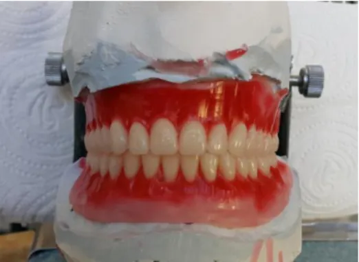
A 65-year-old man was referred after his maxillectomy surgeon to the Department of Prosthodontics at the Selcuk University, Faculty of Dentistry in Konya, Turkey for an evaluation for prosthetic treatment. His medical history revealed that the patient had been operated for ‘‘Squamous cell carcinoma’’ of maxilla one year ago and rehabilitated with radiotherapy. After 6 months the patient revisited the dental center seeking definitive rehabilitation. Intra-oral examination revealed an existing surgical defect with adequate healing and there were no natural teeth remaining in the mandibular and maxillar arch (Figure 1). Treatment plan was formulated with obturator prosthesis for the maxillar arch and mandibular complete denture. The patient’s major complaints were lack of retention, instability of the prosthesis, impaired speech, mastication and liquid leakage into the oral cavity. Extra-oral examination revealed like edentulous patients. Intraoral exami- nation showed resectioning of the hard palate, al- veolar bone, teeth and soft tissue in posterior maxilla until soft palate. The patient did not accept implant surgery that’s why we preferred the conventional obturator prosthesis retained by the defect area. A primary impression was made using irreversible impression material (Soft Hydrogum, Zhermack, Rovigo, Italy) and poured in Type IV dental stone (Dentstone; Pankaj Industries, Mumbai, India). The diagnostic cast was surveyed for fabricating autopoly- merizing acrylic resin special tray. The final impression was made by zinc oxide eugenol (SS White Group, England) for mandibular arch and irreversible impress- ion material (Soft Hydrogum, Zhermack, Rovigo, Italy) for maxillary arch with special tray. Occlusal relationship were secured and mounted in a semi-adjustable articulator. Teeth position and occlusion were checked, and the necessary corrections were made before processing the dentures (Figure 2). A hollow-bulb obturator prosthesis was processed from heat-polymerizing acrylic resin using conventional laboratory procedures (Lucitone 199; Dentsply, Austenal, New York) (Figure 3) and delivered to the patient (Figure 4). Treatment was completed to the aesthetic and functional satisfaction of the patient, who has been using his prosthesis for 3 months with no complaints. The patient was provided with oral
hygiene instruction, and called for follow-up evaluations after 6 months.
Figure 1. Intraoral view of patient.
Figure 2. Teeth position and occlusion.
Atatürk Üniv. Diş Hek. Fak. Derg. KARCI, DEMİR
J Dent Fac Atatürk Uni
Cilt:28, Sayı:4, Yıl: 2018, Sayfa, 570-573
572 Figure 4. Final appearance.
DISCUSSION
The obturator prostheses were generally used in rehabilitating maxillary defect. A well-designed obturator prostheses for maxillary defects were not only to maintain durable and good retention, stability and support, but also to relieve pain, esthetic and psychological problems and result in ease of use. One of the most crucial parts for application of obturator prosthesis is the retention of prosthesis.7 Based on
location and size of defect, health conditions of remaining teeth and bones, available soft tissue undercuts and muscular control, various obturator prostheses with different retentive designs were used to improve oral functions: Conventional designed, attachment enhanced or magnet enhanced.8
Conventional retained obturators, retention can be provided by hollow bulb or buccal extension.9 The
bulb, or part of prosthesis extending into the defect, is generally hollow to reduce the weight of the obturator. Different techniques have been described to fabricate hollow bulb obturators but the simplest method is to grind out the interior of the obturator bulb after acrylisation.10 Bulbs are divided into three
groups according to materials they are made: hard, soft, hard and soft.11 Stud attachments were
economical, easy-replaced, and one of the most important benefit was reduction of the unbalanced stress on the abutment teeth. But the tooth adjacent to the defect suffered many overburden outside forces, resulting in rapid periodontal damages of the tooth. Prosthesis retained by magnetic attachment was widely used in strengthening retention.9 With the
development of research and improved techniques, there were various strategies, designs and materials to achieve enhanced retention, such as perfect
attachment supported by implant retentive obturator prosthesis.12 Osseointegrated implants have been
successful in providing retention, stability, and support of dental and craniofacial prostheses.
Unfortunately, in various situations, the loss of adequate osseous structures and economical and healthy problems makes implant placement difficult and less predictable.13 In these situations, conven-
tional designed obturator prosthesis can compensate patient’s problems.
CONCLUSION
Obturator prostheses mainly depend on out- comes of maxillofacial reconstruction and rehabili- tation involving functions, esthetics, phonetic, psycho- logy acceptance and resocialization. It can be provided by conventional retained obturator prosthesis.
Muhammet KARCI: ORCID ID: 0000-0002-7101-822X Necla DEMİR: ORCID ID: 0000-0003-0927-6962
REFERENCES
1. Taylor TD. Clinical maxillofacial prosthetics. Quintessence Publishing Co: 2000. p. 103–20. 2. Roumanas ED, Nishimura RD, Davis BK. Clinical
evaluation of implants retaining edentulous maxillary obturator prostheses. J Prosthet Dent 1997;77:184-90.
3. Dingman C, Hegedus PD, Likes C, McDowell P, McCarthy E, Zwilling C. A coordinated, multidisciplinary approach to caring for the patient with head and neck cancer. J Support Oncol 2008;6:125-31.
4. Jacob FJ. Clinical management of the edentulous maxillectomy patient. Quintessence Publishing Co: 2000. p. 85-7.
5. Minsley GE, Warren DW, Hinton V. Physiologic responses to maxillary resection and subsequent obturation. J Prosthet Dent 1987;57:338-44. 6. Okay DJ, Genden E, Buchbinder D, Urken M.
Prosthodontic guidelines for surgical reconstruction of the maxilla: A classification system of defects. J Prosthet Dent 2001;86:352-63.
7. Aramany MA. Basic principles of obturator design for partially edentulous patients. Part II: Design principles. J Prosthet Dent 2001;86:562.
Atatürk Üniv. Diş Hek. Fak. Derg. KARCI, DEMİR
J Dent Fac Atatürk Uni
Cilt:28, Sayı:4, Yıl: 2018, Sayfa, 570-573
573 8. Grossmann Y, Madjar D. Resin bonded attachments
for maxillary obturator retention: A clinical report. J Prosthet Dent 2004;92:229-32.
9. Cheng C, Wenhao R, Ling G, Zheng C, Linmei Z, Shaoming L, Pro Ke-qian Z. Function of obturator prosthesis after maxillectomy and prosthetic obturator rehabilitation. Braz J Otorhinolaryngol 2016;82:177-83.
10. Didier M, Laccoureye O, Brasnu D, Vignon M. New surgical obturator prosthesis for hemimaxillectomy patients. J Prosthet Dent 1993;69:520–3.
11. Yazıcıoğlu H, Yaluğ S, Akalın MR. İki farklı obturatör yapım yöntemine göre dişler arasındaki boyutsal farklılığın değerlendirilmesi. Atatürk Üni Diş Hek Fak Derg 2001;11:28-32.
12. Candel-Marti E, Carrillo-Garcia C, Penarrocha-Oltra D, Penarrocha-Diago M. Rehabilitation of atrophic posteriormaxilla with zygomatic implants: review. J Oral Implantol 2012;38:653-7.
13. Roumanas ED, Nishimura RD, Davis BK, Beumer J 3rd. Clinical evaluation of implants retaining edentulous maxillary obturator prosthesis. J Prosthet Dent 1997;77:184-9.
Yazışma Adresi
Muhammet Karcı
Selcuk University, Faculty of Dentistry, Department of Prosthodontics
Campus / KONYA
Phone: +90-332-2410041/1186 Fax: +90-332-2410062