A novel mutation in two cousins with guanidinoacetate
methyltransferase (GAMT) deficiency presented with autism
Halil İbrahim Aydın1, Fatma Müjgan Sönmez2
1Department of Pediatrics, Section of Inborn Errors of Metabolism, Başkent University Faculty of Medicine; 2Developmental Child Neurology Association, Ankara. E-mail: [email protected]
Received: 4th May 2017, 19th September 2017, Accepted: 17th January 2018
SUMMARY: Aydın Hİ, Sönmez FM. A novel mutation in two cousins with guanidinoacetate methyltransferase (GAMT) deficiency presented with autism. Turk J Pediatr 2019; 61: 92-96.
Guanidinoacetate methyltransferase (GAMT) deficiency is a rare autosomal recessive disorder of creatine biosynthesis. Here, we report 9 and 10-year-old cousins with GAMT deficiency caused by a novel mutation who both exhibited neurodevelopmental retardation, seizures, behavioral problems, and autism that began during early infancy. The patients were diagnosed as having only autism and followed for years without a specific diagnosis although they had very low levels of serum creatinine for several times. A novel nonsense mutation in the GAMT gene that caused cessation of synthesis of the protein encoded by this gene was identified in these patients. GAMT deficiency is a treatable inborn error of metabolism and should be considered for all patients with hypotonia, developmental delay, seizures and autism, particularly if low serum creatinine levels are observed.
Key words: creatine deficiency syndromes, guanidinoacetate methyltransferase, autism, epilepsy.
Endogenously produced creatine is synthesized from arginine, glycine, and methionine by arginine: glycine aminotransferase (AGAT) and guanidinoacetate methyltransferase (GAMT) in the renal cortex, pancreas, and liver. Endogenous and dietary creatine are then transported via the bloodstream to muscle and the brain by creatine transporter 1 (CRTR or the SLC6A8 protein) for utilization.1 Creatine acts as an intracellular high-energy phosphate shuttle and participates in energy storage, mainly in muscle and brain. Creatine also appears to play roles in cytoprotection, osmoregulation and neurotransmission. Creatine is excreted via non-enzymatic conversion to creatinine, and creatinine excretion is directly proportional to total body creatine. Inborn errors of creatine synthesis and transport are referred to as creatine deficiency syndromes (CDS) and commonly manifest as intellectual disability and seizures.2 The most common form of this disorder is GAMT deficiency, an autosomal recessively inherited neurometabolic disorder
characterized with developmental delay, epilepsy, movement disorder and behavioral symptoms such as autistic behaviors and self-mutilation. Its carrier frequency was found to be 0.4 % (1:250), and its incidence was estimated to be 1:250,000 in a mixed ethnic group of newborns by direct sequencing of the
GAMT gene.3 Symptoms of GAMT deficiency appear between three months and three years of age.4 Here, we report two cousins whose symptoms began during early infancy although they were diagnosed with GAMT deficiency at 9 and 10 years of age.
Case Reports Case 1
A 10-year-old boy born to a first-degree cousin marriage was admitted to our department with epilepsy and behavioral problems. The patient had a healthy 2-year-old sister, and 9-year-old female cousin with developmental delay, autism and epilepsy (Case 2). Delayed developmental milestones were first noticed by his parents at This study was presented at the 14th National Metabolic Diseases and Nutrition Congress, 26-30 April 2017, Bodrum, Turkey.
the age of nine months. His developmental age was recorded as 9 months when he was first evaluated at another health care facility at the age of 18 months. The patient had generalized seizures at the age of two years and used a lot of different antiepileptic drugs. He had behavioral problems in school and was offered risperidone therapy by a psychiatrist. He also had missed developmental milestones such as spelling and eye contact during childhood. Low serum creatinine levels ranging from 0.01 mg/dl to 0.20 mg/dl were detected at different hospitals. His physical examination was normal and he had no dysmorphic features. Neurological evaluation revealed developmental delay and agitation. Fragile X syndrome was excluded and karyotype was normal. Metabolic tests including plasma and urine amino acids, urine organic acids, and blood carnitine and acylcarnitine were normal. His electroencephalogram showed rare periods of generalized suppression. Brain proton magnetic resonance spectroscopy showed sharply reduced creatine peak in the white matter and basal ganglia, indicating a cerebral creatine deficiency (Fig.1). An analysis of urinary creatine metabolites revealed increased guanidinoacetic acid (GAA) (766.0 mmol/ mol creatinine; normal: 28-180 mmol/mol creatine) and a normal creatine/creatinine ratio (0.071; reference range: 0.01-0.96). These findings suggested GAMT deficiency. A novel homozygous mutation in the GAMT gene (1. exon p.Cys16*; c.C48A) was detected in the patient. Creatine monohydrate therapy 400 mg/ kg/day in addition to a low-protein diet and L-ornithine supplementation (400 mg/kg/day) were initiated. Informed consent was obtained from the parents for mutation analysis and publication.
Case 2
This patient was a 9-year-old female and the first-degree cousin of the index patient. She exhibited the similar clinical signs and a low serum creatinine level (0.01 mg/dl). She had been followed with the diagnosis of developmental delay, autism and epilepsy at another center. Her prior medical records showed that she had been able to walk alone at the age of two and half years and spoke a few meaningful words at five years of age. She had been receiving phenobarbital for her atonic seizures. On admission, she could speak only
a few single words and had a little interest in communication. The rest of the physical examination was completely normal and she had no dysmorphic features. The brain MRI indicated toxicometabolic changes in the globus pallidi, pons, central tegmental tract and reticular formation. All of the metabolic tests were within normal limits although she had a very low serum creatinine level (0.01 mg/dl). Brain MR spectroscopy could not be performed. Clinical findings and low creatinine levels of the patient together with the history of a cousin with the same clinical and biochemical findings led us to the diagnosis of GAMT deficiency. Mutation analysis of this patient showed the same result with the index case. Informed consent was obtained from the parents for mutation analysis and publication. Discussion
GAMT deficiency, which is a rare recessive metabolic disorder in the pathway of creatine synthesis, results in a lack of creatine in both brain and muscle and an accumulation of GAA, an intermediary metabolite in creatine synthesis, in the brain and body fluids5. In all forms of the CDS, creatinine level is low in all body fluids. The accumulation of GAA is characteristic of GAMT deficiency, whereas abnormally low GAA levels are typical of AGAT deficiency, and GAA levels are not altered in CRTR deficiency.2 Common clinical presentations of CDS, such as mental delay and disturbance of expressive and cognitive speech, are believed to be attributable to creatine deficiency in the brain. However, relative to other forms of CDS, GAMT deficiency exhibits a more severe clinical phenotype, including intractable epilepsy, extrapyramidal movement syndromes and abnormalities in the basal ganglia. These additional neurological presentations in GAMT deficiency are believed to arise from the toxic accumulation of GAA in the brain.2
The clinical presentation of GAMT deficiency can vary with respect to age of onset and symptom severity, although this deficiency typically induces developmental delay, epilepsy, and autistic spectrum disorder in infancy or early childhood.2-4 Mildly affected patients with only developmental delay and seizures have been reported.4,6 Both of our patients were first
evaluated for developmental delay at the age of 18 months and two years, respectively. Mental and motor delay are generally the first and most common symptoms of GAMT deficiency, but these symptoms are extremely nonspecific and do not easily led the physician to suspect creatine synthesis defects. Our patients’ serum creatinine levels were found to be extremely low in numerous laboratory tests, however these findings were not recognized as a clue for a creatine synthesis defect. When an inherited metabolic disease is suspected in the diagnosis of a neurological disorder, routine biochemical tests should be performed primarily. Low serum/plasma creatinine levels may suggest a neurometabolic disorder such as GAMT deficiency, although the serum creatinine level
is influenced by other factors like nutritional status. So low-normal creatinine levels do not exclude the presence of CDS.7 In addition to developmental delay, our patients had epilepsy and autistic features, a phenotype similar to previously described phenotypes of GAMT deficiency. In patients with GAMT deficiency, head nodding, drop attacks, and several types of seizures, including myoclonic, generalized tonic-clonic, and sporadic partial complex seizures, typically starts between the ages of 10 months and 3 years and are generally resistant to anticonvulsant therapy.8 Our index case had generalized tonic-clonic seizures resistant to antiepileptic drug therapy. Case 2 experienced many intractable drop attacks. Autism is a common and a major neurological sign of all types of CDS.9 Both of our patients had been diagnosed and followed for years with a diagnosis of autism spectrum disorder with speech delay, lack of eye contact, difficulties with social and communication skills, and aggressive behavior patterns. Progressive extrapyramidal symptoms, especially dyskinetic and dystonic movements, which occur in one-third of patients with GAMT deficiency, were not seen in our patients. It has been suggested that these extrapyramidal symptoms arise from basal ganglia involvement induced by neurotoxic effects of GAA.2
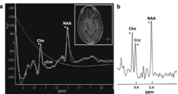
In all CDS, creatine levels detected using in vivo proton MR spectroscopy are low or absent in different regions of brain tissue, whereas Fig. 1. Brain proton magnetic resonance spectroscopic
imaging of the male patient shows severely reduced creatine peak at 3.02 ppm, normal choline peak at 3.2 ppm, and normal N-acethylaspartate peat at 2.0 ppm (a), normal choline, creatine and N-acethylasparte peaks seen in healthy individulas at brain MR spectroscopy (b). Cho: choline; Cre: creatine; NAA: N-acetylaspartate.
Fig. 2. Schematic presentation of GAMT mutation in the patients. c.C48A mutation results in the occurrence of a premature stop codon (TGA) leading to cessation of synthesis of the protein encoded by the GAMT gene.
normal spectral patterns are observed for other metabolites.2 Brain proton MR spectroscopy in our index patient indicated the absence of a creatine peak in the white matter and basal ganglia (Fig. 1). Low creatine levels in brain as detected by MRS may be used in vivo diagnosis of these conditions, but it cannot differentiate the types of CDS. The diagnosis of CDS can be based on measurements of creatine, guanidinoacetate, and creatinine using LC-MS/MS (liquid chromatography–tandem mass spectrometry) or GC-MS (gas chromatography– mass spectrometry) in plasma, urine, or dried blood spots, and DNA sequence analysis of the three genes involved, GAMT, GATM, and SLC6A8. High GAA levels in urine and plasma are characteristic of GAMT deficiency, whereas abnormally low GAA levels in urine and plasma are typical of AGAT deficiency, Normal GAA levels in urine and plasma and increased urine creatine/creatinine ratio is consistent with CRTR deficiency.10 The high GAA levels in urine and normal urine creatine/ creatinine ratio detected in our index patient were characteristic for GAMT deficiency. GAMT deficiency is caused by mutations in the GAMT gene, which is located at 19p13.3; more than 50 types of mutations in this gene have been reported.11 We identified a novel homozygous mutation, 1. exon p.Cys16* (c.C48A), in both patients (Fig. 2). This mutation was a nonsense mutation that caused the cessation of synthesis of the protein encoded by the GAMT gene. No associations between mutations and patterns of GAA accumulation density, such as phenotypic features, have been identified.4 Our patients had a severe phenotype with severe mental delay, intractable seizures, lack of speech, and autistic features.
Late diagnosis and treatment of GAMT deficiency results in severe physical and cognitive disabilities, although certain levels of improvement can be achieved. Excellent outcomes have been achieved for several patients with GAMT deficiency who were diagnosed and treated from birth.4 Research has indicated that oral supplementation with creatine monohydrate could restore cerebral creatine levels, as detected using MR spectroscopy, and lead to improvements in epilepsy and movement disorders. Low arginine intake with
ornithine supplementation is thought to lower neurotoxic GAA concentrations and to favorably affect basal ganglia involvement and movement disorders.2,4 Both of our patients became seizure-free for 18 months after receiving creatine monohydrate therapy (400 mg/kg per day), a low-protein diet and L-ornithine supplementation (400 mg/kg per day), and the index patient’s parents reported improvement in attention and anxiety but not in speech disturbance.
CDS are underdiagnosed because of limited awareness of these treatable conditions. These disorders have nonspecific and variable clinical presentations, and most of the physicians believe that sophisticated tests are required to reach a presumptive diagnosis. For GAMT deficiency, early diagnosis and appropriate therapy are essential to minimize learning disabilities.4,11 To diagnose creatine synthesis defects, serum creatinine levels should be checked carefully in every patient with neurodevelopmental delay, especially if this delay is accompanied by hypotonia, seizures, extrapyramidal symptoms, behavioral impairment, and speech delay. The subsequent diagnostic step should be measurement of urine creatine, creatinine and guanidinoacetate or evaluation of the creatine peak in brain via MR spectroscopy.
REFERENCES
1. Béard E, Braissant O. Synthesis and transport of creatine in the CNS: importance for cerebral functions. J Neurochem 2010; 115: 297-313.
2. Rackayova V, Cudalbu C, Pouwels PJ, Braissant O. Creatine in the central nervous system: From magnetic resonance spectroscopy to creatine deficiencies. Anal Biochem 2017; 15: 144-157.
3. Mercimek-Mahmutoglu S, Pop A, Kanhai W, et al. A pilot study to estimate incidence of guanidinoacetate methyltransferase deficiency in newborns by direct sequencing of the GAMT gene. Gene 2016; 575:127-131. 4. Stockler-Ipsiroglu S, van Karnebeek C, Longo N, et al.
Guanidinoacetate methyltransferase (GAMT) deficiency: outcomes in 48 individuals and recommendations for diagnosis, treatment and monitoring. Mol Genet Metab 2014; 111:16-25.
5. Gordon N. Guanidinoacetate methyltransferase deficiency (GAMT). Brain Dev 2010; 32: 79-81. 6. Pacheva I, Ivanov I, Penkov M, Kancheva D, Jordanova
A, Ivanova M. Creatine Deficiency Syndrome could be Missed Easily: A Case Report of Guanidinoacetate Methyltransferase Deficiency Presented with Neurodevelopmental Delay, Seizures, and Behavioral Changes, but Normal Structural MRI. Ann Clin Lab Sci 2016; 46: 557-561.
7. Stockler-Ipsiroglu S, van Karnebeek CD. Cerebral creatine deficiencies: a group of treatable intellectual developmental disorders. Semin Neurol 2014; 34: 350-356.
8. Schutz PW, Stockler S. Cerebral creatine deficiency disorders. In: Johnston MV, Adams HP. Fatemi A (eds). Neurobiology of Disease (2nd ed). New York: Oxford University Press, 2016: 451-459.
9. Nasrallah F, Feki M, Kaabachi N. Creatine and creatine deficiency syndromes: biochemical and clinical aspects. Pediatr Neurol 2010; 42: 163-171.
10. Sharer JD, Bodamer O, Longo N, Tortorelli S, Wamelink MM, Young S. Laboratory diagnosis of creatine deficiency syndromes: a technical standard and guideline of the American College of Medical Genetics and Genomics. Genet Med 2017; 19: 256-263.
11. Mercimek-Mahmutoglu S, Ndika J, Kanhai W, et al. Thirteen new patients with guanidinoacetate methyltransferase deficiency and functional characterization of nineteen novel missense variants in the GAMT gene. Hum Mutat 2014; 35: 462-469.