Related Surgery Original Article / Özgün Makale doi: 10.5606/ehc.2014.16
An investigation of consistency between posterior condylar axis
+3 degree external rotation line and clinical transepicondylar axis line
techniques in primary total knee arthroplasty
Primer total diz artroplastide arka kondiler aks +3 derece dış rotasyon çizgisi ve
klinik transepikondiler aks çizgisi teknikleri arasındaki tutarlılığın araştırılması
Hakan Boya, M.D.,1 Özal Özcan, M.D.,2 Gökhan Maralcan, M.D.2
1Department of Orthopedics and Traumatology, Başkent University Zübeyde Hanım Practice and Research Center, İzmir, Turkey 2Department of Orthopedics and Traumatology, Medical Faculty of Afyon Kocatepe University, Afyonkarahisar, Turkey
• Received: July 05, 2013 Accepted: March 13, 2013
• Correspondence: Hakan Boya, M.D. Çatalkaya Mah., Günaydın Sok., Seymen Apt., No: 3/11, 35320 Narlıdere, İzmir, Turkey, Tel: +90 506 - 343 36 68 Fax: +90 232 - 336 94 21 e-mail: [email protected]
Amaç: Bu çalışmada primer total diz protezi uygulamala-rında posteriyor kondiler aks (PKA)+3 derece dış rotasyon (DR) çizgisi ve klinik transepikondiler aks (kTEA) çizgisi arasındaki farklılık ve her iki teknik arasındaki tutarlılık araştırıldı.
Hastalar ve yöntemler: Yirmi dört hastanın [3 erkek (%12.5), 21 kadın (%87.5); ortalama yaş 67 (59-80 yaş)] 36 dizi [12 hastada her iki diz aynı seansta eşzamanlı olarak ameliyat edildi (%50)] çalışmaya alındı. Cerrahi sırasında distal femur kesisini takiben kesi yüzeyine koter ucu ile PKA+3° DR çizgisi ve kTEA çizgisi çizil-di. Distal femur üzerinde bulunan her iki çizgi dijital kamera ile kaydedildi ve PKA+3° DR çizgisi referans alınarak çizgiler arasındaki ilişki araştırıldı [paralel, iç rotasyon (İR), DR]. İstatistiksel analiz McNamara ki kare testi ve Kappa (k) değeri ile yapıldı.
Bulgular: Görüntüler değerlendirildiğinde, kTEA çizgisi-nin PKA+3° DR çizgisine kıyasla 22 dizde (%61.2) paralel olduğu, 14 dizde ise paralel olmadığı (%38.8) [10 dizde İR (%71.5), 4 dizde DR (%28.5)] tespit edildi. Çizgiler arasında belirgin bir farklılık (McNamara ki kare=12.7±1; p<0.001) ve teknikler arasında düşük oranda tutarlılık (k=0.00055) vardı.
Sonuç: Cerrahi sırasında femoral komponent rotasyonunun tespitinde kullanılan PKA+3° DR çizgisi ve kTEA çizgisi arasındaki farklı sonuçlar, olasılıkla tekniklerin uygulama dezavantajlarına ve distal femurun anatomik farklılıklarına bağlıdır. Bu nedenle, iki teknikten birinin diğerinin sonucunu kontrol etmek için kullanılması güvenli değildir.
Anahtar sözcükler: Femoral komponent; femoral transepikondiler
aks; arka kondiler aks; rotasyonel hizalanma; total diz protezi.
Objectives: This study aims to investigate discrepancy between posterior condylar axis (PCA)+3 degree external rotation (ER) line and clinical transepicondylar axis (cTEA) line and consistency between the both techniques in primary total knee arthroplasty.
Patients and methods: Thirty-six knees [Bilateral knees were operated simultaneously in 12 patients (50%)] in 24 patients [3 men (12.5%), 21 women (87.5%); average age 67 (59-80 age)] were included in the study. During surgery, PCA+3° ER line and cTEA line were drawn on the distal femoral cutting surface by electrocautery pencil following distal femoral cut. The both lines on distal femur were recorded by digital camera and relationship between lines was ascertained in reference to PCA+3° ER line [parallel, internal rotation (IR), ER]. Statistical analysis was performed by the McNamara chi square test and Kappa (k) value.
Results: Assessment of the images revealed that cTEA line in comparison to PCA+3° ER line was parallel in 22 knees (61.2%), but not parallel in 14 knees (38.8%) [IR in 10 knees (71.5%), ER in 4 knees (28.5%)]. There was a significant difference (McNamara chi square=12.7±1; p<0.001) and poor consistency (k=0.00055) between both lines and techniques, respectively.
Conclusion: For determination of femoral component rotation in surgery setting, different results between cTEA and PCA+3° ER techniques possibly may due to disadvantages of techniques and anatomic variation of distal femur. Thus, using both techniques to check each other’s results seems unsafe.
Key words: Femoral component; femoral transepicondylar axis;
Rotational alignment of femoral component (FC) is crucial for long-term survival of total knee arthroplasty (TKA).[1,2] Proper placement of the FC reduces
abnormal patella femoral tracking and tibia femoral articulation.[3] There are several different methods/
references described for identification of rotational alignment of FC in primary TKA; Whiteside's line,[4]
transepicondylar axis (TEA),[3,5] posterior condylar
axis (PCA)+3° external rotation (ER),[6] ligament
balancing,[5,7] and tibial mechanic axis.[8]
Flexion-extension movements of the knee result in an around fixed axis which lies between the origins of collateral ligaments.[9-11] Transepicondylar
axis and flexion-extension axis are not completely interchangeable.[12] As both of them are rather nearby,
TEA can be used instead of flexion-extension axis of the knee.[10,11,13] Therefore, placement of FC in
axial plane should be in parallel with TEA.[10,11,13]
In addition, PCA lies in 3° internal rotation (IR) compare to TEA.[14]
Today, modern instrumentation guides which use PCA (+3° ER) are popular for determination of FC rotation in surgery setting.[15] In our clinical practice,
we use these instruments routinely in primary TKA except valgus knees.
This study aims to investigate discrepancy between posterior condylar axis (PCA)+3 degree ER line and clinical TEA (cTEA) line and consistency between the both techniques in primary total knee arthroplasty.
PATIENTS AND METHODS
This prospective study was approved by Afyon Kocatepe University, Medical Faculty Ethics Committee. Written informed consents were obtained from all participants before surgery. Thirty-six knees in 24 patients (3 males, 21 females; mean age 67 years; range 59 to 80 years) which
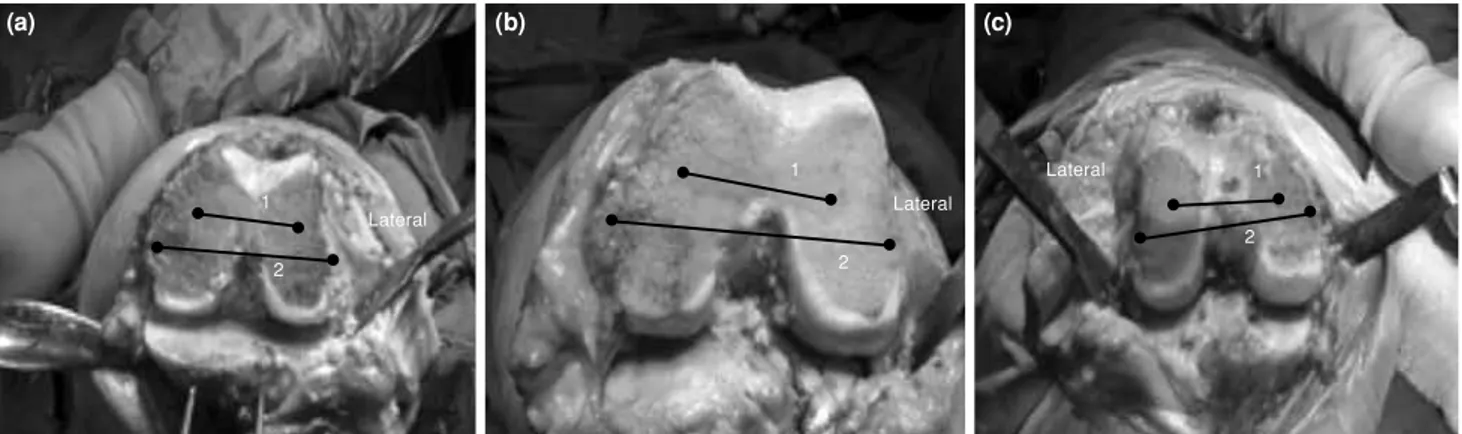
were subsequently operated for severe varus gonarthrosis were included. Both knees were operated simultaneously in 50% of the patients. During surgery, following distal femoral bone cut, PCA+3° ER line and cTEA line were drawn on the distal femoral cutting surface by electrocautery pencil. The both lines on distal femur were recorded by digital camera and relationship between lines was ascertained in reference to PCA+3° ER line [parallel, IR and ER] (Figure 1). Statistical analysis was performed using the McNamara chi square test for discrepancy between both lines and Kappa (k) value for consistency between both techniques.
Femoral components were placed in certain rotational alignment in the axial plane determined with guide instrument using PCA for all cases.
RESULTS
Assessment of the images revealed that cTEA line in comparison to PCA+3° ER line was parallel in 22 knees (61.2%), but not parallel in 14 knees (38.8%). Of the non-parallel group, there were 10 knees (71.5%) with IR and four knees (28.5%) with ER, respectively. There was a significant difference (McNamara chi square=12.7±1; p<0.001) and poor consistency (k=0.00055) between both lines and techniques, respectively.
DISCUSSION
Total knee arthroplasty is a standard treatment for various disabling disorders of the knee and has proven long-term success.[16] However, it is likely
to encounter various complications related with TKA.[17,18] The vast majority of the complications are
related to the patellar-femoral joint.[2] Although the
implant design is considered effective, the primary reason for failure is improper rotation of FC in the absence of frontal plane misalignment.[16,19,20] In TKA,
Figure 1. Position of clinical transepicondylar axis line in reference to posterior condylar axis +3 degree external rotation line. (a) Parallel, (b) Internal rotation, (c) External rotation. 1: PCA +3 degree external rotation line; 2: cTEA line.
(a) (b) (c) Lateral 1 1 1 2 2 2 Lateral Lateral
FC rotation should be proper for long-term function and survival.[1,2]
Transepicondylar axis is a valid reference for determination of the rotation of FC.[3,11,20] Surgical (the
most prominent point of lateral epicondyle-medial femoral sulcus) and clinical (the most prominent point of lateral epicondyle - the most prominent point of medial epicondyle) TEAs are distinct concepts (Figure 2).[14] Surgical TEA (sTEA) is accepted as
a more accurate reference to determine neutral rotation of FC.[3,11] However, determination of sTEA is
challenging.[13,20-23] As the epicondylar peaks obscured
by soft tissues, it is difficult to identify precisely in the surgical setting.[9,21,24] In addition, medial sulcus
becomes obscure as a result of disease progression in osteoarthritis; it can be detected in only 20% severely damaged knees.[25] However, most prominent
point of the medial epicondyle is detectable in all knees independent of the severity of osteoarthritis.[25]
Thus, we used cTEA in this study. Furthermore, the variability between surgeons is high in determination of TEA and establishing femoral rotational alignment is influenced by an individual surgeon’s skill.[22]
Moreover, PCA lies in 3° IR compared to sTEA.[14] Thus, PCA+3° ER line in the surgical setting
is determined using guide instruments. It is accepted that the line is in parallel with sTEA.[14,15] Improper
placement of the guide instrument, anatomic variability of distal femoral condyle, bone and cartilage wearing of posterior femoral condyle due to severe osteoarthritis and valgus knee are common problems related with this method.[2,4,26] Also, there are studies
that suggested and refused any relationship between sex and the anatomic angle in PCA and TEA.[3,14]
Surgical TEA is more consistent than PCA+3° ER line to produce balanced flexion gap.[7,27] Preoperative
alignment of the knee directly influences the accuracy of these techniques.[24,27] In addition, TEA is the most
viable reference used for this purpose; however,
it is unlikely to produce flexion gap stability in neutral/varus knees and valgus knees in 10% and 14%, respectively.[27] Any single method used to
determine rotation carries at least a 10% change of creating flexion gap asymmetry, therefore, it is better to use more than one method of femoral rotation alignment.[7,27] There were also reports indicating a
wide variety between PCA and cTEA/sTEA.[3,14,21,24]
This variety is possibly due to the disadvantages of each technique including anatomic variation of the distal femur primarily.[12] Although knowing details
of more than one technique for determination of FC rotation has been suggested, almost all techniques have a wide error interval.[28] Therefore, it still remains
unknown how much variability in the axial femoral alignment can be tolerated.[29] In surgery setting,
reproducing of FC absolute rotation should be the main purpose.
In our study, a statistically significant discrepancy was determined between cTEA and PCA+3° ER lines (p<0.001), and consistency between these techniques was extremely poor (k=0.00055). At first, it is believed that this was caused by the disadvantages of the techniques, however, possibly anatomic variation of distal femur was the main cause. Another cause of differences between techniques may be an external rotation position of cTEA in respect to sTEA.
On the other hand, limitations of our study include small sample size and lack of quantitative assessment of the relationship between cTEA and PCA+3° ER lines. Prospective studies in a larger patient population with severe gonarthrosis will provide valuable clinical information regarding determination of femoral component rotation in surgery setting. We determined a relationship between the lines with qualitative assessment, therefore, it is impossible to know the certain degrees of incoordinate.
There is at least 10% change of creating improper femoral component rotation if one of the methods developed to determine the rotation use alone, therefore, it is better to use more than one method simultaneously intraoperatively.[7,27] In addition, TEA
and PCA+3° ER techniques are frequently used to determine the rotation in surgery setting.[3,5,6]
In conclusion, we focused on the both methods in our study. Previous studies evaluated angular relationship between s/cTEA and PCA with only mean values and standard deviation or compared different axial references to PCA statistically, however, our study statistically ascertained consistency and discrepancy between the both (cTEA/PCA+3° ER) techniques and lines, respectively (Table I). For determination Figure 2. Surgical transepicondylar axis (sTEA) and clinical
transepicondylar axis (cTEA).
The most prominent point of lateral epicondyle
The most prominent point of medial epicondyle
Medial epicondylar sulcus Lateral epicondyle
Medial epicondyle cTEA sTEA
of FC rotation in surgery setting, different results between cTEA and PCA+3° ER techniques possibly may be due to disadvantages of techniques and anatomic variation of distal femur. Thus, using both techniques for check each other’s results seems unsafe. Undoubtedly, it is possible to determine the angular relationship between PCA and surgical/clinical TEA radiologically (computed tomography) preoperatively. Carrying this knowledge to intraoperative setting by using adjustable instrumentation guide rather than standard instrumentation guide which has fixed +3° ER may be an ideal method of choice.
Declaration of conflicting interests
The authors declared no conflicts of interest with respect to the authorship and/or publication of this article.
Funding
The authors received no financial support for the research and/or authorship of this article.
REFERENCES
1. Akagi M, Matsusue Y, Mata T, Asada Y, Horiguchi M, Iida H, Nakamura T. Effect of rotational alignment on patellar tracking in total knee arthroplasty. Clin Orthop Relat Res 1999;336:155-63.
2. Anouchi YS, Whiteside LA, Kaiser AD, Milliano MT. The effects of axial rotational alignment of the femoral component on knee stability and patellar tracking in total knee arthroplasty demonstrated on autopsy specimens. Clin Orthop Relat Res 1993;287:170-7.
3. Berger RA, Rubash HE, Seel MJ, Thompson WH, Crossett LS. Determining the rotational alignment of the femoral component in total knee arthroplasty using the epicondylar axis. Clin Orthop Relat Res 1993;286:40-7.
4. Whiteside LA, Arima J. The anteroposterior axis for femoral rotational alignment in valgus total knee arthroplasty. Clin Orthop Relat Res 1995;321:168-72.
5. Insall JN, Binazzi R, Soudry M, Mestriner LA. Total knee arthroplasty. Clin Orthop Relat Res 1985;192:13-22.
6. Laskin RS. Flexion space configuration in total knee arthroplasty. J Arthroplasty 1995;10:657-60.
7. Olcott CW, Scott RD. Determining proper femoral component rotational alignment during total knee arthroplasty. Am J Knee Surg 2000;13:166-8.
8. Stiehl JB, Abbott BD. Femoral component rotational
TABLE I
Review of study characteristics from published articles
References Berger et al.[3]
Arima et al.[21]
Poilvache et al.[24]
Olcott and Scott[27]
Griffin et al.[14] Katz et al.[12]
The study
Study type Anatomic cadaver study
Anatomic cadaver study
Clinical
(Intraoperative measurement) Clinical
(Intraoperative measurement)
Radiological (MRI) Anatomic cadaver study
Clinical
(Intraoperative measurement)
Statistics Mean values with standard
deviation
Mean values with standard deviation Student t-test Mean values with standard
deviation Mean values with standard
deviation Paired t-test
Mean values with standard deviation Mean values with standard
deviation ANOVA, Student’s t-test
McNamara Chi square test Kappa (k) value
Outcome sTEA method is useful to
orient FC
AP axis method appears to be more reliable and is certainly easier to use than
TEA method Angle between cTEA and PCA is important to orient
FC
Do not use only one method for rotational alignment of
FC
PCA+3° ER method is unsafe cTEA provide more external rotation in reference
to PCA+3° ER for FC than other methods Using the both techniques
for check each other’s results seems unsafe Evaluation
Angle between cTEA and PCA 4.7°±3.5°
5.2°±4.1°
Angle between sTEA and PCA 3.5°±1.2°
0.3°±1.2° Angle between cTEA and PCA
4.4°±2.9°
Discrepancy between cTEA and Whiteside line reference to PCA
Angle between cTEA and PCA 3.51°±2.03° Varus-neutral knees Consistency of PCA+3° ER, Whiteside line, cTEA methods reference to tibial mechanic axis
Varus-neutral knees cTEA> Whiteside line> PCA+3° ER
Angle between sTEA and PCA 3.11°±1.7°
Angle between cTEA and PCA 6.1°±3.3°
cTEA, Whiteside line, LB methods results discrepancy reference to
PCA
Discrepancy between cTEA and PCA+3° ER lines Consistency between the both
technique
cTEA: Clinical transepicondylar axis; PCA: Posterior condylar axis; sTEA: Surgical transepicondylar axis; FC: Femoral component; TEA: Transepicondylar axis; ER: External rotation; MRI: Magnetic resonance imaging; LB: Ligament balancing.
alignment using the extramedullary tibial shaft axis: A technical note. J Orthop Rheumatol 1995;8:93-6.
9. Churchill DL, Incavo SJ, Johnson CC, Beynnon BD. The transepicondylar axis approximates the optimal flexion axis of the knee. Clin Orthop Relat Res 1998;356:111-8. 10. Hollister AM, Jatana S, Singh AK, Sullivan WW, Lupichuk
AG. The axes of rotation of the knee. Clin Orthop Relat Res 1993;290:259-68.
11. Yoshioka Y, Siu D, Cooke TD. The anatomy and functional axes of the femur. J Bone Joint Surg [Am] 1987;69:873-80. 12. Katz MA, Beck TD, Silber JS, Seldes RM, Lotke PA.
Determining femoral rotational alignment in total knee arthroplasty: reliability of techniques. J Arthroplasty 2001;16:301-5.
13. Akagi M, Yamashita E, Nakagawa T, Asano T, Nakamura T. Relationship between frontal knee alignment and reference axes in the distal femur. Clin Orthop Relat Res 2001;388:147-56.
14. Griffin FM, Math K, Scuderi GR, Insall JN, Poilvache PL. Anatomy of the epicondyles of the distal femur: MRI analysis of normal knees. J Arthroplasty 2000;15:354-9. 15. Berger RA, Rubash HE. Rotational instability and
malrotation after total knee arthroplasty. Orthop Clin North Am 2001;32:639-47.
16. Insall JN, Kelly M. The total condylar prosthesis. Clin Orthop Relat Res 1986;205:43-8.
17. Ozcan O, Boya H, Ateş A, Doğruöz F. Bilateral periprosthetic distal femoral fractures following total knee arthroplasty. Eklem Hastalik Cerrahisi 2013;24:178-81.
18. Erkan S, Yercan HS, Okcu G, Ozalp RT. Factors causing stiff knee after total knee arthroplasty. [Article in Turkish] Eklem Hastalik Cerrahisi 2011;22:16-21.
19. Dennis DA. Patellofemoral complications in TKA: A literature review. Am J Knee Surg 1992;5:156-66.
20. Siston RA, Patel JJ, Goodman SB, Delp SL, Giori NJ. The variability of femoral rotational alignment in total knee arthroplasty. J Bone Joint Surg [Am] 2005;87:2276-80. 21. Arima J, Whiteside LA, McCarthy DS, White SE. Femoral
rotational alignment, based on the anteroposterior axis, in total knee arthroplasty in a valgus knee. A technical note. J Bone Joint Surg [Am] 1995;77:1331-4.
22. Jenny JY, Boeri C. Low reproducibility of the intra-operative measurement of the transepicondylar axis during total knee replacement. Acta Orthop Scand 2004;75:74-7.
23. Jerosch J, Peuker E, Philipps B, Filler T. Interindividual reproducibility in perioperative rotational alignment of femoral components in knee prosthetic surgery using the transepicondylar axis. Knee Surg Sports Traumatol Arthrosc 2002;10:194-7.
24. Poilvache PL, Insall JN, Scuderi GR, Font-Rodriguez DE. Rotational landmarks and sizing of the distal femur in total knee arthroplasty. Clin Orthop Relat Res 1996;331:35-46.
25. Yoshino N, Takai S, Ohtsuki Y, Hirasawa Y. Computed tomography measurement of the surgical and clinical transepicondylar axis of the distal femur in osteoarthritic knees. J Arthroplasty 2001;16:493-7.
26. Griffin FM, Insall JN, Scuderi GR. The posterior condylar angle in osteoarthritic knees. J Arthroplasty 1998;13:812-5. 27. Olcott CW, Scott RD. A comparison of 4 intraoperative
methods to determine femoral component rotation during total knee arthroplasty. J Arthroplasty 2000;15:22-6.
28. Yau WP, Chiu KY, Tang WM. How precise is the determination of rotational alignment of the femoral prosthesis in total knee arthroplasty: an in vivo study. J Arthroplasty 2007;22:1042-8.
29. Stöckl B, Nogler M, Rosiek R, Fischer M, Krismer M, Kessler O. Navigation improves accuracy of rotational alignment in total knee arthroplasty. Clin Orthop Relat Res 2004;426:180-6.