In neonatal medicine, it is important to assess tissue oxygen-ation due to hypoxia, as hyperoxia can have adverse effects. Usually, measurements of arterial oxygen tension (PaO2) and
arterial oxygen saturation (SaO2) are used for clinical
deci- sions regarding oxygen therapy in neonates. However, this ap-proach cannot explain the complete physiological economy of oxygen (1-3). Some recommend measuring venous oxygen to evaluate tissue perfusion and response to therapy (1, 3-8). Umbilical venous catheters are widely used in neonatal in- tensive care units for medication, fluid and nutrition adminis- tration. Mixed venous oxygenation can be measured by insert-ing an umbilical venous catheter into the inferior vena cava (IVC) (3). The advantages of measuring IVC oxygenation are not affected by atrial right-left shunting (3, 4).
Monitoring of mixed venous oxygen saturation (Svo2) shows the residual oxygen after tissue oxygen extraction and also represents the combined sufficiency of arterial oxygen content, cardiac output and tissue oxygen consumption (2, 9, 10). Right atrial SvO2 was used for monitoring, in contrast to us-ing SaO2 alone in an animal model (7). True mixed venous blood is derived from a pool of blood entering the pulmonary artery via the great veins in the chest. It contains blood that has passed through all systemic capillary beds capable of ex-tracting oxygen, and is thoroughly mixed in the right ventricle (11). However, catheterization of the pulmonary artery or right atrium is hazardous for neonates and is not routinely ap-plied (3, 12). Umbilical venous catheters are frequently used in neonatal intensive care units due to their lower complica- Background: Inferior vena cava (IVC) oxygen saturation as an indi- cator of mixed venous oxygenation may be valuable for understand-ing postnatal adaptations in newborn infants. It is unknown how this parameter progresses in critically ill premature infants. Aims: To investigate IVC oxygen saturation during the first three days of life in preterm infants with and without patent ductus arte-riosus (PDA). Study Design: Case-control study. Methods: Twenty-seven preterm infants were admitted to the Neo- natal Intensive Care. Preterm infants with umbilical venous catheter-ization were included in the study. Six umbilical venous blood gas values were obtained from each infant during the first 72 hours of life. Preterm infants in the study were divided into two groups. Hae-modynamically significant PDA was diagnosed by echocardiography in 11 (41%) infants before the 72nd hour of life in the study group and ibuprofen treatment was started, whereas 16 (59%) infants who didn’t have haemodynamically significant PDA were included in the control group. Results: In the entire group, the highest value of mean IVC oxygen saturation was 79.9% at the first measurement and the lowest was 64.8% at the 72nd hour. Inferior vena cava oxygen saturations were significantly different between the study and control groups. Post-hoc analysis revealed that the first and 36th hour measurements made the difference (p=0.01). Conclusion: Inferior vena cava oxygen saturation was found to be significantly different between preterm infants with and without PDA. Further studies are needed to understand the effect of foetal shunts on venous oxygenation during postnatal adaptation in new-born infants.
(Balkan Med J 2014;31:230-4).
Key Words: Inferior vena cava oxygen saturation, mixed venous oxygen saturation, patent ductus arteriosus, prematurity
Inferior Vena Cava Oxygen Saturation during the First Three
Postnatal Days in Preterm Newborns with and without Patent Ductus
Arteriosus
1Division of Neonatology, Department of Pediatrics, Başkent University Faculty of Medicine, Ankara, Turkey 2Division of Cardiology, Department of Pediatrics, Başkent University Faculty of Medicine, Ankara, Turkey 3Department of Biostatistics, Başkent University Faculty of Medicine, Ankara, TurkeyEce Yapakçı
1, Ayşe Ecevit
1, Deniz Anuk İnce
1, Mahmut Gökdemir
2, M. Agah Tekindal
3, Hande Gülcan
1,
Aylin Tarcan
1Address for Correspondence: Dr. Deniz Anuk İnce, Division of Neonatology, Department of Pediatrics, Başkent University Faculty of Medicine, Ankara, Turkey Phone: +90 312 215 75 97 e-mail: [email protected]
tion rates. Umbilical venous catheterization is not affected by intracardiac shunting that leads to mixing of systemic and pul-monary venous blood (3, 4, 13).
The measurement of venous oxygen saturation has been used in several studies of sepsis and septic shock, and dur-ing the perioperative period of major surgery because it shows major derangements in oxygen balance (13-16). Umbilical venous oxygen saturation pointed a great similar-ity to arterial oxygen saturation in some premature infants in our clinical practice that it was hypothesized as a sign of se-vere ductal shunt predicting patent ductus arteriosus (PDA). In this study, on the basis of this clinical observation, the course of IVC oxygen saturation was measured in preterm infants as an indicator of mixed venous oxygenation in the first three days of life; and differences between infants with and without PDA were investigated.
MATERIAL AND METHODS
Preterm infants who were admitted to the Neonatal Inten-sive Care Unit within 12 postnatal hours between June 2007 and December 2008, were included in the study. Umbilical venous catheters were inserted in all of them for medical reasons. The study was approved by the institutional review board (IRB) and ethics committee. Written informed consent was obtained from the parents of all study infants. The placement of each umbilical venous catheter and the catheters’ position were confirmed with a chest radiography. If the venous catheter tip was not at the right place it was re-positioned. The exclusion criteria were infants who had in-correct catheter position and infants whose catheters did not allow them to receive blood during the 72 hours.
An umbilical venous blood gas analysis was obtained after each infant stabilized and then other blood gas analyses were obtained at postnatal 12th, 24th, 36th, 48th and 72nd hours. In total,
six umbilical venous blood gas samples were obtained from the neonates. A physical examination and arterial tension measure-ment were performed before each assessment and urine output was noted. Complete blood counts were studied at the 24th, 48th
and 72nd hours. A blood gas analyser (Gem Premier 3000 Blood
Gas/Electrolyte Analyzer Model 5700; Bedford, Massachu-setts, United States of America) was used for the gas analyses. During the process of obtaining blood gases, we prioritized that the baby was quiet and the oxygen saturation was above 85%. The blood specimens from all preterm infants who par-ticipated in this study were obtained in a standardized manner. The gestational age, birth weight, gender, antenatal steroid usage and surfactant dose of each infant were noted. In the physical examination general condition, evaluation of periph-eral circulation, presence of a murmur, wide pulse pressure (>30 mmHg) or hyperactive precordial pulsation,
hypoten-sion, need for an increase in PIP of more than 2 cm H2O or
FiO2 more than 0.2, presence of pulmonary congestion or
cardiomegaly (cardiothoracic index >%60) on teleradiogra-phy, heart rate and blood pressure of each patient were noted. If PDA was confirmed by echocardiography before the 72nd hour in patients who had haemodynamic symptoms, ibuprofen treatment was started. Oral ibuprofen was given at an initial dose of 10 mg/kg followed by 5 mg/kg at 24 and 48 hours. Echocardiography was performed after the postnatal 72nd hour in infants with no clinical suspicion of PDA. Left atrial to aor- tic root ratio (LA: AO) and ductus diameter in mm per birth-weight of the baby in kg were recorded. Infants who had haemodynamically significant PDA before the 72nd
hour of life were included in the study group and in-fants who did not have haemodynamically significant PDA were included in the control group.
For statistical evaluation, SPSS for Windows SPSS soft-ware (Statistical Package for Social Sciences, version 15.0, SPSS Inc., Chicago, IL, USA) was used. IVC venous oxygen saturation levels in the study and control groups of patients were compared bymixed designed ANOVA [analysis of two-level mixed (one factor repeated and one factor nonrepeated) variance analysis] in the first 3 days. A Mouchly sphericity test was done, sphericity (p>0.05) was provided. Inferior vena cava oxygen saturation levels were found to be significantly different in the two groups (p<0.01). Multiple comparisons with Bonferroni multiple comparison test were done in order to understand which level was different from the others.
RESULTS
We enrolled 27 babies in the study. The demographic char-acteristics of the patients are shown in Table 1.
Haemodynamic symptoms were observed during the first 72 hours in the study group - hypotension was noted in 3 of 11, peripheral hypoperfusion in 2 of 11, and pulmonary conges-tion in 2 of 11 babies.
Echocardiography was performed on day-of-life one for three (27%), on day-of-life two for three (27%), and on day-of-life three for five (46%) of the 11 babies in the study group; on day-of-life one for one (6%), on day-of-life two for one (6%), and day-of-life three for 14 (88%) of the 16 babies in the control group.
Treatment was started within 12 to 24 hours in 3 (27%), within 25 to 36 hours in 1 (9%), within 37 to 48 hours in 2 (18%) and within 49 to 72 hours in 5 (46%) of 11 babies in the study group. Ten (91%) of the 11 infants in the study group had at least one dose of surfactant, while one infant (9%) did not receive surfactant treatment. Eleven (69%) of 16 infants in the con-trol group had at least one dose of surfactant, while 5 infants (31%) did not receive surfactant treatment.
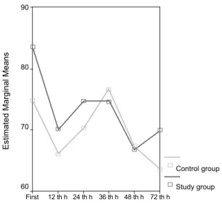
The mean ductal diameter was reported as 2.1±0.8 (1.2-3.7) mm/kg in the study group and as 0.7±0.9 (0-2.9) mm/kg in the control group. The difference was significant (p=0.001). The mean LA:AO was noted as 1.41±0.33 (1.07-1.90) in the study group and as 1.11±0.19 (0.86-1.51) in the control group. The difference was significant (p=0.02). The means and standard deviations of IVC oxygen satura- tion in the infants (study and control group) and in all the in-fants are given in Table 2. The changes in mean blood pressure, urine output and hae-moglobin concentration in whole infants during the first 72 hours are shown in Figure 1. Inferior vena cava venous oxygen saturation was found to differ significantly between the two groups. Post-hoc analysis revealed that the first and 36th hour measurements made the difference (p=0.01) (Figure 2). DISCUSSION In this study, the course of IVC oxygen saturation was mea- sured in preterm infants as an indicator of mixed venous oxy-genation in the first three days of life and the effect of PDA on venous oxygenation was investigated. Unlike previous stud-ies, this study investigated intermittent IVC oxygen saturation in the first 72 hours of life. The inferior vena cava oxygen saturation of the whole group decreased over time. The highest value of mean IVC oxygen saturation was 79.9% at the first measurement and the lowest was 64.8% at the measurement done at the 72nd hour. O’Connor et al. (5) found that the mean mixed venous oxy-gen saturation was 83.3% in 1 to 3 day old newborn infants that required mechanical ventilation for respiratory diseases, but who were haemodynamically stable and in normal acid-base balance. A prospective clinical observational study was made with 10 preterm infants breathing room air after the acute phase of respiratory distress syndrome. A fiberoptic catheter was placed via the umbilical vein and remained in the right atrium
All infants Control group Study group Number of patients [n (%)] 27 (100) 16 (59) 11 (41) Gestational age, mean±SD 28.15±2.78 28.94±2.43 27.00±2.97 (min-max; median) (24-34; 28) (24-34; 28.5) (24-32; 26) Birthweight (g), mean±SD 1160±336 1239±261 1046±409 (min-max; median) (540-1860; (770-1860; (540-1730; 1160) 1240) 950) Gender Female [n (%)] 13 (48.1) 7 (44) 6 (55) Male [n (%)] 14 (51.9) 9 (56) 5 (45) TABLE 1. Demographic parameters of infants
All the infants Control group Study group mean±SD mean±SD mean±SD (min-max; (min-max; (min-max; median) median) median) First analysis 79.9±13.4 77.1±14.9 83.7±10.7 (53-99; 83) (53-99; 77) (62-97; 86) 12th hour 67.3±19.1 65.2±18.2 70.5±20.9 (32-97; 72) (32-87; 72) (33-97; 72) 24th hour 70.6±18.6 70.4±16.8 70.8±21.9 (31-99; 72) (35-92; 73) (31-99; 72) 36th hour 75.4±16.5 77.6±13.3 72.4±20.6 (26-97; 72) (49-96; 75.5) (26-97; 68) 48th hour 67.6±16.8 69.4±16.5 64.9±17.8 (39-98; 66) (40-98; 68.5) (39-95; 61) 72nd hour 64.8±17.2 63.2±17.4 66.7±17.6 (36-96; 60) (36-92; 60.5) (43-96; 60) TABLE 2. The IVC oxygen saturation levels according to postnatal time FIG. 1. a-c. Changes in mean blood pressure (a), urine output (c) and haemoglobin concentration (c) in all the infants during the first 72 hours
a b c
12th h 24th h 36th h 48th h 72nd h 12th h 24th h 36th h 48th h 72nd h 24th h 48th h 72nd h
First
Mean blood pressure (mm/Hg) (95% Cl)
Urine output (mL/kg/h) (95% Cl) Hemoglobin (g/dL) mean (95% Cl) 60 50 40 30 20 15.5 15.0 14.5 14.0 13.5 13.0 12.5 12.0 7 6 5 4 3 2
for continuous measurement of oxygen saturation. The mean age of the infants during the monitoring of central venous oxygen saturation was 52 hours (range 18-168). In this study stable preterm infants had a central venous oxygen saturation ranging from 65% to 82% (5th and 95th
percentile), which cor-responded to arterial oxygen saturation > than or = to 86% (2). In the study of O’Connor et al. (5), newborn infants par-ticipated in the study, while preterm infants breathing room air were included in the study of van der Hoeven MA et al. (2). In both studies the measurements of oxygen saturation were performed from the right atrium. Although the results are close to those in the mentioned studies, in our study the infants were of similar age and birth weight and were followed during their first 72 hours. Furthermore, in our study the intermittent measurements of oxygen saturation were done from the IVC, unlike the other studies where measurements of oxygen satu-ration were done from the right atrium. In our study, the mean of the first measurements of IVC oxy-gen saturations in all the infants were higher than those from consecutive measurements. Some factors like foetal shunts, car- diopulmonary, haemodynamic, vascular and inflammatory re-sponses may influence venous oxygenation during the postnatal adaptation period (5, 6). All of these factors may be the reason for the progressive decrease in venous oxygen saturation. The average rise in blood pressure and the fall in haemoglo-bin levels within hours, and also the increase in urine output at the 48th hour, are expected results during the postnatal adapta-tion of premature babies. It is clear that iatrogenic blood loss also contributed to the fall in haemoglobin levels. In our study IVC oxygen saturation was found to be sig-nificantly different between the study and control groups, and the first and 36th hour measurements made the difference. The higher venous oxygen levels in the first sample from the study group might be due to the shunt between the arterial and ve-nous systems at the level of the ductus arteriosus due to PDA. This may be a result of the wide ductus diameter or severe pressure gradient between the two systems. To confirm this, the shunt index was calculated using SaO2 and Svo2. A portion of this study was also about a shunt index in the early hours of postnatal life, which we have shown predicts haemodynami-cally significant PDA (17). The converging levels of venous oxygen in the two groups in the 36th hour samples might be
due to ductal closure, as a response to treatment started as soon as diagnosis of PDA was established. Our study is the first in which the course of inferior vena cava oxygen saturation, as an indicator of mixed venous oxy-genation, in preterm newborns with and without PDA during the first three postnatal days was shown. Inferior vena cava oxygen saturation, as a parameter that can be easily measured, may be valuable in understanding postnatal adaptation period problems in newborn infants. It is unknown how this parameter progresses in critically ill premature infants. Inferior vena cava oxygen saturation as an indicator of mixed venous oxygen-ation, used together with arterial oxygenation measurement, may give more accurate results toward understanding the early postnatal adaptation process. Inferior vena cava oxygen satu-ration should also be tested in larger patient populations in order to understand more accurately the effect of PDA on venous oxygenation and its use in diagnosis and decision of treatment.
Ethics Committee Approval: Ethics committee approval was received for
this study from the institutional review board.
Informed Consent: Written informed consent was obtained from patients’
parents who participated in this study.
Author contributions: Concept - A.T.; Design - E.Y.; Supervision - A.T.,
H.G.; Materials - A.T., H.G.; Data collection&/or Processing - E.Y., A.E., M.G.; Analysis&/or Interpretation - E.Y., A.T., M.A.T.; Literature search - E.Y., D.A.İ.; Writing - E.Y.; Critical Reviews - D.A.İ, A.T. Conflict of interest: No conflict of interest was declared by the authors. Financial Disclosure: No conflict of interest was declared by the authors. REFERENCES 1. Whyte RK. Mixed venous oxygen saturation in the newborn. Can we and should we measure it? Scand J Clin Lab Invest Suppl 1990;203:203-11. [CrossRef]
2. Van der Hoeven MA, Maertzdorf WJ, Blanco CE. Continuous cen-tral venous oxygen saturation (ScvO2) measurement using a fiber
optic catheter in newborn infants. Arch Dis Child Fetal Neonatal Ed 1996;74:177-81. [CrossRef]
FIG. 2. Estimated marginal means of IVC oxygen saturation according
to group
IVC oxygen saturation
Control group 90 80 70 60 Study group First 12 th h 24 th h 36 th h 48 th h 72 th h
3. Plotz FB, van Lingen RA, Bos AP. Venous oxygen measurements in the inferior vena cava in neonates with respiratory failure. Crit Care 1998;2:57-60. [CrossRef]
4. Hart J, Vemgal P, Cocks-Drew S, Harrison C, Andersen C. The relation between inferior vena cava oxygen saturation, superior vena cava flow, fractional oxygen extraction and haemoglobin affinity in sick newborns: A pilot study. Acta Paediatr 2006;95:50-5. [CrossRef]
5. O’Connor TA, Hall RT. Mixed venous oxygenation in critically ill neo-nates. Crit Care Med 1994;22:343-6. [CrossRef]
6. Dudell G, Cornish JD, Bartlett RH. What constitutes adequate oxygen-ation? Pediatrics 1990;85:39-41.
7. Hirschl RB, Palmer P, Heiss KF, Hultquist K, Fazzalari F, Bartlett RH. Evaluation of the right atrial venous oxygen saturation as a physiologic monitor in a neonatal model. J Pediatr Surg 1993;28:901-5. [CrossRef]
8. van der Hoeven MA, Maertzdorf WJ, Blanco CE. Feasibility and accu-racy of fiberobtic catheter for measurement of venous oxygen saturation in newborn infants. Acta Paediatr 1995;84:122-7. [CrossRef]
9. van der Hoeven MA, Maertzdorf WJ, Blanco CE. Mixed venous oxygen saturation and biochemical parameters of hypoxia during progressive hypoxemia in 10- to 14-day-old piglets. Pediatr Res 1997;42:878-84.
[CrossRef]
10. van der Hoeven MA, Maertzdorf WJ, Blanco CE. Relationship between mixed venous oxygen saturation and markers of tissue oxygenation in
progressive hypoxic hypoxia and in isovolemic anemic hypoxia in 8- to 12-day-old piglets. Crit Care Med 1999;27:1885-92. [CrossRef]
11. Edwards JD. Oxygen transport in cardiogenic and septic shock. Crit
Care Med 1991;19:658-63. [CrossRef]
12. MacDonald MG, Chou MM. Preventing complications from lines and tubes. Semin Perinatol 1986;10:224-33.
13. Martin J, Shekerdemian LS. The monitoring of venous saturations of oxygen in children with congenitally malformed hearts. Cardiol Young 2009;19:34-9. [CrossRef]
14. Goodrich C. Continuous central venous oximetry monitoring. Crit Care
Nurs Clin North Am 2006;18:203-9. [CrossRef]
15. Liakopoulos OJ, Ho JK, Yezbick A, Sanchez E, Naddell C, Buckberg GD, et al. An experimental and clinical evaluation of a novel central ve-nous catheter with integrated oximetry for pediatric patients undergoing cardiac surgery. Anesth Analg 2007;105:1598-604. [CrossRef]
16. Kissoon N, Spenceley N, Krahn G, Milner R. Continuous central venous oxygen saturation monitoring under varying physiological conditions in an animal model. Anaesth Intensive Care 2010;38:883-9.
17. Yapakçı E, Ecevit A, Törer B, Ince DA, Gökdemir M, Gülcan H, et al. “Shunt index” can be used to predict clinically significant patent ductus arteriosus in premature neonates in early post-natal life. Cardiol Young 2013;2:1-5.