A . Ü. Veteriner Fakültesi, II.
Ş
irurji Kürsüsü Direktörü
Prof Dr. B. S
ı
tk
ı
Artun
B
İ
R DANADA SUPRAKOND
İ
LER FEMUR
KIRIGININ TEDAVISI
Küçüker, N.*
Özet: Femur'un suprakondiler kırıkları, danalarda çok seyrek olarak görülmekte-dir. Küçük evcillerde bu tür kırıkların tedavisi için çeşitli yöntemler bulunmasına karşılık, literatürde danalardan söz edilmemektedir. Bu yazıda, köpeklerde uygulanan bir yöntemle, bir danadaki suprakondiller femur kırığının fiksasyonu anlatılmaktadır.
The menagement of the Supracondylary Femoral Fracture in a calf
Summary: in this report, the menagement of the supracondylary femoral fracture in a calf has been represented.
Introduct
ı
on
The incidence of the supracondylary femoral fracture in calves is very few. Trauma is common responsible or contributory in some kind of mineral substances.
Numerous technics have been described for the correction of the supracondylary femoral fractures in small animals but not in calves (1,2, 3 , 4 , 5) .
Material And Method
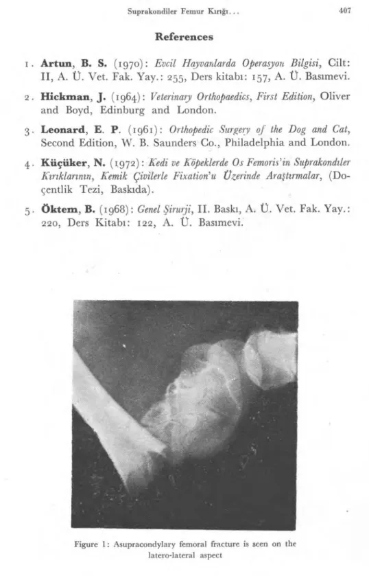
A three months old male calf was presented shortly after being struck. And a supracondylary fracture diagnosis was based on the left femur bone by physical and radiological examinations. (Figure: I).
Modified PETTİT-WHEAT method by using bone pin was considered for the treatment of this case. The patient was brought to a surgical plane of anesthesia by ROMPUN** and placed in lateral recumbency with the affected leg uppermost.
* A. Ü. Veteriner Fakültesi, II. Şirurji Kürsüsü Doçenti. ** A product of Bayer Farmacetical Company.
406 Küçüker, N.
Surgical Procedure :
A skin incision approximately ı o centimeters long is started lateral to the tendon of quadriceps and extended across the lateral aspect of the stifle to a point opposite the tibial crest. After incising the subcutaneous tissue, a stab incision was made into the the stifle joint through the lateral part of the joint capsule just beside the patella. Enough capsule and overlying fascia was left attached to the recto-patellar ligament to simplfy suturing it to the free portion.The capsular incision was extended with scissor both proximally and distally from the stabbed site, and the patella was reflected medially therefore the fracture site was exposed using aseptic pro-cedure. Obstructive blood clots and tissue debris were removed only as necessary to clean the fracture site.
A hole following a channel have been opened by using a hand chuck just over the İntercondylar fossa through the proximal frag-ment of the fracture obliquely.
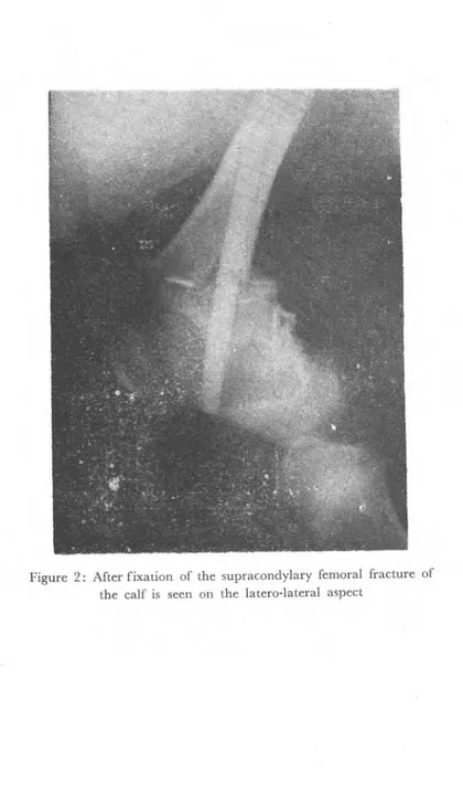
Priorly produced bone pin was inserted into the channel. After the fragments were brought into the close apposition, the pin was nailed by using metalic hammer. (Figure: 2).
First row suture was placed on the joint capsule and its over-lying tissue with the absorbable material. In order to close subcu-taneous tissue, second row was performed with the absorbable materi-al materi-also and the skin was clossed verticmateri-al mattress suture used nonab-sorbable material.
Conclusion
A method of fixation supracondylary femoral fracture of the calves have not been described in the literature, but many technics already been documented for small animals.
The method of PETTİT-WHEAT which is performed by using bone pine has been considered tha is the convenient method for the fixation of the supracondylary femoral fracture of the calves. The re-duction was not clearly perfect in this case but the fixation was firm and the calf has began bearing weight on the 15 postoperative day. Further follow-up on this case was not possible.
Suprakondiler Femur Kırığı... 407
References
. Artun, B. S.
(197o):
Evcil Hayvanlarda Operasyon Bilgisi,
Cilt:
II, A. Ü. Vet. Fak. Yay.: 255, Ders kitab
ı
: 157, A. Ü. Bas
ı
mevi.
2. Hickman, J.
(1964):
Veterinary Orthopaedics, First Edition,
Oliver
and Boyd, Edinburg and London.
3. Leonard, E. P.
(1961):
Orthopedic Surgery of the Dog and Cat,
Second Edition, W. B. Saunders Co., Philadelphia and London.
4. Küçüker, N. (1972):
Kedi ve Köpeklerde Os Femoris'in Suprakond
ı
ler
K
ı
r
ı
klar
ı
n
ı
n, Kemik Çivilerle Fixation'u Czerinde Ara
ş
t
ı
rmalar,
(Do-çentlik Tezi, Bask
ı
da).
5. Öktem, B.
(1968):
Genel
Ş
irurji, II. Bask
ı
, A. Ü. Vet. Fak. Yay.:
220,
Ders Kitab
ı
:
122,A. Ü. Bas
ı
mevi.
Figure 1: Asupracondylary femoral fracture is seen on the latero-lateral aspect
408 Küçüker, N.
Figure 2: After fixation of the supracondylary femoral fracture of the calf is seen on the latero-lateral aspect