Research Article
Oncol Res Treat 2018;41:444–448 DOI: 10.1159/000488082Evaluation of Factors Affecting Lymph Node Metastasis
in Clinical Stage I–II Epithelial Ovarian Cancer
Baki Erdem
aIlkbal T. Yüksel
aNuri Peker
bSuat C. Ulukent
cOsman A ˛sıcıo ˘glu
aIpek Y. Özaydin
dVolkan Ülker
aOzgur Akbayir
aa Department of Gynecologic Oncology, Kanuni Sultan Süleyman Training and Research Hospital, Istanbul, Turkey; b Department of Obstetrics and Gynecology, Istinye University Bahçe ¸sehir Liv Hospital, Istanbul, Turkey;
c Department of General Surgery, Kanuni Sultan Süleyman Training and Research Hospital, Istanbul, Turkey; d Department of Pathology, Kanuni Sultan Süleyman Training and Research Hospital, Istanbul, Turkey
patients with grade 3 disease (p = 0.016). Conclusion: Comprehensive lymphadenectomy increases the detec-tion rate for metastatic lymph nodes in patients with clinically early-stage EOC. The rate of lymph node in-volvement is significantly higher in grade 3 tumors, se-rous cytology, bilateral adnexal involvement, and posi-tive intraabdominal fluid cytology.
© 2018 S. Karger GmbH, Freiburg
Introduction
Ovarian cancer is the second most common gynecologic cancer worldwide, and it is the leading cause of death associated with gy-necologic malignancies in women. The life-long risk of developing epithelial ovarian cancer (EOC) is 1.3%, and about 70% of patients are already at an advanced disease stage at the time of initial diag-nosis; only 30% of patients are diagnosed at an early stage (stages I, II) [1].
EOC spreads primarily through exfoliation of the tumor cells into the peritoneal cavity, but lymphatic and hematogenous spread can also occur [2]. According to the International Federation of Gynecology and Obstetrics (FIGO), the presence of lymph node involvement in the early stages of ovarian cancer raises the disease stage to 3A1 [3]. Systematic lymphadenectomy is useful for accu-rate stage assessment of early-stage ovarian cancer and has a proven prognostic value. Accurate staging in very early-stage dis-ease may prevent unnecessary postoperative chemotherapy. How-ever, there is still a debate on the therapeutic role of lymphadenec-tomy [4]. Lymphadeneclymphadenec-tomy is the major surgical procedure
asso-Keywords
Lymph node dissection · Lymph node metastases · Ovarian cancer
Summary
Background: Systematic lymphadenectomy is useful for
accurate staging of early-stage ovarian cancer and has obvious prognostic value. Accurate staging may prevent unnecessary postoperative chemotherapy. The aim of this study was to evaluate the rate of lymph node in-volvement and factors affecting it in clinically early-stage epithelial ovarian cancer (EOC; stages I, II). Patients and
Methods: The study included 163 patients who
under-went surgery at our hospital between January 2004 and April 2017 and who were diagnosed with early-stage EOC based on preoperative and intraoperative examina-tion. Patient data were retrospectively analyzed. The rate of lymph node involvement and factors affecting it were analyzed. Results: Of 163 patients, 21 (12.9%) had lymph node metastasis, whereas 16 (16.3%) of 98 patients who underwent comprehensive lymphadenectomy had lymph node metastasis. According to the univariate re-sults for patients undergoing any type of lymphadenec-tomy, the rate of positive lymph nodes was significantly higher (37.1%) in those with bilateral ovarian involve-ment (p < 0.001). The rate was significantly higher in pa-tients with positive intraabdominal fluid cytology (25.9%; p < 0.001), serous histology (20.5%; p = 0.02), and grade 3 disease (33.3%; p < 0.001). In multivariate logistic re-gression analysis, the rate was significantly higher in pa-tients with bilateral adnexal involvement (p = 0.012). The risk of positive lymph nodes was significantly higher in
Received: August 21, 2017 Accepted: March 02, 2018 Published online: July 6, 2018
ciated with other morbidities such as lymphedema, lymphocyst, ileus, blood loss, nerve or vascular injury, blood transfusion, pro-longed length of hospital stay, and increased treatment costs [5–7].
In the present study, we aimed to evaluate the rate of lymph node involvement and factors affecting lymph node involvement in clinically early-stage (stages I, II) EOC.
Patients and Methods
This retrospective study was approved by the Kanuni Sultan Süleyman Training and Research Hospital Ethics Committee and was conducted in ac-cordance with the principles of the Declaration of Helsinki. Written informed consent was obtained from each participant.
The study included a total of 163 patients who underwent surgery in the Gynecologic Oncology Clinic of the Kanuni Sultan Süleyman Training and Re-search Hospital between January 2004 and April 2017 and were diagnosed with early-stage EOC based on preoperative and intraoperative findings. Patient data in the database of the Gynecologic Oncology Clinic were analyzed retrospec-tively. According to the standard procedures at the time of surgery, the patients underwent intraperitoneal fluid or intraabdominal lavage fluid sampling, total hysterectomy, bilateral salpingo-oophorectomy, omentectomy, appendectomy, careful exploration of the pelvic and upper abdominal cavity, random perito-neal biopsy from suspected or seemingly benign areas, and pelvic paraaortic lymphadenectomy for the purpose of disease staging. Demographic characteris-tics of the patients, CA-125 marker levels, surgical procedure, presence of as-cites, results of cytological examination of the intraabdominal lavage fluid, tumor diameter, laterality, tumor grade, histological type, performance of lym-phadenectomy, number and sites of lymph nodes, involvement of lymph nodes, and final surgical stage of the patients were recorded. Patients receiving neoad-juvant chemotherapy, those with a concomitant primary malignancy, those who had extrapelvic spread at the initial surgical exploration, and those with non-EOC were excluded from the study.
As described by Ayhan et al. [7], systematic bilateral pelvic and paraaortic lymphadenectomy removing 25 or more pelvic lymph nodes and 15 or more paraaortic lymph nodes was considered comprehensive lymphadenectomy. Cases that did not meet these conditions were considered sampling lymphad-enectomies. Systematic lymphadenectomy involved the removal of external iliac, internal iliac, obturator, and presacral lymph nodes, as well as those re-lated to the aorta and vena cava up to the level of the renal vein. The rate of pa-tients with positive lymph nodes was identified, and possible factors affecting lymph node positivity were evaluated.
Statistical Analysis
Statistical analysis was performed using SPSS version 22.0 software (IBM Corp., Armonk, NY, USA). Descriptive data were expressed as mean, standard deviation, median, frequency, and percentage. In the univariate analysis, the Pearson’s chi-square and Fisher’s exact tests were used to evaluate the factors associated with lymph node positivity. Significant risk factors included in the univariate analysis underwent further multivariate analysis using logistic re-gression, and variables were selected with the enter method. The odds ratio was calculated by taking the first categories as reference. A p value of < 0.05 was considered statistically significant with a 95% confidence interval.
Results
The overall characteristics of the 163 patients included in the analysis are shown in table 1. The median age was 49 years (range 19–86 years). Of these patients, 79 (48.5%) were postmenopausal.
The applied surgical procedures are summarized in table 2. Over the period studied, 154 patients underwent hysterectomy and 157 underwent salpingo-oophorectomy; 9 patients had previously un-dergone hysterectomy and 6 had previously unun-dergone salpingo-oophorectomy. Of the study patients, 98 (60.1%) underwent com-prehensive bilateral pelvic and paraaortic lymphadenectomy and 65 (39.9%) underwent sampling lymphadenectomy during the pe-riod studied, while 162 (99%) patients underwent pelvic lymphad-enectomy and 124 (76%) underwent paraaortic lymphadlymphad-enectomy.
Table 1. Patient and tumor characteristics
Median age (range), years 49 (19–86)
Median parity (range), n 2 (0–13)
Menopausal status, n (%)
Postmenopausal 79 (48.5)
Premenopausal 84 (51.5)
CA-125, median (range), U/ml 269 (5–5,500)
Tumor diameter, median (range), cm 14 (4–45)
Ascites present, n (%) 32 (19.6)
Final assigned stage, n (%)
1B 55 (33.7) 1B 2 (1.2) 1C1 6 (3.7) 1C2 29 (17.8) 1C3 17 (10.4) 2A 8 (4.9) 2B 11 (6.7) 3A1 18 (11) 3A2 17 (10.4) Histology, n (%) Serous 73 (44.8) Mucinous 47 (28.8) Endometrioid 25 (15.3) Clear cell 9 (5.5) Mixed 4 (2.4) Undifferentiated 3 (1.8) Squamous 2 (1.2) Grade, n (%) 1 42 (25.8) 2 67 (41.1) 3 54 (33.1)
Lymph node count, mean ± standard deviation, n
Pelvic 23.7 ± 11.8
Paraaortic 9.7 ± 9.6
Table 2. Surgical procedures
Procedure n (%)
Cytology collected 163 (100)
Omentectomy 163 (100)
Random peritoneal biopsies 154 (94.5)
Apendectomy 70 (42)
Comprehensive bilateral pelvic and paraaortic lymphadenectomy
98 (60.1)
Pelvic lymph node assessment 162 (99)
The mean number of removed pelvic lymph nodes was 23.72 ± 11.85, and the mean number of removed paraaortic lymph nodes was 9.718 ± 9.603. Of the patients, 55 (33.7%) had stage 1A disease, 29 (17.8%) had stage 1C2 disease, and 18 (11%) had stage 3A1 dis-ease. Pathological examination showed that 73 (44.8%) patients had serous, 47 (28.8%) mucinous, 25 (15.3%) endometrioid, and 9 (5.5%) clear cell tumors, while 9 (5.5%) patients had other types of tumors (mixed, undifferentiated, squamous) (table 1).
Of 163 patients, 21 (12.9%) were found to have positive lymph nodes. The rate of pelvic lymph node metastasis was 7.4% (12/162). The rate of paraaortic lymph node metastasis was 9.6% (12/124). 3
patients had both pelvic and paraaortic lymph node metastasis. Of 98 patients who underwent comprehensive lymphadenectomy, 16 (16.3%) were found to have lymph node metastasis. The risk fac-tors for lymph node metastasis were evaluated and the results are shown in tables 3 and 4.
Among the patients who underwent any type of lymphadenec-tomy, the rate of positive lymph nodes was significantly higher in those with bilateral ovarian involvement (37.1%; p < 0.001). The rate of lymph node positivity was significantly higher in the patients with positive intraabdominal fluid cytology (25.9%; p < 0.001). There was no significant difference in lymph node
posi-Total, n (%) LN-negative, n (%) LN-positive, n (%) p value Number of patients 163 142 (87.1) 21 (12.9)
Laterality of adnexal involvement unilateral 128 (78.5) 118 (92.2) 10 (7.8) < 0.001a
bilateral 35 (21.5) 22 (62.9) 13 (37.1) Cytology negative 105 (64.4) 99 (94.3) 6 (5.7) < 0.001b positive 58 (35.6) 43 (74.1) 15 (25.9) Ascites absent 131 (80.4) 114 (87) 17 (13) 0.604a present 32 (19.6) 28 (87.5) 4 (12.5) Histology serous 73 (44.8) 58 (79.5) 15 (20.5) 0.02b mucinous 47 (28.8) 45 (95.7) 2 (4.3) other 43 (26.4) 39 (90.7) 4 (9.3) FIGO grade 1 42 (25.8) 40 (95.2) 2 (4.8) < 0.001b 2 67 (41.1) 66 (98.5) 1 (1.5) 3 54 (33.1) 36 (66.7) 18 (33.3) CA-125 ≤ 35 U/ml 33 (20.2) 31 (93.9) 2 (6.1) 0.153a > 35 U/ml 130 (79.8) 111 (85.4) 19 (14.6)
aFisher’s exact test. bPearson’s chi-squared test.
LN = Lymph node.
Table 3. Risk factors
for lymph node metas-tasis among patients with any lymph node assessment (univariate) Total, n (%) LN-negative, n (%) LN-positive, n (%) p value Number of patients 98 82 (83.7) 16 (16.3)
Laterality of adnexal involvement unilateral 75 (76.5) 69 (92) 6 (8) < 0.001a
bilateral 23 (23.5) 13 (56.5) 10 (43.5) Cytology negative 58 (58.9) 39 (90.7) 4 (9.3) 0.014a positive 40 (41.1) 22 (73.3) 8 (26.7) Ascites absent 75 (76.5) 63 (84) 12 (16) 0.549a present 23 (23.5) 19 (82.6) 4 (17.4) Histology serous 53 (54.1) 40 (75.5) 13 (24.5) 0.05b mucinous 19 (19.4) 18 (94.7) 1 (5.3) other 26 (26.5) 24 (92.3) 2 (7.7) FIGO grade 1 20 (20.4) 18 (90) 2 (10) 0.001b 2 38 (38.8) 37 (97.4) 1 (2.6) 3 40 (40.8) 27 (67.5) 13 (32.5) CA-125 ≤ 35 U/ml 15 (15.3) 14 (93.3) 1 (6.7) 0.247a > 35 U/ml 83 (84.7) 68 (81.9) 15 (18.1)
aFisher’s exact test. bPearson’s chi-squared test.
LN = Lymph node.
Table 4. Risk factors
for lymph node metas-tasis (univariate) in 98 patients with compre-hensive lymph node assessment
tivity between the patients with and without ascites (p = 0.604). The rate of lymph node involvement was significantly higher in patients with serous histology (20.5%; p = 0.02) and in those with grade 3 disease (33.3%; p < 0.001). There was no significant differ-ence in lymph node positivity between patients with CA-125 values ˯ 35 and those with CA-125 values > 35 (p = 0.153).
Among patients who underwent comprehensive lymphad-enectomy, the rate of lymph node positivity was significantly
higher among patients with bilateral ovarian involvement (43.5%; p < 0.001). The rate was significantly higher in patients with posi-tive intraabdominal fluid cytology (26.7%; p = 0.014). There was no significant difference in lymph node positivity between patients with and without ascites (p = 0.549). The rate of lymph node in-volvement was significantly higher in patients with serous histol-ogy (24.5%; p = 0.05) and in those with grade 3 disease (32.5%; p = 0.001). There was no significant difference in lymph node positiv-ity between patients with 125 values ˯ 35 and those with CA-125 values > 35 (p = 0.247).
The variables that showed significant difference in univariate analysis in patients who underwent any type of lymphadenectomy were further evaluated via multivariate analysis (table 5). Accord-ing to multivariate logistic regression analysis, the rate of lymph node positivity was significantly higher in patients with bilateral adnexal involvement (p = 0.012). The effect of positive intraab-dominal fluid cytology on lymph node involvement was not statis-tically significant (p = 0.219). The risk of positive lymph nodes was significantly higher in patients with grade 3 disease (p = 0.016).
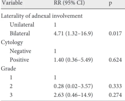
The variables that showed significant difference in univariate analysis in patients who underwent comprehensive lymph node as-sessment were further evaluated via multivariate analysis (table 6). Multivariate logistic regression analysis showed that the rate of lymph node positivity was significantly higher in patients with bi-lateral adnexal involvement (p = 0.017). The effects of grade and positive intraabdominal fluid cytology on lymph node involvement were not statistically significant (p > 0.05).
Discussion
In gynecologic malignancies, cancer spreads mostly through the lymphatic vessels, which has a prognostic value [8]. In EOC, the rate of lymph node metastasis is lower in early-stage tumors [9]. Powless et al. [10] reported that the rate of lymph node metastasis was 13.2% in patients with clinically early-stage EOC who
under-Variable RR (95% CI) p
Laterality of adnexal involvement
Unilateral 1 Bilateral 4.14 (1.37–12.5) 0.012 Cytology Negative 1 Positive 2.07 (0.64–6.66) 0.219 Grade 1 1 2 0.44 (0.03–5.39) 0.527 3 7.02 (1.43–34.3) 0.016
RR = Risk ratio; CI = confidence interval.
Table 5. Risk factors
for lymph node metas-tasis (multivariate)
Variable RR (95% CI) p
Laterality of adnexal involvement
Unilateral 1 Bilateral 4.71 (1.32–16.9) 0.017 Cytology Negative 1 Positive 1.40 (0.36–5.49) 0.624 Grade 1 1 2 0.28 (0.02–3.57) 0.333 3 2.63 (0.46–14.9) 0.274
RR = Risk ratio; CI = confidence interval.
Table 6. Risk factors
for lymph node metas-tasis (multivariate) among those with comprehensive staging
Study [ref.] Serous, % Mucinous, % Endometrioid, % Clear cell, % Other, %
Desteli et al. [9] 14.2 0.0 0.0 0.0 11.1 Harter et al. [16] 15.4 0.0 7.1 0.0 0.0 Morice et al. [6] 30.8 0.0 0.0 – 64.3 Negeishi et al. [11] 14.3 4.1 20.0 17.4 20.0 Nomura et al. [15] 50.0 0.0 7.4 5.6 – Onda et al. [17] 33.3 6.7 0.0 31.3 0.0 Suzuki et al. [18] 30.8 0.0 0.0 11.1 0.0 Tsumura et al. [12] 8.7 3.4 – 19.0 –
Table 7. Incidence of lymph node metastasis
went any type of lymphadenectomy and 16.5% in patients who un-derwent comprehensive lymphadenectomy. Negishi et al. [11] re-ported a rate of 12.7% for lymph node involvement in patients who underwent systematic lymphadenectomy, whereas Tsumura et al. [12] reported a rate of 8.6% for lymph node involvement in 81 pa-tients who underwent systematic lymphadenectomy. In a meta-analysis of 14 studies in the literature, Kleppe et al. [13] reported a rate of 14.2% for lymph node metastasis. The present study reports a rate of 12.9% for lymph node metastasis in patients with clinically early-stage (stages I, II) EOC who underwent any type of lymphad-enectomy and a rate of 16.3% in patients who underwent extended lymphadenectomy.
The present study evaluated via univariate analysis the variables that were shown to affect early-stage EOC; among patients who underwent any type of lymphadenectomy, bilaterality, malignant intraabdominal fluid cytology, serous histological type, and the presence of FIGO grade 3 tumors were found to increase the risk of lymph node metastasis. In multivariate analysis, laterality and tumor grade were found to be independent risk factors for lymph node metastasis.
Among patients who underwent comprehensive lymphadenec-tomy, bilaterality, malignant intraabdominal fluid cytology, serous histological type, and presence of FIGO grade 3 tumors were found to increase the risk of lymph node metastasis. In multivariate anal-ysis, bilateral adnexal involvement was found to be an independent risk factor for lymph node metastasis. The majority of patients who underwent comprehensive lymphadenectomy had grade 3 disease (40.8%); we therefore concluded that the effect of grade on lymph node metastasis did not show statistical significance in the multi-variate analysis.
Powless et al. [10] reported that tumor grade, histological type, laterality of adnexal involvement, and presence of ascites were
in-dependent risk factors. Sakuragi et al. [14] reported an 11.1% lymph node involvement rate in grade 3 tumors, whereas Nomuro et al. [15] reported a rate of 33.3%. In a meta-analysis of 7 studies in the literature, Kleppe et al. [13] reported a rate of 20% for lymph node involvement in grade 3 tumors. The present study found a significantly higher rate of lymph node involvement in grade 3 tu-mors (33.3%). Table 7 shows the studies in the literature that ex-amined lymph node involvement according to histological type. In the present study, the rate of lymph node involvement was higher in serous tumors when compared with mucinous tumors and tu-mors of other histological types.
Our study has some limitations, such as its retrospective design and the small number of affected lymph nodes. Because our clinic is a tertiary referral gynecologic oncology center, we are primarily referred patients with low performance status. In addition to bad performance status, morbid obesity is another major problem. De-pending on these factors, the number of removed pelvic and par-aaortic lymph nodes should be less than the minimum number of lymph nodes described in the literature.
In conclusion, based on our study results, comprehensive lymphadenectomy increases the detection rate of metastatic lymph nodes in patients with clinically early-stage EOC. In addi-tion, the rate of lymph node involvement significantly increases in grade 3 tumors, in tumors with serous histology, in the presence of adnexal involvement, and in cases with positive intraabdominal fluid cytology.
Disclosure Statement There is no disclosure of funding.
References
1 Kuznia AL, Roett MA: Genital cancers in women:
ovarian cancer. FP Essent 2015;438:24–30.
2 Tan DS, Agarwal R, Kayse SB: Mechanisms of transcoelomic metastasis in ovarian cancer. Lancet
Oncol 2006;7925–7934.
3 Prat J; FIGO Committee on Gynecologic Oncology: FIGO’s staging classification for cancer of the ovary, fallopian tube, and peritoneum: abridged
republica-tion. J Gynecol Oncol 2015;26:87–89.
4 Maggioni A, Benedetti Panici P, Dell’Anna T, et al.: Randomised study of systematic lymphadenectomy in patients with epithelial ovarian cancer macroscopically
confined to the pelvis. Br J Cancer 2006;95:699–704.
5 Legge F, Petrillo M, Adamo V, et al.: Epithelial ovarian cancer relapsing as isolated lymph node disease: natural
history and clinical outcome. BMC Cancer 2008;8:367.
6 Morice P, Joule F, Camatte S, et al.: Lymph node in-volvement in epithelial ovarian cancer: analysis of 276 pelvic and paraaortic lymphadenectomies and surgical
implications. J Am Coll Surg 2003;197:198–205.
7 Ayhan A: Panoramic approach to ovarian cancer; in Ayhan A, Özdemir H (eds): Textbook of Gynaecologi-cal Oncology. Ankara, Gunes Publishing, 2013, p. 482.
8 Signorelli M, Guearra L, Pirovano C, et al.: Detection of nodal metastases by 18F-FDG PET/CT in apparent early stage ovarian cancer: a prospective study.
Gy-necol Oncol 2013;131:395–399.
9 Desteli D, Gultekin M, Usubutin A, et al.: Lymph node metastasis in grossly apparent clinical stage 1a epithe-lial ovarian cancer: Hacettepe experience and review of
literature. World J Surg Oncol 2010;8:106.
10 Powless CA, Aletti GD, Bakkum-Gamez JN, Cliby WA: Risk factors for lymph node metastasis in appar-ent early- stage epithelial ovarian cancer: implications
for surgical staging. Gynecol Oncol 2011;122:536–540.
11 Negishi H, Takeda M, Fujimoto T, et al.: Lymphatic mapping and sentinel node identification as related to the primary sites of lymph node metastasis in early
stage ovarian cancer. Gynecol Oncol 2004;94:161–166.
12 Tsumura N, Sakuragi N, Hareyama H, et al.: Distribu-tion pattern and risk factors of pelvic and para-aortic lymph node metastasis in epithelial ovarian carcinoma.
Int J Cancer 1998;79:526–530.
13 Kleppe M, Wang T, Van Gorp T, et al.: Lymph node metastasis in stages 1 and 2 ovarian cancer: a review.
Gynecol Oncol 2011;123:610–614.
14 Sakuragi N, Yamada H, Oikawa M, et al.: Prognostic significance of lymph node metastasis and clear cell histology in ovarian carcinoma limited to the pelvis
(pT1M0 and PT2M0). Gynecol Oncol 2000;79:251–
255.
15 Nomuro H, Tsuda H, Susumu N, et al.: Lymph node metastasis in grossly apparent stages 1 and 2 epithelial
ovarian cancer. Int J Gynecol Cancer 2010;20:341–345.
16 Harter P, Gnauert K, Hils R, et al.: Pattern and clinical predictors of lymph node metastases in epithelial
ovar-ian cancer. Int J Gynecol Cancer 2007;17:1238–1244.
17 Onda T, Yoshikawa H, Yasugi T, Taketani Y: Assess-ment of metastases to aortic and pelvic lymph nodes in epithelial ovarian carcinoma. A proposal for essential
sites for lymph node biopsy. Cancer 1996;78:803–808.
18 Suzuki M, Ohwada M, Yamada T, et al.: Lymph node metastasis in stage 1 epithelial ovarian cancer. Gynecol