573
52
52.1 INTRODUCTION
The dramatic rise in obesity is unavoidably linked to globally changing social trends toward increased energy intake and reduced energy expenditure. With the rise in obesity, a cluster of pathologies including diabetes, atherosclerosis, steatohepa-titis, hypertension, dyslipidemia, and some cancers, collec-tively known as the metabolic syndrome, has been climbing to disturbing proportions. The obesity-related disorders have become the leading public health challenge for not only the developed countries but also in the developing ones, and with-out sparing the children. Hence, the research efforts to identify the causative molecular mechanisms and effective therapeutic targets to combat this epidemic have gained great momentum in the last decade. Genetic studies that investigated the individual differences in predisposition to obesity and related disorders could identify a few obesity susceptibility genes in humans that in isolation could explain the disease phenotype. These stud-ies clearly demonstrated that the genetic makeup of the host is an important parameter in the complex interactions between energy intake, expenditure, and deposition in fat stores that underlie weight gain. However, the phenotypic outcomes related to genetic variation with smaller effect size are a func-tion of dynamic interacfunc-tions between complex dietary input and bewildering arrays of microbes, the natural and diverse occupants of the gut, determining the metabolic health or dis-ease of the host and contributing to the pathogenesis of obesity, insulin resistance, diabetes, and steatohepatitis. In this chapter, we present an overview of the complex molecular interactions that take place between the environment, diet, genetic, and microbial participants at the metabolic and immune interface to incite metaflammation, the low-grade, chronic, and meta-bolically driven inflammatory changes underlying chronic metabolic disorders associated with obesity.
52.2 OBESITY AND METABOLIC DISEASE: INTEGRATION OF THE HOST, PATHOGENS, AND DIET
Genetic studies in humans and experimental findings from mice suggest that satiety, energy intake, expenditure, and deposition are regulated by the integration of central nervous system and peripheral organs, particularly the hypothalamus and through the hypothalamic actions of factors derived from metabolic organs such as the adipose tissue. The outcome of the studies on family-based linkage analyses of monogenic (Mendelian) disorders (with severe and early-onset obesity) and common forms of population-based obesity studies links excess body weight and adiposity to an extreme tilting of an “adipostatic set point” at which body fat stores are normally stabilized.1 For example, mutations associated with extreme
and early-onset obesity were discovered in leptin (LEP), leptin receptor (LEPR), and melanocortin 4 receptor (MC4R) genes, all of which target the hypothalamic regulatory circuits.2–5
Genome-wide population genetic studies contributed to the identification of additional mutations in genes that also tar-get the central nervous system or other endocrine pathways, although the effect size in general is rather small.6–10 While the
majority of genetic studies support a strong genetic component, the target pathways remain to be established in obesity in con-cert with environmental influences and interactions between peripheral endocrine signals. There is yet a lack of evidence of significant associations between obesity and genetic varia-tions related to intrinsic factors such as the basal metabolic rate, energy expenditure, or the drive to exercise.10,11 These are
important areas of current research efforts in experimental sys-tems as well as in humans, and they indicate the power of the homeostatic drive to establish equilibrium, which is resistant to most single assaults.
Inflammatory Causes of Obesity
and Metabolic Diseases
Ebru Erbay and Gökhan S. Hotamıs¸lıgil
CONTENTS52.1 Introduction ... 573
52.2 Obesity and Metabolic Disease: Integration of the Host, Pathogens, and Diet ... 573
52.3 Evolutionary Connections between Metabolism and Immunity ... 574
52.4 Features and Mechanisms of Metabolic Inflammation ... 575
52.5 Origins of Inflammation in Obesity ... 576
52.6 Impact of Microbiome on Systemic Metabolism ... 577
52.7 Organelle Function in Inflammatory Signaling and Metabolic Deterioration ... 579
52.8 Metaflammation: Obesity-Induced Inflammation ... 581
52.9 Conclusions ... 584
References ... 584
Copyright © 2014. CRC Press. All rights reserved. May not be reproduced in any form without permission from the publisher, except fair uses permitted under U.S. or applicable copyright law.
The dietary components that give rise to obesity and associated metabolic pathologies are discussed in detail else-where in this book and hence not covered here. However, the critical contribution, as well as the complexity, of the diet needs to be emphasized here. In addition to the dietary input and the genetic contribution of the host, which is unequivocal, trillions of accompanying gut microbiota (with overwhelmingly more cells, genes, and metabolites than the host) participate in meta-bolic homeostasis, through their direct products or by modula-tion of the dietary environment. The present chapter discusses this aspect in further detail below. Hence, the metabolic health and disease in free-living humans, as well as in experimental models, are functions of the integration of three major compo-nents: diet, genetic variation, and microbiome.
52.3 EVOLUTIONARY CONNECTIONS
BETWEEN METABOLISM AND IMMUNITY Overwhelming evidence supports a close functional and molecular integration between metabolic and immune sys-tems that is crucial for systemic homeostasis and whose deregulation is causally linked to obesity and associated dis-eases such as insulin resistance, diabetes, fatty liver disease, and atherosclerosis.12–15 Evolutionarily, survival in the face of
insufficient or irregular food supply and abundant new patho-gens may have been the catalysis for the coevolution of nutri-ent- and pathogen-sensing and response systems. Integration of these systems may once have ensured energy efficiency and storage in preparation for times of food deprivation or fighting off infections. Indeed, mounting a potent immune response is energetically costly; fever, expansion of immune cells, and their recruitment, phagocytosis, and humoral responses can all place a large bioenergetic demand on the organism.16,17 For
example, sepsis increases the basal metabolic rate by 40%. The increases of 1°C in body temperature during fever requires around 10% caloric expansion.18 On the contrary,
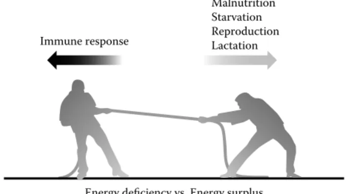
malnutri-tion and starvamalnutri-tion as well as obesity all severely impair the integrity and proper regulation of the immune response. In rodents, significant reductions in total energy depots com-promise the humoral immune response. The induction of an immune response in starving insects reduces their survival. Also, biological processes like reproduction, lactation, and thermoregulation have to compete with immune defense, par-ticularly in conditions of energy limitations or deficit. Taken together, the immune system cannot operate properly when an organism is deprived of nutrients and energy or fails to deploy energy in proper temporal and spatial order (Figure 52.1).
Modern human reality, however, is not only one of nutri-ent deficiency but also pertains to energy and nutrinutri-ent excess as evidenced by the obesity pandemic. Many components of the immune system including macrophages, mast cells, and T-cell-mediated immune responses, as well as neutrophil and natural killer cell activities, are altered in the obese state.19,20
The energy surplus in obesity is associated with impaired immune responses and drives a chronic, sterile inflamma-tory state referred to as metaflammation (referring to meta-bolically orchestrated chronic inflammation).21 Metabolic
inflammation is now recognized as a major underlying fac-tor for reduced insulin sensitivity, abnormal glucose and lipid metabolism, and the development of type 2 diabetes and ste-atohepatitis, particularly, but not exclusively, in the context of obesity.21,22 Historically, the earliest indication of the
associa-tion between inflammaassocia-tion and insulin resistance goes back to the observations made in the late 1950s when an elusive “insulin antagonist” activity was described in the serum of a patient suffering from foot gangrene and infection; when this patient’s serum was transferred to mice, it could antagonize the hypoglycemic effects of insulin.23–25 In the 1980s, Feingold
and Grunfeld demonstrated that infection or administration of tumor necrosis factor α (TNFα), a pro-inflammatory cytokine, caused dyslipidemia and metabolic abnormalities.26,27 Bagby
and Lang also demonstrated the impact of TNFα administra-tion in inducing insulin resistance.28,29
The first evidence regarding the presence of inflammation in obesity and its contribution to the pathogenesis of insu-lin resistance and type 2 diabetes came in the early 1990s by the discovery of adipose tissue inflammatory changes associated with obesity in experimental models30 and
sub-sequently in humans.31,32 Other studies showed that exposure
to inflammatory cytokines in adipocytes or liver cells caused insulin resistance by blocking postinsulin receptor signal-ing.33–35 Importantly, a large number of independent studies
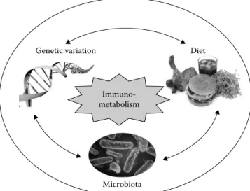
Immuno-metabolism
Genetic variation Diet
Microbiota
FIGURE 52.1 Immunometabolism—a network of complex
inter-actions between metabolic and immune pathways in physiology and pathology. The complex molecular interactions that take place between genetic variation, diet and environment, and microbial occupants (of the gut) at the metabolic and immune interface incite metabolically driven, low-grade, chronic inflammatory changes named metaflammation underlying metabolic syndrome. This interface represents a rapidly growing field of research known as immunometabolism. The focus of immunometabolism research is on the close functional and molecular integration between meta-bolic and immune systems that is crucial for systemic homeostasis as well as these systems’ deregulation that has been causally linked to obesity and associated diseases such as insulin resistance, dia-betes, and fatty liver disease. The consideration of each component and their integrated output is necessary to link potential mecha-nisms with outcomes in a context-dependent manner.
Copyright © 2014. CRC Press. All rights reserved. May not be reproduced in any form without permission from the publisher, except fair uses permitted under U.S. or applicable copyright law.
demonstrated that insulin sensitivity could be improved in obese mice or rats by neutralization of TNFα using molecu-lar or genetic approaches to block the function of this path-way.30,36–43 Many more studies since have provided support
that chronic inflammation in metabolic tissues plays a cen-tral role in the pathogenesis of insulin resistance, diabetes, steatohepatitis, and cardiovascular disease.13–15,44,45 Important
advances also came from understanding the immune compo-nents that contribute to metabolic inflammation (discussed later in Section 52.8 in further detail) and discovery of key signaling pathways that produce metaflammation.13,15,46–49
Today, the studies starting with the obesity-induced adipose tissue inflammation have greatly expanded, leading to the establishment of the field of immunometabolism.
52.4 FEATURES AND MECHANISMS OF METABOLIC INFLAMMATION
It is critical to note that obesity leads to an aberrant form of immunity or metaflammation in a specific and unique energetic context; the inflammation in obesity occurs in the presence of excess nutrients and energy in metabolic tissues, predominantly the adipose tissue but also in many other criti-cal organs such as liver, pancreas, and the central nervous system.21,50 However, this inflammation does not feature an
increase in energy expenditure or basal metabolic rate and raises the possibility that this may in fact have originated from an adaptive mechanism, perhaps under intermittent exposure to food, to prevent sustained insulin sensitivity, hence promotion of adipose expansion and obesity.34 Many
cytokines, including TNFα, interleukin-6 (IL-6), IL-1β, and monocyte chemoattractant protein-1 (MCP-1), are increas-ingly expressed and secreted from the adipose tissue of obese subjects as well as from other metabolic target cells and tis-sues.51,52 Their local concentrations can be high with strong
autocrine/paracrine influence (such as inhibiting insulin receptor signaling and recruitment and activation of immune cells), but their levels in systemic circulation remain low in comparison to classic inflammatory situations such as sepsis, infection, or trauma.51 Furthermore, signaling pathways that
may be responsible for the production of these cytokines, such as inhibitor of kappa B kinase (IKK), c-jun terminal kinase (JNK), and protein kinase RNA-activated (PKR) pathways, are also upregulated in the metabolic tissues in obesity, and their ablation through genetic or chemical approaches proved to be beneficial for insulin sensitivity or metabolic homeosta-sis in obese mice.15,53–56 Unlike classic inflammation,
obesity-induced metaflammation is uniquely characterized by energy conservation. Of note, blocking JNK, IKKε, and PKR dere-presses energy expenditure while preventing the inflamma-tory responses, thereby generating the most consistent and substantial impact on systemic metabolic improvements.53–55
The intricate links between nutrient-sensing and pathogen-sensing systems have been weaved into the architecture of metabolic organs. For example, the fruit fly’s fat body is a single organ that coordinates both the immune and meta-bolic responses.57 Similar functional, temporal, and physical
contiguity of energy and nutrient stores for normal func-tion of immune cells can be found in higher organisms too. Metabolically active hepatocytes are found side by side with macrophage-like Kupffer cells in the liver.21 The mesenteric
adipose tissue is embedded with lymph nodes and harbors various immune effector cells. The activation of local immune response triggers selective lipolysis of the perinodal fat tis-sues, suggesting the purposeful colocalization of immune cells with energy stores in this example may be to meet the excessive energy demands of mounting an immune response.58
While the interaction between resident immune cells and the stromal components is essential for tissue homeostasis in gen-eral, this kind of proximity to rich local energy sources can become particularly important during infection, coupled to suppression of appetite, weight loss, and the consequent sys-temic deficit in deliverable fuels.20 Additionally, the perinodal
adipose tissue has a high content of polyunsaturated fatty acids and could supply them to neighboring immune cells for the generation of lipid-based immune mediators such as pros-taglandins and leukotrienes.59 The intimate and dynamic
rela-tionship between immune cells and local energy depots could thus influence both the immune response and metabolism as is abundantly evident in chronic metabolic diseases such as obesity and diabetes. These intricate links also appear to be emphasized in some chronic inflammatory diseases, which are accompanied by adipose tissue remodeling such as the panniculitis associated with inflammatory bowel disease or the HIV-associated adipose tissue redistribution.60,61 It is thus
not surprising that metabolic abnormalities are also common in many chronic inflammatory diseases. For example, patients with psoriasis, a systemic autoimmune disease influencing the skin, or rheumatoid arthritis, an autoimmune disease mainly affecting the joints, carry markedly elevated risk of develop-ing insulin resistance, diabetes, and atherosclerosis. There is also compelling emerging data that blocking inflammatory pathways, most efficiently by TNFα neutralization, can result in a marked reduction in the incidence of diabetes among sub-jects with psoriasis and rheumatoid arthritis.62
In addition to colocalizing anatomically, the genetic simi-larities and functional overlap that can be seen in metabolic and immune cells are striking and further accentuated with obesity. Despite originating from distinct lineages, macrophages and adipocytes resemble each other in relation to specific functions and genomic expression profiles. For example, preadipocytes, similar to macrophages, express nicotinamide adenine dinu-cleotide phosphate oxidase and can support phagocytosis.63
Adipocytes express the pattern recognition receptors, toll-like receptor (TLR), PKR, inflammasome components, and T-cell receptors that can be activated by nutrients, pathogens, and lipopolysaccharide (LPS); activate inflammatory signal-ing cascades in a cell autonomous manner; and secrete vari-ous cytokines and chemokines when metabolically stressed and regulate the responses of immune cells.64 The
transdif-ferentiation of preadipocytes into macrophage-like cells has also been observed. Consistently, transcriptional profiling reveals a striking resemblance between preadipoctyes, adipo-cytes, and macrophages. The list of shared genes is further
Copyright © 2014. CRC Press. All rights reserved. May not be reproduced in any form without permission from the publisher, except fair uses permitted under U.S. or applicable copyright law.
increased when macrophages are transformed to lipid-laden, pro-atherogenic foam cells.12,63,65 Similar to adipocytes,
intes-tinal epithelium was also found to reactivate an ancient capac-ity to respond to pathogens. In a recent study, it was shown that intestinal epithelium upregulates its interferon-inducible immune response pathways at the expense of its metabolic functions when faced with microbiota under conditions when the adaptive immune system is impaired.66
On the contrary, immune cells also express metabolic programs and are equipped to actively monitor nutrients and energy sources to dictate the inflammatory capacity of the effector cells. For example, macrophages express the recep-tor for advanced glycation end products that recognize lipids and nucleic acids produced as a result of oxidative stress and hyperglycemia; activate lipid synthesis and storage programs through the activity of transcription factors like peroxisome proliferator-activated receptor (PPAR), liver X receptor (LXR), and sterol regulatory element-binding protein (SREBP); and generate lipid droplets as they transform to foam cells.12,67
Furthermore, PPAR and LXR not only activate transcription of genes involved in lipogenesis, but they also limit inflammatory output.68,69 PPARγ regulates the phenotypic switch between
M1 and M2 in macrophages recruited to the adipose tissue in obesity. These studies also showed that PPARγ-induced M2 polarization is protective against insulin resistance in diet-induced obesity; mice with macrophage-specific deletion of PPARγ exhibit increased insulin resistance and obesity.70
A recent study also demonstrated an immunoregulatory role for SREBP-1a through upregulating components of the masome complex, further supporting the link between inflam-mation and lipid metabolism.71 These observations underscore
the coevolution of transcriptional networks to respond to a wide range of challenges from nutrient status to pathogens. Whether functional or genetic similarities exist between other metabolic cells and other immune effectors is not as well characterized. However, all immune cells rely on active metabolism in prep-aration for and during immune response. Glucose and lipids are important for fueling the proliferation of immune cells like lymphocytes.72 In addition, glucose and lipids energize
an immune attack by macrophages and neutrophils, particu-larly phagocytic activity, which consumes copious amounts of energy and requires a high rate of lipid turnover.73 Lipids
can also be important in the generation of lipid-based immune mediators secreted from these cells, and by altering membrane organization and domains, they can impair immune func-tion.74,75 Finally, lipids and other nutrients can serve as signals
or even ligands to engage specific immune pathways.12,54,76,77
In summary, the immune and the metabolic systems intercept at many levels, from molecules to cells to organs, and this can serve for the benefit or the detriment of the organism, depend-ing on the context within which they interact.
52.5 ORIGINS OF INFLAMMATION IN OBESITY The traditional initiators of inflammatory response are patho-gens (such as microbes, viruses, and parasites) or tissue damage. These components engage inflammatory signaling
pathways and induce a response that could contain the hazard and establish tissue homeostasis. The proximal mechanisms responsible for obesity- or high-fat diet-induced metaflam-mation leading to metabolic pathologies are not fully under-stood and remain an important area of research. Nutrients may initiate inflammation from within the metabolic cells (such as adipocytes, hepatocytes, myocytes, and pancre-atic cells) or in immune cells (such as macrophages, mast cells, and lymphocytes) recruited to metabolic organs or both. One potential mechanism involves nutrients directly engaging inflammatory responses on the cell membrane through immune receptors. For instance, high concentra-tions of saturated fatty acids stimulate signaling through the TLR, a pattern recognition receptor pathway in adi-pocytes and macrophages. However, whether this is based on a direct interaction with the TLR receptor or indirectly through fatty acid metabolites like ceramide has not been determined.78 Recently, a role for fetuin in lipid–ligand
inter-action with the TLR has been described, lending support to direct engagement of innate immune pathways by nutrients or some specialization for nutrients, however, distinguishing it from other signals.76 Obesity, with high levels of saturated
fatty acids, is associated with heightened TLR signaling, and the genetic ablation of TLR4 in mice was found protective against weight gain on chow diet.79 Bone marrow
transplan-tation chimeras for the TLR4–/– and the Myd88–/– genotype in mice display protection against insulin resistance on a high-fat diet.80 Later studies presented a mixed picture, however,
where either TLR4–/– on a different background or mice lack-ing the primary mediator of TLR and IL-1R, the myeloid dif-ferentiation primary response protein 88 (Myd88), were not protected against insulin resistance when compared to wild-type, control mice on a high-fat diet.81–84 In light of recent
findings, these conflicting results are likely because of the colonization of the gut microbiota that differs among facili-ties, handling, and specific diets, as directly demonstrated in a recent report.85 In fact, divergent metabolic outcomes
in animal models with single-gene or pathway modifications are not uncommon and most likely reflect the contribution of diet and microbiota to the impact of the underlying genetic manipulation. Further studies are needed to determine the contribution of specific pathways downstream of TLRs and other sensing and signaling molecules that are triggered by nutrients, how and which combination of these factors deter-mines the outcomes.
In addition to membrane-based activation of inflamma-tion, nutrients could engage inflammatory signal transduc-tion pathways in metabolic and immune cells. For example, obesity leads to increased ceramide synthesis and produc-tion of reactive oxygen species (ROS) from the endoplas-mic reticulum (ER) and the mitochondria. Both ceramide and ROS may activate the cytosolic inflammasome, a multiprotein complex that consists of nucleotide-binding domain (NOD)–, leucine-rich repeats–, and pyrin domain– containing family member protein, the adaptor molecule apoptosis-associated speck-like protein containing a caspase recruitment domain, and pro-caspase-1. This complex is
Copyright © 2014. CRC Press. All rights reserved. May not be reproduced in any form without permission from the publisher, except fair uses permitted under U.S. or applicable copyright law.
normally activated by pathogen-derived molecular patterns and cleaves pro-caspase 1, thereby activating it. The active caspase-1 then cleaves pro-IL-18 and pro-IL-1β.86 Indeed,
the activation of the inflammasome and elevated IL-1β levels are well documented in obesity in human and experimen-tal models. Furthermore, genetic deficiency for caspase-1 and NLRP3 (and other inflammasomes including NOD1 and NOD2) improves systemic glucose homeostasis and insulin sensitivity.87 Recently, a novel and critical role for the
double-stranded RNA-dependent protein kinase (PKR) was defined in inflammasome activation.88 Importantly, PKR deficiency
in mice also leads to major metabolic phenotypes including protection against high-fat-diet-induced weight gain, inflam-mation, and insulin resistance and preservation of metabolic health in obesity.54 The discovery of PKR’s integral role in
the inflammasome may provide an important advance in the understanding of coupling innate immune responses to metabolic stimulus and may lead to further understanding of the signals that initiate immune responses in a metabolic context and integrate insulin signaling, translational control, and immune response.
The integration between nutrients and inflammation may also potentially occur in the nucleus. Obesity is associated with an accelerated aging phenotype of the adipose tissue that impacts inflammation and insulin action. It was recently shown that cellular tumor antigen p53 is not only activated in response to shortening telomeres that happens during aging but also by excessive caloric intake and high-fat diet characterized by oxidative stress, senescence-like changes, and elevated p53 levels in the adipose tissue. Adipose tissue– specific p53 deficiency (TRP53–/–) in mice protected against
insulin resistance and adipose tissue inflammation induced by genetic or diet-induced obesity.89 Overexpression of p53
in the adipose tissue resulted in inflammation and insulin resistance. Bone marrow transplantation from wild-type to adipose tissue–specific TRP53–/– mice partially improved
insulin sensitivity, demonstrating the important contribu-tion of macrophage p53 to systemic glucose homeostasis. The authors of this particular study showed that telomere shortening (that occurs in aging) is linked to metabolic deterioration; using telomerase-deficient mice, the authors demonstrated that increased DNA damage and senescence markers in the adipose tissue correlate with insulin resistance and inflammation.89 These observations suggest that
obesity-induced, aging-like changes in the adipose tissue are linked to inflammation, insulin resistance, and metabolic dysfunc-tion through an important nuclear regulator of senescence, p53, and thus offer an additional mechanism by which meta-bolic derangements can trigger immune response, such as is the case in obesity.
52.6 IMPACT OF MICROBIOME ON SYSTEMIC METABOLISM
In recent years, a large body of evidence emerged demonstrat-ing the critical importance of microbial communities in the regulation of systemic metabolism. The gut microbiota that
play a critical role at the juncture of the environment and the host belong to four major bacterial phyla: the gram- negative Bacteroidetes and Proteobacteria and the gram- positive Actinobacteria and Firmicutes.90 Early studies showed
genetic obesity (in leptin-deficient mice) correlates with a reduced ratio of Bacteroidetes to Firmicutes.91 Diet
(high-fat and high-polysaccharide) generates a similar change that can be reversed with antibiotics or weight loss.92 In most but
not all human studies, a reduced Bacteroidetes/Firmicutes ratio in obesity has also been observed.93–95 Some of the
dis-crepancies may have been due to confounding factors such as varying diets, antibiotics usage, housing conditions, and environmental factors between the breeding facilities that could impact the initial colonization of the experimental groups as well as phenotypic outcomes.85,96 Future studies
based on the analysis of metagenomic-derived functional biomarkers rather than phylogenetic ones could help refine the data and define stronger associations with specific popu-lations. What is consistent among the studies is that both diet, the primary nutritional source for the intestinal bacteria, and the genetic makeup of the host directly influence the micro-bial populations and their metabolic consequences, adding yet another level of complexity. In other words, in addition to the direct effects of diet and microbiota on their own, the energy extraction from the diet, as well as the byproducts of metabolism, could be influenced by the interactions between them. Remarkably, the metabolic phenotype associated with the microbiota could be transferred to germ-free mice in both genetic and diet-induced obesity.92,97 Furthermore, a dietary
shift from low to high fat swiftly altered the gut micro-biota in humanized mice (colonized with human intestinal microbiota).98 In the future, it may be possible to directly test
the metabolic consequences of the disappearing human gut microbial complexity in mice by utilizing these humanized models. More studies in humans are also needed to deter-mine the relevance to human disease of these conclusions derived from mouse studies.
How do the changes in the gut microbiota contribute to obesity and metabolic deterioration? Again, this paradigm is a prime example of the interactions between the genetic makeup of the host, diet, and the identity, composition, func-tion, and output of the pathogen populations. For example, it is possible that certain microbial species are more efficient than others in extracting energy and contributing to weight gain.97 The gut microbiota can convert nondigestible
carbo-hydrates (fibers) into short-chain fatty acids (SCFA). These diet-derived SCFA products can be oxidized to provide energy for the host and delivered directly to the liver through the portal vein.99 The increased flux of fatty acids could lead
to steatohepatitis, followed by insulin resistance in the liver and gradual impairment of systemic glucose metabolism.100
Whether these products are directly transported or need to interact with trafficking proteins is not known, and their exploration presents formidable experimental challenges.
In an alternative mechanism, high-fat diet and certain pathogens instigate inflammatory changes and epithelial dysfunction and thus elevate the overall permeability in
Copyright © 2014. CRC Press. All rights reserved. May not be reproduced in any form without permission from the publisher, except fair uses permitted under U.S. or applicable copyright law.
the gut. The resulting leakiness of the gut could lead to an increased delivery of gut microbiota or their metabolites to the mesenteric fat bordering on the gut.101–103 What these
microbial-derived factors are and which host metabolic tar-gets they modify remain unanswered questions of profound interest. Recent studies elaborated angiopoietin-like pro-tein 4 (Angptl4) as one host target, intestinal expression of which can be inhibited by the gut microbiota.104 Angptl4 can
increase plasma triglyceride levels by inhibiting lipoprotein lipase-mediated lipolysis of lipoproteins and consequent fatty acid mobilization toward adipose tissue.105,106 Furthermore,
hypothalamic actions of Anglpltl4 also suggests a potential role in central regulation of energy balance.107 Another host
target could be the endocannabinoid (EC) system engaged through LPS released by the gut microbiota.103 A greater EC
tone affected by the gut microbiota may negatively influence appetite, satiety, and adiposity.
Exposure to microbial products such as lipopolysaccha-ride, flagellin, or others can also engage innate immune response through the TLR or other sensing molecules. Because of inflammation and increased gut permeability, the bacterial flagellin can engage its specific TLR5 receptor on the basolateral surface of the gut epithelia to initiate an immune response against the pathogen, which is necessary for containment. Indeed, the TLR5-deficient (TLR5–/–) mice displayed increased chronic inflammation, adiposity, insulin resistance, and hyperlipidemia, whereas antibiotic treatment could reduce the bacterial load and reverse the metabolic parameters to healthy levels. In this particular study, even the lean mice exhibited insulin resistance due to excess inflam-matory responses in metabolic tissues.108 Furthermore, both
the gut microbiota and the associated metabolic dysfunction could be transferred from the TLR5–/– mice to germ-free,
TLR5+/+ mice.108 The gut microbiota from the TLR5–/– mice differed in composition but not in the relative proportions between the major phyla.108 However, these observations
were recently challenged by another study, which did not observe inflammation of the gut or systemic metabolic dys-function in two separate colonies of TLR5–/– mice. This latter study did confirm improper immune response to flag-ellated bacterial species.109 These disparate results may have
been because of the colonization of the gut microbiota that differs among facilities, handling, and specific diets and may illustrate the complexities of these experimental sys-tems and their sensitivity to each component in place that can modify the phenotype. For example, earlier studies in germ-free mice showed that TRL2 deficiency (TLR2–/–) is protective against insulin resistance and obesity. However, when grown in conventional facilities, these mice devel-oped insulin resistance, metabolic dysfunction, and obesity. These changes in the metabolic phenotype of the TLR2–/– mice in conventional facilities accompanied changes in the gut microbial populations that resembled, at least in part, those found in obese mice and humans. In addition, the met-abolic phenotype could be reversed by antibiotic treatment and transferred to wild-type mice with fecal transplant of the
TLR2–/– mice’s gut microbiota.85 Collectively, these results
underscore the metabolic impact of the gut microbiota that can override or modify the genetic preconditioning of an organism, at least under certain conditions, drawing atten-tion to these interacatten-tions between dietary input and meta-bolic and immune responses that possibly underlie many conflicting phenotypic outcomes of similar genetic models in different studies.
Finally, the mesenteric fat, embedded with lymph nodes and in close contact with the resident immune cells and fac-tors, is essentially an innate immune barrier and is capable of producing an inflammatory response to the incoming microbial challenge from the gut.110 The proinflammatory
cytokines, adipokines, and fatty acids released from the mes-enteric fat could enter the liver through the portal vein and in turn destabilize hepatic metabolism. Consequently, steatosis and insulin resistance develop in the liver. Interestingly, the liver phenotype driven by dysbiosis, a microbial imbalance within the digestive tract, and inflammation appears to be heavily, if not completely, driven by TNFα action and could be rescued by blocking this pathway.111 The development of
extrahepatic insulin resistance and the growing metabolic pressure resulting from insulin resistance on the pancreas can bring about full-blown diabetes. What these microbial metabolites or factors are and their molecular targets respon-sible for this kind of a profound metabolic shift will become an important area for future research.
In conclusion, the three central factors contributing to the nutrient-driven metabolic and immune responses and the phenotypic outcomes include the genetic heritage of the host and genetic variation between subjects, host–pathogen interactions, and dietary exposure and diet composition (Figure 52.2). Immune response Malnutrition Starvation Reproduction Lactation
Energy deficiency vs. Energy surplus
FIGURE 52.2 The competing functions of the organism at times
of energy deficit and surplus. The mounting of a potent immune response is an energetically costly endeavor. Conditions such as malnutrition, reduction in energy depots, and starvation severely impair the immune system. Conditions of energy deficit lead to sig-nificant competition between immune defense and biological pro-cesses of central importance for the organism like reproduction, lactation, and thermoregulation. On the other side of the coin, the immune system is also not equipped to adapt to chronic energy sur-plus and associated molecular and cellular alterations, which cause it to exhibit malfunction. The best example for the latter is illus-trated in obesity and associated metabolic inflammation setting the grounds for a cluster of metabolic diseases.
Copyright © 2014. CRC Press. All rights reserved. May not be reproduced in any form without permission from the publisher, except fair uses permitted under U.S. or applicable copyright law.
52.7 ORGANELLE FUNCTION IN
INFLAMMATORY SIGNALING AND METABOLIC DETERIORATION
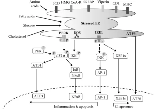
An important primer for metaflammation in obesity is the chronic metabolic overloading of anabolic and catabolic organelles leading to impairment of their function. One such organelle, the ER, serves as a critical intracellular metabolic hub for protein, lipid, and calcium metabolism and lipid droplet formation.112 The vital functions of ER are
main-tained by a conserved, adaptive stress response that emanates from its membranes and is known as the unfolded protein response (UPR). Several diverse stimuli including accumu-lation of unfolded proteins, hypoxia, and toxins can induce the UPR.113 UPR can also be triggered by acute or chronic
excess of nutrients (including fatty acids and free cholesterol) or their deficiency (such as glucose). ER communicates its distress by engaging three signaling branches initiated by the pancreatic ER kinase (PERK), inositol-requiring trans-membrane kinase/endonuclease 1 (IRE1), and activating
transcription factor 6 (ATF6).21,114,115 IRE1 possesses dual
activity consisting of a kinase, which autophosphorylates itself, and an endoribonuclease, which leads to mRNA pro-cessing. The major target of the IRE1 endoribonuclease activity is an important transcriptional regulator of the UPR, the X-box-binding protein 1 (XBP1).116–118 In synergy with
IRE1, the ATF6 branch leads to transcriptional upregulation of XBP1 expression.119 Together, XBP1 and ATF6 maintain
a complex transcriptional program vital for the execution of UPR that involves upregulation of ER-resident chaperones to promote folding and components of the protein degrada-tion apparatus.113,115 If ER stress cannot be resolved, the UPR
can engage apoptotic pathways and lead to cell death48,120
(Figure 52.3).
The ER and the UPR play a significant role in both the physiological and pathological responses of immune cells. For example, some aspects of the UPR are important for the maturation of immune cells such as dendritic cells, lympho-cytes, and plasma cells, XBP1 being a central regulator for the latter.115 ER stress can impair adaptive immune responses
XBP1s
Stressed ER
ATF6 PERK ROS IRE1
AP-1 ATF6 eIF2 αP IKK JNKP AP-1 IκB NFκB NFκB XBP1s
Inflammation & apoptosis Glucose Amino acids Fatty acids Cholesterol ATF4 ATF3 Chaperones CD1 MHC Viperin PKR SCDHMG CoA-R SREBP P P P P P P P
FIGURE 52.3 (See color insert.) The endoplasmic reticulum as a potential hub for metabolism, energy, immune, and stress responses.
In addition to its well-known roles in protein quality control, folding, secretion, and calcium homeostasis, the endoplasmic reticulum (ER) also plays a critical role in lipid metabolism and lipid droplet formation through harboring central players such as the cholesterol-sensitive transcription factor sterol regulatory element-binding protein-1 (SREBP-1); the key regulating enzyme in cholesterol synthesis 3-hydroxy-3-methylglutaryl-CoA reductase (HMG-CoAR); and in fatty acid desaturation through stearoyl CoA reductase (SCD) on its membranes. Studies have now shown that lipid droplets are released from specialized regions of the ER and regulated by proteins like viperin, also associated with ER membranes. Many links also exist between ER responses and glucose metabolism (not depicted here). These myriads of functions are maintained by an adaptive stress response system that emanates from ER membranes, known as the unfolded protein response (UPR). Various stressful situations including accumulation of unfolded proteins, hypoxia, toxins, and acute or chronic excess of nutrients (including fatty acids and free cholesterol) or their deficiency (such as glucose) can activate the UPR. Upon induction, the UPR engages three signaling branches initiated by the pancreatic ER kinase (PERK), inositol-requiring transmembrane kinase/endonuclease 1 (IRE1), and activating transcription factor 6 (ATF6). The major target of the IRE1 endoribonuclease activity is the X-box binding protein 1 (XBP1), which together with the ATF6 branch mounts a complex transcriptional program vital for the execution of UPR. The UPR can also engage inflammatory cascades such as JNK and the IKK–NF-κB pathway, leading to the production of cyclooxygeneases and other pro-inflammatory mediators. Induction of the double-stranded RNA-dependent protein kinase (PKR) can activate the inflammasome, leading to pro-inflammatory responses, and interfere with insulin action.
Copyright © 2014. CRC Press. All rights reserved. May not be reproduced in any form without permission from the publisher, except fair uses permitted under U.S. or applicable copyright law.
by reducing the processing and presentation of peptides by major histocompatibility complex (MHC)–associated antigen presentation.121 Chronic ER stress, induced by overnutrition
in obesity, for example, can also potentially serve as a primer for inflammatory and stress responses in both immune cells (such as macrophages) and metabolic cells (such as adipo-cytes or beta cells). During the course of obesity, unresolved stress in the ER can engage inflammatory response systems through several mechanisms. To begin with, the stressed ER and the mitochondria can produce significant amounts of ROS that can lead to oxidative damage.122 ROS, whether
originating from the mitochondria or the ER, has been shown to activate several stress and inflammatory pathways and contribute to the development of metabolic deterioration in obesity.123,124 Regardless of the origin, in a chronic disease
process, the stress can spread from one organelle to another through inter-organelle contact sites (such as the ER mito-chondria encounter sites [or ERMES] found between the ER and the mitochondria), which can transmit vital information in the form of proteins or lipids or through the endomem-brane system.21,125 Thus, the ER and the mitochondria can
have a strong impact on each other’s function; however, it remains to be seen whether these interactions have any role in the functional deficiencies of these organelles seen in obe-sity and the metabolic pathologies.
Alternatively, the UPR can directly engage inflammatory cascades and stimulate cytokine production. For example, the IRE branch can stimulate JNK kinase activity, through its association with the adaptor protein TNF receptor– associated factor 2 (TRAF2), leading to production of pro-inflammatory gene expression by the transcription factor activator protein-1.118 Active JNK1 can also phosphorylate
the serine residues on IRS proteins and inhibit postinsulin receptor signaling. Consistent with these findings, JNK1-deficient mice exhibit reduced inflammatory cytokine levels and were protected from developing insulin resistance on a high-fat diet.53 Inhibiting JNK by chemical or peptide
inhibi-tors also improved insulin sensitivity in cultured cells such as macrophages, adipocytes, and whole animals.126–130
In another mechanism, IRE1 and PERK arms activate the IKK–nuclear factor-κB (NF-κB) pathway, leading to the production of cyclooxygenases and other pro-inflammatory mediators.131,132 PERK activation has been associated with
the degradation of IκB and nuclear transport of NF-κB. On the other hand, IRE1 activates IκB-kinaseβ [IκKβ] indirectly, through association with TRAF2. Signals through TLR2 and TLR4, but not intracellular TLRs, were recently shown to activate IRE1, through TRAF6, to induce XBP1 process-ing and the secretion of pro- inflammatory cytokines such as IL-6, TNFα, and interferon-β.133 This study and an earlier
one also showed that spliced XBP1 could bind the promoter of TNFα and inducible nitric oxide synthase (iNOS) genes, implicating XBP1 in the regulation of these pro-inflammatory cytokines.133,134 However, clear mechanistic links between
inflammatory pathways and UPR remain to be established in metabolic diseases. Recently, obesity was found to be associ-ated with the activation of an important pathogen sensor, the
double-stranded RNA-dependent protein kinase (PKR), in concert with the induction of UPR in metabolic tissues. PKR can be responsive to ER stress, but the mechanisms of its activation remain unclear. PKR deficiency in mice that fed a high-fat diet is protective against weight gain, metabolic dete-rioration, and insulin resistance. Lipid-induced insulin resis-tance also requires PKR, both in vitro and in vivo, and PKR interacts with and leads to IRS1 phosphorylation.54 PKR can
interact with and modulate the inflammasome activation and potentially contribute to inflammasome-mediated metabolic deterioration. Moreover, recent studies point out regulation of the inflammasome by a proximal ER stress sensor, IRE1 itself.135 Collectively, these studies emphasize the integral role
played by the ER in connecting the pathogen-sensing, inflam-matory, and metabolic systems.54
The UPR is highly receptive of the nutrient status of cells. Historically, the deletion of yeast IRE1 is associated with aux-otrophy for inositol.113 Another set of UPR proteins, namely,
the glucose-regulated proteins, were discovered by their induction in glucose-deprived conditions, a first implication of the nutrient-sensing function of the ER.136 Either lowering
or increasing glucose levels can activate the UPR. The PERK arm plays an important role in systemic glucose homeostasis, particularly through its actions in the liver and pancreas.137,138
Glucose acutely regulates IRE1 activity, and when this becomes chronic, IRE1 can impinge on inflammatory cas-cades mediated by JNK or IKK.113 UPR-induced
transcrip-tional programs are also directly linked to glucose synthesis and breakdown.139 Moreover, the ER assumes a central role in
the synthesis of phospholipids and cholesterol. The UPR con-tributes to the lipogenic program, for example, through the activities of ATF6 and XBP1, although a great deal of uncer-tainty exists among reported phenotypes of the genetic mouse models with respect to lipid metabolism.140,141 An essential
sensor for intracellular cholesterol levels, the SREBP, is situ-ated on the ER membrane.113 Cholesterol deprivation activates
this transcription factor, which upregulates key enzymes in the cholesterol synthetic pathway. Furthermore, elevated free cholesterol or free fatty acids induce ER stress and initiate all three arms of the UPR, but the molecular mechanisms for how the ER senses these changes remain unclear.142,143
In obesity or dyslipidemic conditions, exposure to high levels of nutrients such as saturated fatty acids and free cholesterol leads to ER stress and activation of the UPR in several metabolically active sites including the liver, hypo-thalamus, atherosclerotic plaque, and adipose tissue.144
Recent studies have elaborated the causal role of ER stress in the liver and adipose tissues to the development of systemic insulin resistance and type 2 diabetes. A genetic haploinsuf-ficiency model for the Xbp1 gene in mice leads to elevated ER stress levels and promotes glucose intolerance, insulin resistance, and obesity. In contrast, treating obese mice with chemical chaperones has been shown to release ER stress and improve all of these metabolic parameters.145,146 Moreover,
prolonged ER stress can spur a homeostatic mechanism called autophagy to recycle damaged cellular components and organelles. In recent studies, the failure of autophagy by
Copyright © 2014. CRC Press. All rights reserved. May not be reproduced in any form without permission from the publisher, except fair uses permitted under U.S. or applicable copyright law.
genetically altering master regulators of this mechanism in obese or atherosclerotic mouse models was associated with increased inflammation, insulin resistance, and lesion prog-ress, respectively.147–149 Recently, autophagy was also linked
to the metabolic benefits of exercise, particularly on insulin action, providing an additional layer of integration between this response and metabolic homeostasis.150
How do nutrients stress the ER in the first place? Recently, the metabolically stressed ERs from obese mice were care-fully examined to understand the mechanisms of nutrient-induced ER stress. These studies showed that in the stressed ER from obese mice, when compared to control nonobese groups, protein synthesis is suppressed and chaperone con-tent is not significantly changed. However, the metabolically stressed organelle exhibits prolific lipid synthetic capac-ity, and the specific alterations in ER lipid composition are associated with the inhibition of sarco/ER ATPase (SERCA) activity. Furthermore, reversing the lipid compositional changes or hepatic overexpression of SERCA resolved ER stress in vivo and improved glucose homeostasis and insulin sensitivity in mice.151,152 In a recent study coupling polysome
profiling to microarray analyses, the mammalian translatome was recaptured in vivo. This study showed the steady-state liver translational profile from an obese animal represented a fasting profile of liver from lean animals, suggesting the liver may become insensitive to the excessive flux of nutrients dur-ing obesity. The liver, in particular, also displays aberrations in alternative pathways of bile acid metabolism. Both the liver and the muscle profiles also emphasize mitochondrial defects. These findings suggest that translational dysfunction is another important aspect of obesity that may contribute to the associated metabolic dysregulation.153
Several studies elaborated on the mechanisms of ER stress-induced insulin resistance and weight gain. For exam-ple, the IRE1 arm of the UPR can inhibit insulin receptor signaling through activating JNK1, which then phosphory-lates serine residues on IRS1.145 JNK can also be activated
through PKR and calcium/calmodulin-dependent kinase sig-naling, each of which has been shown to disrupt metabolism and insulin signaling.154 Another mechanism has been
pro-posed where XBP1 interacts with forkhead box protein O1, leading to its proteosomal targeting and degradation.155 ER
stress can also lead to leptin resistance in the hypothalamus that is associated with increased weight gain in mice on a high-fat diet, which can be reversed by the administration of chemical chaperones.156 These findings highlight ER’s
important position as an interface between metabolism and immunity whose dysfunction plays a causal role in the devel-opment of obesity and chronic metabolic disease.145,157–159 In
summary, organelle stress and inflammation both contribute to the development of obesity-associated insulin resistance and atherosclerosis. However, it remains to be determined which is the preceding factor and how the metabolic con-sequences are determined through each of these potential mechanisms. Furthermore, a critical remaining question is whether inflammation itself can be proximal to disruption of ER function or its adaptive capacity and if so, in what
capacity? Future studies chemically or genetically targeting intermediate genes in both UPR and inflammatory pathways should be instrumental in delineating the order of events and their isolated or combined effects. Regardless, thera-peutic targeting of organelle dysfunction offers potential new opportunities for the management of chronic metabolic diseases and rare genetic diseases that display defective ER function and diabetes, such as the Wolfram syndrome (also called DIDMOAD; Diabetes Insipidus, Diabetes Mellitus, Optic Atrophy, and Deafness) and the Wolcott–Rallison syn-drome (WRS).160,161 The Wolfram syndrome occurs because
of a rare mutation leading to defective wolframin gene, ER function, and insulin secretion. The WRS is a rare autoso-mal recessive disease characterized by neonatal/early-onset, nonautoimmune, insulin-requiring diabetes associated with skeletal dysplasia and growth retardation, and it is caused by mutations in the PERK gene. Two chemical chaperones that are known for their ability to improve insulin sensitivity and systemic glucose homeostasis in obese mice, taurourso-deoxycholic acid (TUDCA) and phenylbutyric acid (PBA), were recently evaluated in humans. One study with TUDCA demonstrated it could increase insulin sensitivity in the liver and the muscle in obese humans.162 PBA treatment in humans
also prevented lipid-induced insulin resistance and pancre-atic beta-cell dysfunction.163 These promising outcomes
war-rant studies of therapeutic agents with capacity to improve ER function and folding capacity in wider clinical trials and other settings.164
52.8 METAFLAMMATION: OBESITY-INDUCED INFLAMMATION
The involvement of the immune system in obesity lacks resemblance to acute inflammation that is characterized by five classic signs including pain, heat, redness, swelling, and loss of function. The acute inflammatory reaction of the vas-cular tissues is a robust response of the immune cells to con-fine the injury or infection at its origin and resolves rapidly when the trigger is removed. However, none of these attri-butes of classic inflammation prevail in obesity-induced met-abolic inflammation. Obesity leads to an atypical immune activation, also referred to as metabolic inflammation or “metaflammation,” for it is triggered by metabolic cues and involves both immune (macrophages, mast cells, T cells, and eosinophils) and metabolic cells (such as adipocytes, beta cells, and hepatocytes).21,22 This immune activation is almost
undetectable at a systemic level, and the obesity-induced inflammation appears to be limited to metabolic tissues and remains unresolved over time. In particular, the obese adipose tissue shows signs of elevated inflammation, which includes infiltration of various immune effector cells and production of a variety of inflammatory mediators. Studies have shown that the stressed adipose tissue can alter the met-abolic functions of the liver and muscle by releasing fatty acids, adipokines, and inflammatory cytokines. For example, the genetic ablation of JNK in the adipose tissue enhanced systemic insulin sensitivity and glucose metabolism, whereas
Copyright © 2014. CRC Press. All rights reserved. May not be reproduced in any form without permission from the publisher, except fair uses permitted under U.S. or applicable copyright law.
overexpression of MCP-1 in the adipose tissue resulted in systemic metabolic deterioration.165–167 These findings thus
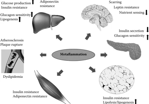
argue that the adipose tissue is important for systemic meta-bolic deterioration as a primary site and through sustaining obesity-induced inflammation. It is, however, important to note that inflammatory alterations in obesity are prominent in many other critical metabolic sites, such as hypothala-mus, pancreatic islets, and liver, and impact a wide variety of metabolic parameters such as insulin action and secretion, glucagon production, hepatic glucose production, and leptin and adiponectin sensitivity (Figure 52.4).48 Finally,
disrup-tion of inflammatory or metabolic pathways in immune cells can also block inflammation and improve metabolism, dem-onstrating the critical role of immune response for metabolic control and illustrating the importance of the interactions between immune and metabolic cells.168 It is, however,
essen-tial to note that inflammatory response is an integral compo-nent of adaptation, defense, repair, and homeostasis. Hence, it may be overly simplistic to label it “friend or foe” in an absolute manner, but rather consider this metabolic–immune interface an equilibrium where the outcome is highly depen-dent on where the organism resides in this continuum.
Obesity leads to enlargement of adipocytes, which becomes an abundant source of inflammatory cytokines including TNFα, IL-6, IL-1β, and MCP-1 as well as many lipid products.169 Immune cells are recruited into the
adi-pose tissue (as well as other metabolic organs).43,170 Several
immune signaling cascades are activated including TLR, IKK, JNK, and PKR and may be responsible for the cyto-kine production from the adipocytes and/or immune cells
infiltrating adipose tissue.48 The adipocytes are overburdened
in their effort to metabolize the excess nutrients, and the resident immune cells are devoid of any significant meta-bolic capacity to deal with this level of nutrient assault and may die. When these stressed cells die, they invite further involvement of the immune system and thus sustain a chronic inflammatory reaction without chance for resolution.34 The
inability to resolve metaflammation in obesity is not mecha-nistically understood. The presence of macrophages in the obese adipose tissue was first recognized in 2003 and found in crown-like structures that form around the necrotic adi-pocytes.46,47,171 Time course analysis of mice on a high-fat
diet showed that macrophages arrive before the first signs of adipocyte necrosis. The adipocyte apoptosis in an inducible mouse model of lipoatrophy leads to macrophage recruitment into the adipose tissue. However, blocking adipocyte necro-sis through the deletion of cyclophilin D did not stop macro-phage infiltration or inflammation in the adipose tissue.172,173
Furthermore, it was also found that human obesity did not increase adipocyte death.174 Hence, these findings suggest
that complex interactions beyond adipocyte necrosis may be required, such as genotoxic events, activation of other stress signaling cascades, or production of immunomodulators and layers of immune activation and cellular complexity for immune cell infiltration and other inflammatory responses of the adipose tissue during obesity, and these mechanisms are not yet fully understood.
Up to 60% of the adipose tissue in obesity can be popu-lated with cells that stain positive for the macrophage marker F4/80 compared to about 10% in adipose tissue from lean
Metaflammation Insulin resistance Adiponectin resistance Insulin secretion Glucagon sensitivity Glucose production Insulin resistance Scarring Dyslipidemia Glucagon sensitivity Lipogenesis Adiponectin resistance Atherosclerosis Plaque rupture Insulin resistance Lipolysis/lipogenesis Nutrient sensing Leptin resistance Insulin secretion Glu G cagon sensitivity
FIGURE 52.4 The impact of metaflammation on multiple organs leading to metabolic syndrome. The adipose tissue is important for
systemic metabolic deterioration as a primary site of sustained obesity-induced inflammation. However, inflammatory alterations are promi-nent in many other critical metabolic sites including the hypothalamus, pancreas, liver, and skeletal muscle in obesity, impacting a wide variety of metabolic parameters such as insulin action and secretion, glucagon production and sensitivity, hepatic glucose production, and leptin and adiponectin sensitivity in the respective targets.
Copyright © 2014. CRC Press. All rights reserved. May not be reproduced in any form without permission from the publisher, except fair uses permitted under U.S. or applicable copyright law.
animals.47 Interestingly, the macrophages in adipose tissue
in obesity display characteristics of classical M1 pheno-type (proinflammatory; nitric oxide synthase 2 and TNFα expression), while macrophages from adipose tissue of lean animals appear to belong to the alternatively activated M2 phenotype (anti-inflammatory; arginase 1 [ARG1]+, c-type lectin domain family member receptor [CD] 206+, CD301+, and IL-10 secreting).175,176 The M1 type macrophages align
around necrotic adipocytes, presumably to scavenge the lip-ids and necrotic material, and form crown-like structures.171
The pro-inflammatory M1 macrophages and the immune mediators they secrete are thought to contribute to the patho-genesis of insulin resistance and systemic metabolic dete-rioration. Consistent with these observations, depletion of M1 macrophages in CD11c-DTR mice or preventing their chemotaxis to tissues in chemokine (C-C motif) recep-tor 2–deficient mice reduced adipose tissue inflammation and improved insulin sensitivity.177,178 Furthermore, myeloid
deficiency of IKKβ or JNK1 prevented myeloid cell–derived inflammation in adipose tissue and improved insulin action or glucose metabolism, although there are differences in the extent of contribution in different studies.165,179–181 Adipose
tissue–driven improvement in inflammatory and metabolic status is best explained in blocking multiple isoforms of JNK in the stroma rather than in the immune cells.165,166,181 This is
also evident in IKKε-deficient as well as in PKR-deficient models.54,55 Taken together, these studies demonstrate how
the recruitment and activation of macrophages to the adipose tissue contribute to systemic glucose homeostasis, but the interactions with the metabolic and stromal cellular compo-nents are also critical.
Recent studies show infiltration of the adipose tissue in obe-sity by several other types of immune cells, which also have systemic effects on metabolism. During obesity, the number of T helper-1 lymphocytes (TH1; secrete pro- inflammatory
cytokines) and CD8+ T cells (cytotoxic T cells; secrete pro-inflammatory cytokines) are increased in the adipose tissue, while the T helper-2 (TH2; secrete anti-inflammatory
cyto-kines) and regulatory T helper-2 cells (Treg; secrete anti-inflammatory cytokines) are decreased.182,183 However, some
recent studies report contradictory findings regarding the type of T cells that are increased or reduced in obesity. What is the role of T lymphocytes in obesity-induced inflammation and metabolic deterioration? One study in mice that lacked T lym-phocytes (mice deficient in recombination-activating genes-1 [RAG-1]) showed they developed worse inflammation and insulin resistance when compared to control mice on a high-fat diet, arguing that certain populations of lymphocytes may play a protective role against metabolic inflammation and dis-ease.182 In fact, adoptive transfer experiments in RAG-1 mice
showed Treg and TH2 lymphocytes but not TH1, and CD8+
cytotoxic T cells suppressed metaflammation and restored glucose metabolism.182 Furthermore, targeted induction of
Treg cells (by IL-2/anti-IL-2 complex) reduces adipose tis-sue inflammation and enhances systemic insulin sensitivity in obese mice.183 Consistently, depleting Treg cells (in the
Foxp3-DTR mice) worsened inflammation and insulin resistance. It
is generally accepted that Treg and TH2 cells improve insulin
sensitivity, while TH1 and CD8+ cytotoxic T cells promote
insulin resistance. In addition to the changes in T cells, the adipose tissue B lymphocytes are found to be elevated, as is serum immunoglobulin levels, over the course of weight gain.184 B cells function in antigen presentation to T cells and
are important for humoral immunity. Mice deficient in B cells or depleted from B cells by CD20-specific immunotherapy exhibit enhanced insulin sensitivity compared to wild-type mice on a high-fat diet.184 This positive response may be due
to reduction in the number of pro-inflammatory T cells and macrophages as seen in the adipose tissue or other sites in these mice. Since adipocytes express unique T-cell receptors and MHC, it is possible that these aspects of metaflammation are also orchestrated by metabolic cells and signals. Either B-cell transfer or serum IgG from obese mice, but not from the lean control mice, to the B-cell-deficient mice induced insulin resistance.184 In summary, obesity leads to activation of both
the innate immune system and the adaptive immune response during the progression of metabolic disease. An important question remains unanswered: What are these antigens recog-nized by the T cells and from where do they originate?
Mast cells and eosinophils are two other types of immune cells that have attracted research interest because they are found in higher numbers in adipose tissue in obesity. Recent studies showed that either genetic depletion or pharmaco-logical stabilization of the mast cells reduces adipose tissue inflammation and improves systemic insulin sensitivity in obese mice.185 Furthermore, the IL-4- and IL-12-producing,
anti-inflammatory M2 macrophage-promoting eosinophils are reduced in the adipose tissue during obesity. The eosinophil-deficient mice exhibit enhanced inflammation and altered insulin sensitivity, suggesting that these cells play a protective role against chronic inflammation and metabolic disease.186
Another important feature of metaflammation is that it is a low-grade immune response. Several immune organs, including primarily the adipose tissue, as well as liver, pan-creas, muscle, and brain, display increased inflammation and local cytokine concentrations. The reflection of these inflam-matory changes to circulating cytokines levels is small, if any, in obese mice and humans.51,187 Furthermore,
metaflam-mation does not increase energy expenditure or basal meta-bolic rate. This is a central issue in the exploring the role of individual immune pathways on systemic metabolism and why these systems were so closely positioned in the first place. These features of metaflammation are in contrast to the classic systemic inflammatory reaction to pathogens or trauma, which uniformly increases metabolic rate and energy expenditure, suggesting that obesity-induced inflam-mation remains a local but effective response that alters systemic metabolic homeostasis but preserves weight. This cycle can also be disrupted, at least in genetic models such as in PKR- and IKKε-deficient mice, which exhibit reduced metabolic inflammation and increased energy expenditure, hence resulting in systemic metabolic improvement as well as better weight control in experimental models.54,55
Copyright © 2014. CRC Press. All rights reserved. May not be reproduced in any form without permission from the publisher, except fair uses permitted under U.S. or applicable copyright law.