Antimycobacterial Activity Some
Different Lamiaceae Plant Extracts
Containing Flavonoids and Other
Phenolic Compounds
Tulin Askun, Gulendam Tumen, Fatih Satil,
Seyma Modanlioglu and Onur Yalcin
Balikesir University
Turkey
1. Introduction
Mycobacterium tuberculosis is a pathogenic bacteria species of the genus Mycobacterium, first discovered in 1882 by Robert Koch, which causes tuberculosis (TB) (Ryan & Ray, 2004). The disease is characterized by symptoms such as sepsis, septic shock, multiple organ failure (Muckart & Bhagwanjee, 1997). It may spread to the central nervous system and cause TB meningitis, intracranial tuberculomas, or abscesses (Harisinghani et al., 2000; Hwang et al., 2010).
After the late 1980s, tuberculosis morbidity and mortality rates became a major health problem for industrialized countries (Raviglione et al., 1995; Heym & Cole, 1997). Multidrug-resistant tuberculosis (MDR TB) and extensively drug resistant tuberculosis (XDR TB) has become a common phenomenon, which cause drugs to be ineffective. MDR-TB results from either primary infection or may develop in the course of a patient's treatment. MDR TB is resistant to at least two first-line anti-TB drugs, isoniazid (INH) and rifampicin (RIF), which are most powerful anti-TB drugs; XDR TB is resistant to INH and RIF, plus fluoroquinolone and at least one of three injectable second-line drugs such as capreomycin, kanamycin, and amikacin. Treatment of XDR-TB is not possible by first-line anti-TB drugs, which are less effective, expensive and toxic; in addition treatment takes two years or more (WHO, 2011a; WHO, 2011b).
Mycobacteria are resistant to most common antibiotics and chemotherapeutic agents due to the mycobacterial cell wall composition of bacterial peptidoglycans (Slayden & Barry, 2000; Lee et al., 1996; Brennan et al., 1995), a lipophilic layer of long-chain fatty acids, and mycolic acids (Barry et al., 1998). The rich lipids of the cell wall has an important role in their virulence (Murray, Rosenthal and Pfaller, 2005). This structure provides a highly hydrophobic and efficient barrier to antibiotics and chemotherapeutic agents (Jarlier & Nikaido 1994). Thus, this cell wall composition restricts the choice of drug treatment. Compounds capable of blocking efflux pumps so that antibiotics can gain access to their targets are of obvious importance (Viveiros et al, 2003). Increased activity of existing efflux
pumps were caused by ineffective therapy of TB patients, which is develops bacterial resistancy to one or more drug. Recent researches showed that mycobacteria have multiple putative efflux pumps which is a key factor for gaining resistance (Braibant, 2000; De Rossi et al., 2002). In addition to, chromosomal gene mutation and then accumulation of these mutations also one of the origine of multidrug-resistant (Ramaswamy & Musser, 1998; Gillespie, 2002; Viveiros et al., 2003).
Some well-known drugs and their mechanism of actions affect bacteria in different ways. Streptomycin (STR) has been used to treat tuberculosis patients since the 1940s; INH was used to treat tuberculosis in the 1960s; RIF was first used at the beginning of the 1970s (Toungoussova et al., 2006); and ethambutol (EMB) was introduced in 1961 as a bacteriostatic first-line drug (Perdigão et al., 2009). RIF inhibits transcription to RNA and translation to proteins by binding its' beta subunit of RNA polymerase in bacteria; however, if bacteria produce a different beta subunit, they are not affected by the drug (O'Sullivan et al., 2005). STR is a protein synthesis inhibitor. STR interacts with a 30S subunit of ribosome and disrupts protein synthesis (Sharma et al., 2007; Springer et al., 2001). Its mechanism of action starts with binding tightly to the phosphate backbone of 16S rRNA in different domains and making contact with the S12 ribosomal protein; finally it causes misreading of the bacterial genetic code during translation (Carter et al., 2000; Hosaka et al., 2006). INH is activated by an enzyme, catalase-peroxidase, called KatG in
M. tuberculosis. KatG, isonicotinic acyl and NADH form a complex that binds enoyl-acyl carrier protein reductase (InhA) and affects fatty acid synthase. The identification of an enoyl-acyl carrier protein (ACP) reductase plays a role in INH resistance named InhA. In this way, mycolic acid synthesis and cell wall development are inhibited (van Veen & Konings, 1998; Slayden & Barry, 2000; Suarez et al., 2009). As a result, when exposed to INH, Mycobacteria lose their acid-fastness and viability. Changes in the catalase-peroxidase gene (katG) and the inhA genes have been defined as one of the mechanisms of drug resistance in M. tuberculosis (Morris et al., 1995; Heym et al., 1995; Mohamad et al., 2004). EMB is a potent synthetic antimycobacterial agent that may cause optic neuropathy in patients (Kozak et al., 1998).
EMB has a bacteriostatic effect and interferes with mycolic acid synthesis, phospholipid metabolism, and arabinogalactan synthesis (Kilburn et al., 1977; Takayama & Kilburn, 1989) and affects nucleic acid metabolism (Forbes et al.,, 1965). EMB has synergistic actions, when combined with other agents, against Mycobacterium avium (Inderlied and Salfinger, 1995). TB is currently one of the most serious infectious diseases all over the world. Antimycobacterial drugs cause unpleasant side effects and trigger changes in the antibiotic target, thereby reducing the efficacy of drug therapies. Mycobacteria have recently increased their virulence and tuberculosis (TB) is the most lethal infection in the world. Between 1980 and 2005, 90 million cases of TB worldwide were reported to the WHO (World Health Organization) and over three in every thousand people die of TB, which is the highest rate in the world (Lall and Meyer, 1999). Yang et al. (2010) also reported that the prevalence of MDR-TB among the Chinese people has increased since 1985. The WHO stated, ‘‘The global incidence of TB was estimated to be 136 cases per 100,000 population per year in 2005. In addition, the WHO region of the Americas and the WHO African region represent a total of 8.8 million new cases of TB and 1.6 million deaths from TB every year” (World Health Organization, 2008a). There were 9.5 million TB-related child deaths globally in 2006 (World Health Organization,
2008b). Today, one of the most important global health problems is changes in behavior of TB, such as resistance to anti-TB drugs and the influence of the HIV epidemic (World Health Organization, 2008a). WHO Global TB Control (2009) reported that there were approximately 0.5 million cases of MDR-TB in 2007. The World Health Organization (2010) reported that there were 9.4 million new TB cases globally and approximately 1.7 million people died from TB. The organization also reported that 1.2 million people were living with HIV and 76% of these people were residing in the African region while 14% were living in the South East Asian region in 2009 (World Health Organization, 2010). In South Africa, TB is the most commonly notified disease and the fifth largest cause of death among the black population. The prevalence of TB continues to increase all over the world. Although the main reasons are known to be the human immunodeficiency virus (HIV) and the emergence of drug-resistant strains of TB (WHO, 2009), the other factors include poverty, drug addiction, inadequate health conditions and migration (Antunes et al., 2000; Merza et al., 2011). WHO reports (2011a) estimated that the risk of developing tuberculosis (TB) is between 20 and 37 times greater in people living with HIV than among the general population. In addition, infection with Human immunodeficiency virus type 1 (HIV-1) disrupts immunological control of Mycobacterium infections due to the loss of CD4+ T cells. Salte et al. (2011) reported that Mycobacterium avium is one of the most common opportunistic infections among AIDS patients. Snider et al. (1985) examined the transmission of MDR-TB strains from adult to child contacts and confirmed the progression of the disease by DNA fingerprint studies. INH-resistant strains caused much infection in children who were in contact with adults.
Mycobacteria are Gram-resistant non-motile pleomorphic rods with a waxy cell wall. These bacteria include high lipid content within the cell wall (Wilbur et al., 2009; Jackson et al., 2007), the complex lipids esterified with long-chain fatty acids. Myobacteria are referred to as acid fast Gram-positive due to their resistance to dilute acid and ethanol-based de-colorization procedures and their lack of an outer cell membrane. When they are stained using concentrated dyes, combined with heat, they do not give up the color by the dilute acid and ethanol-based de-colorization procedures (Ryan & Ray, 2004).
Some medicinal plants have been used to treat the symptoms of TB including Acacia nilotica,
Cassine papillosa, Chenopodium ambrosioides, Combretum molle, and Euclea natalensis from Africa (Watt and Breyer-Brandwijk, 1962; Pujol, 1990; Lall & Meyer, 2001; Bryant, 1966). Natural products are an important source of new chemical compounds and, hopefully, therapeutic agents for many bacterial diseases. Lall and Meyer (1999) reported antimycobacterial activity of Euclea natalensis (Ebenaceae), which is rich in naphthoquinones, against drug-sensitive and drug-resistant strains of M. tuberculosis. Gordien et al. (2009) studied two terpenes, sesquiterpene and longifolene; and two diterpenes, totarol and trans-communic acid, obtained from the aerial parts and roots of Juniperus communis. They reported that totarol showed the highest activity against Mycobacterium tuberculosis H37Rv and that longifolene and totarol exhibited the most activity against rifampicin-resistant variants. Phenolic compounds have some effects on microbial metabolism and growth, depending on their concentration and active compounds (Alberto et al., 2001; Reguant et al., 2000).
Many studies have shown that phenolic compounds inhibit the growth of a wide range of Gram-positive and Gram-negative bacteria (Davidson et al., 2005; Estevinho et al., 2008)
Flavonoids are the most common group of polyphenolic compounds. Flavonoids are plant secondary metabolites with a fused ring system, which are found as glycosides in plants. Of the well-known flavonoids, apigenin has a calming effect, while quercetin and kaempferol have a sedative effect (Jäger & Saaby, 2011).
In previous studies, flavonoids were reported to show antimicrobial (Cushnie & Lamb, 2006, 2011), allergic (Chen et al., 2010), inflammatory (Seo et al., 2000), and anti-carcinogenic (Lee et al., 2008) activities. Until 2004, it was suggested (Cushnie and Lamb, 2005, 2011) that their antibacterial efficacy was dependent upon cytoplasmic membrane damage by perforation (Ikigai et al., 1993), inhibition of nucleic acid synthesis (Mori et al., 1987) and disruption of energy metabolism due to NADH-cytochrome c reductase inhibition (Haraguchi et al., 1998). Currently, some other supporting mechanisms have emerged to indicate the role of flavonoids in antibacterial activity; these mechanisms include damage to the cytoplasmic membrane by generating hydrogen peroxide (Tamba et al., 2007; Kusuda et al., 2006; Sirk et al., 2008), inhibition of nucleic acid synthesis (Gradisar et al., 2007; Wang et al., 2010) and inhibition of ATP synthase (Chinnam et al., 2010). While Puupponen-Pimiä et al. (2001) reported that catechin, rutin and quercetin did not affect the growth of E. coli, Vaquero et al., (2007) reported that quercetin was the strongest inhibitor active against bacteria, dependent on concentration.
Lamiaceae, also known as mint, is a family of flowering plants that includes 250 to 258 genera and approximately 6,000 to 6,970 species across the world (Zomlefer, 1994; Mabberley, 1997). The family has a cosmopolitan distribution and contains many plant species with culinary and medicinal purposes; examples of the former are basil, mint, rosemary, sage, savory, marjoram, oregano, thyme, lavender, and perilla (Naghibi et al., 2005). The Lamiaceae family of plants have been used since ancient times as folk remedies for various health problems such as common cold, throat infections, acaricidal, psoriasis, seborrheic eczema, hemorrhage, menstrual disorders, miscarriage, ulcer, spasm and stomach problems (Takayama et al., 2011; Loizzo et al., 2010;. Ribeiro et al., 2010). Their constituents, particularly diterpenoids and triterpenoids, have been found to have antiseptic, antibacterial, anti-inflammatory, cytotoxic, cardio-active and other properties (Ulubelen, 2003).
In our previous studies, we tested more than 100 plant extracts, some of which showed antimycobacterial activity against Mycobacterium tuberculosis. In this study, in the light of our past experiences, we present a continuation of the testing of some of the plant extracts and the efficacy of their antimycobacterial properties.
2. Materials & methods
2.1 Plant materialsAerial parts (herbs in the flowering stage) of plants, Origanum acutidens (Hand.-Mazz.) Ietswaart, Origanum sipyleum L., Salvia viridis L., Salvia microstegia Boiss&Bal., Satureja boissieri Hausskn. ex Boiss., Stachys byzantina C.Koch., Stachys cretica L., Stachys cretica subsp. smyrnaea Rech. fil., Thymus syriacus Boiss., and Thymus cilicicus Boiss&Bal.(endemic) were collected from different parts of Turkey between 2009 and 2010. The plants were identified by Assoc. Prof. Dr. F. Satil at Balıkesir University, Turkey. Voucher specimens were deposited in the herbarium of Balikesir University Department of Biology. Herbarium plant data, such as locality, altitude, and collection time and identification number of species are given in Table 1.
2.2 Preparation of plant extracts
The plants [O. acutidens (60 g), O. sipyleum (66 g), Salvia viridis (12 g), S. microstegia (100 g),
Satureja boissieri (101 g), Stachys byzantina (65 g), S. cretica (37 g), S. cretica subsp smyrnaea (71 g), T. syriacus (44 g), and T. cilicicus (85 g) (endemic)], were air-dried at room temperature. Extracts of dried plants were prepared by the sequential extraction method (Chan et al., 2008) using 1 L of chloroform (CL), ethyl acetate (EA) and methanol (ME) at room temperature over a period of fifteen days. Finally, three extract fractions were obtained from each plants. The extracts were filtered through filter paper concentrated using a rotary evaporator and dried in vacuo at 40 ºC. They were stored at −20◦C until use. The total yields from chloroform (CL), ethyl acetate (EA) and methanol (ME) extracts were O. acutidens (0.57, 0.74, 4.88g), O. sipyleum (2.14, 1.61, 5.50g), Salvia viridis (0.22, 0.15, 1.56 g), S. microstegia (8.17, 0.77, 6.70g), Satureja boissieri (4.05, 1.08, 8.82g), Stachys byzantina (7.69, 1.10, 5.15g), S. cretica
(1.16, 0.59, 3.47g), S. cretica subsp. smyrnaea (1.92, 1.43, 6.92g), T. syriacus (1.80, 1.08, 2.85g), and T. cilicicus (2.37, 3.18, 5.35g) respectively. All stocks were stored at -20 ºC. To conduct antimicrobial activity tests, samples were dissolved in dimethyl sulfoxide (DMSO) and prepared at a concentration of 100 mg/mL. All the extracts used were sterilized by passing through a syringe filter (Sartorius, Ø 0.22 µm.) before use.
2.3 Chemicals and samples
Gradient grade MeOH and acetonitrile were purchased from MERCK. Gradient grade water (18m) was prepared using a Purelab Option-Q elga dv25 system. All standard stock solutions (1 mg/mL) were prepared by dissolving each compound in MeOH. Standards, rosmarinic acid, trans cinnamic acid, and ferulic acid were purchased from Aldrich, caffeic acid and gallic acid from Sigma-Aldrich and all other chemicals used were obtained from Sigma. All solutions were filtered through a membrane filters (Sartorius, Ø 0.22 µm.) before injection into the capillary.
2.4 LC-MS conditions
Analyses were performed with Agilent LC-MS system (1200 LC with a single quadrupole) with ESI source negative mode. Source parameters were optimized to provide highest sensitivity. The source parameters are: Gas temperature 350 °C, drying gas flow 12 l/min, nebulizer pressure 50 psi, capillary voltage 3500 V., seperation was carried by a C-18 column (EC-C18 4,6x50mm 2.7um). Mobile phases are A: Water (5 mM ammonium formate+ 0.5 % formic acid) and B (acetonitrile). The gradient program is: 5 % B for starting condition and increased up to 45 % B in 1 min, hold 2 min, increase % B to 95 from 3 to 6 min, hold 1 min and decrease % B to 5% at final step. Total run time is 12 min. Injection volume is 5 µl. The detection was accomplished using MS SIM mode. Scan mode is also used. The LC–MS analysis was based in a method described by Pérez-Magariño et al. (1999).
2.5 Preparation of standards
Twenty standards were used for quantitative and qualitative determination: trans-cinnamic acid [(Rt) 4.98 min], ρ-coumeric acid (Rt 3.95 min), vanillic acid (Rt 3.79 min), gallic acid (Rt
1.89 min), caffeic acid (Rt 3.72 min), ferulic acid (Rt 3.99 min), ), apigenin (Rt 4.83 min),
min), carnosic acid (Rt. 8.55 min), chlorogenic acid (Rt 3.59 min), rosmarinic acid (Rt 3.97
min), apigenin 7-glucoside (Rt 3.89 min), oleuropein (Rt 3.969 min), amentoflavone (Rt 5.16
min), naringin (Rt 3.83 min), rutin hydrate (Rt 3.69 min), hesperidin (Rt 3.85 min).
Calibration concentrations were 1,4,5 and 20 ppm except one, apigenin 7-glucoside, was 0.9, 1.8, 4.5, 9, and 18 ppm and injection volume was 5 µL for all standards.
2.6 Organisms
The extracts were screened against four strain, M. tuberculosis H37Ra (ATCC 25177), M.
tuberculosis H37Rv (ATCC 25618) and two-positive M. tuberculosis isolates obtained from patient from hospital, for antibacterial activity.
2.7 Preparation of Mycobacterium tuberculosis inocula
Bacterial suspensions of M. tuberculosis were prepared either from Lowenstein–Jensen slants or from complete 7H9 broth cultures. To prepare an inoculum that was less than 15 days old from a culture grown on Lowenstein-Jensen medium, a suspension was prepared in Middlebrook 7H9 broth. The turbidity of the suspension was adjusted to a 1.0 McFarland standard. The suspension was vortexed for several minutes and was allowed to stand for 20 min for the initial settling of larger particles. The supernatant was transferred to an empty sterile tube and was allowed to stand for an additional 15 min. After being transferred to a new sterile tube, the suspension was adjusted to a 0.5 McFarland turbidity standard by visual comparison. One mL of the adjusted suspension was diluted in 4 mL of sterile saline solution.
No Genus species authority
(Lamiaceae) Locality Altitude (m) Collection Time Herbarium Number 1 Origanum acutidens (Hand.-Mazz.) Ietswaart. Between Elazig-Erzincan 1230 15.Jul.2009 FS 1605 2 Origanum sipyleum L. Between
Balıkesir-Savastepe 200 02.Jul.2009 FS1561 3 Salvia viridis L. Balikesir-Cagis 160 02. Jun.2010 FS1560 4 Salvia microstegia
Boiss&Bal. Van, Gurpinar 1100 26.Jun.2009 FS 1559 5 Satureja boissieri Hausskn.
ex Boiss.
Adiyaman-Yazibaşı
village 980 20.Sep.2010 FS1562 6 Stachys byzantine
C.Koch. Bursa, Mezitler 860 08.Jul.2009 FS1602 7 Stachys cretica L. Balikesir-Edremit,
Kazdagi, 350 23.Jun.2009 FS1603 8 Stachys cretica subsp.
smyrnaea Rech.fil.
Balikesir-Edremit,
Kazdagi, 1260 17.Jul.2009 FS1604 9 Thymus syriacus Boiss. Gaziantep-Burc
forest 850 03.Aug.2009 FS1558 10 Thymus cilicicus
Boiss&Bal.(endemic) Antalya, Belek 1000 12.Jul.2010 FS1556 Table 1. Herbarium data of plants
To prepare M. tuberculosis inoculum using a BACTEC MGIT tube with positive growth, the positive tubes were used beginning from the day after the sample first became positive (day-1 positive), up to and including the fifth day (day-5 positive). The positive tubes that were older than five days were subcultured into fresh growth medium. Tubes that were day-1 and day-2 positive were used in the inoculation procedure for the susceptibility tests. The tubes that were between day-3 and day-5 positive were diluted using 1 mL of the positive broth and 4 mL of sterile saline solution; the 5 mL diluted suspension samples were used for the inoculation procedures.
2.8 Antimycobacterial activity test
Antimycobacterial bioassay was performed using the Microplate Alamar Blue Assay (MABA) method (Collins and Franzblau, 1997). MIC was recorded as the lowest drug concentration that prevented to turn blue to pink colour by adding Alamar blue. MBC was also recorded the minimum extract concentration that do not cause any color changing in cultures reincubated in fresh medium.
2.8.1 Determination of Minimal Inhibitory Concentrations (MICs) for Mycobacterium
tuberculosis
Microplates were inoculated with the bacterial suspension (20 μL per well except for the negative control wells) and incubated at 37 °C for 6 days. Alamar blue (15 μL, Trek Diagnostic system) was then added to the bacterial growth control wells (without extract) and the microplates were incubated at 37 °C for an additional 24 hours. If the dye turned from blue to pink, (indicating positive bacterial growth) then Alamar blue solution was added to the other wells to determine the MIC values. All tests were performed in triplicate.
2.8.2 Determination of mycobactericidal activity
All the extracts prepared from aerial parts of plants, the herbarium data of these species shown in Table 1, were analyzed by LC-MS. The quantity of chemicals in the methanol extracts are given in Table 2. Chromatograms of phenols in all extracts were compared to chromatograms of standards (Figs. 1–3).
The plant extracts described above were used in mycobactericidal activity tests. Two-fold dilution series in triplicate sets of parallel microplate wells were used for each extract. To determine the minimum bactericide concentrations (MBCs), fresh Middlebrook 7H9 culture broth (185 μL) was transferred to each well. A fifteen microliter of an Mycobacterial suspension, from MIC concentration and higher concentration wells obtained from the MIC test described above was added to each well, in order to determine the minimum bactericide concentration (MBC).
Two microplate wells were used as positive and negative controls, and each test was repeated in triplicate. For the negative controls, 200 mL of fresh broth (Middlebrook 7H9 culture medium and OADC) was used. For positive controls, including 185 μL and inoculums from former positive control wells (15 μL) was used. After 24 hours of incubation and colour development using the Alamar blue solution, MBCs were recorded as the minimum extract concentration that did not cause any colour change in cultures when reincubated in fresh medium.
3. Results
3.1 Phenolics determined by LC-MS analyses
The ten samples and selected standards were analyzed by MS in ESI negative ion mode. Scan mode is also used. In this method, trans-cinnamic acid, p-coumaric acid, vanillic acid, gallic acid, caffeic acid, ferulic acid, apigenin, naringenin, luteolin, epicatechin, quercetin, carnosic acid, chologenic acid, rosmarinic acid, apigenin 7-glucoside, amentoflavone, oleuropein, naringin, rutin hydrate and hesperidin were chosen as standard phenolics to determine the phenolic structures of the samples according to ionization response in ESI mass spectrometry and chromatographic retention time.
Ion profile of negative ion electrospray LC/MS analysis experimental conditions are given above, from plants CL, EA and ME extracts is shown Fig. 1-2 and Table 2. Phenolics of samples were identified by comparing standard phenolic data such as retention times, main ions observed under fragmentation voltage of 80 Volt.
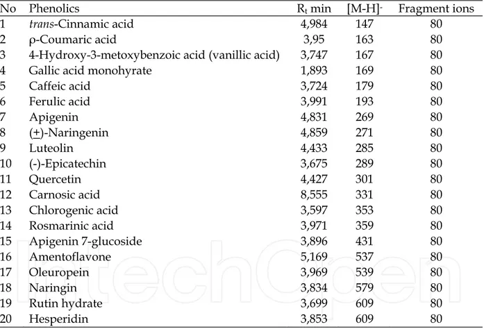
No Phenolics Rt min [M-H]- Fragment ions
1 trans-Cinnamic acid 4,984 147 80
2 ρ-Coumaric acid 3,95 163 80
3 4-Hydroxy-3-metoxybenzoic acid (vanillic acid) 3,747 167 80 4 Gallic acid monohyrate 1,893 169 80
5 Caffeic acid 3,724 179 80 6 Ferulic acid 3,991 193 80 7 Apigenin 4,831 269 80 8 (+)-Naringenin 4,859 271 80 9 Luteolin 4,433 285 80 10 (-)-Epicatechin 3,675 289 80 11 Quercetin 4,427 301 80 12 Carnosic acid 8,555 331 80 13 Chlorogenic acid 3,597 353 80 14 Rosmarinic acid 3,971 359 80 15 Apigenin 7-glucoside 3,896 431 80 16 Amentoflavone 5,169 537 80 17 Oleuropein 3,969 539 80 18 Naringin 3,834 579 80 19 Rutin hydrate 3,699 609 80 20 Hesperidin 3,853 609 80
Table 2. LS-MS characteristics of phenolic compounds
The major phenolic compounds of T. cilicicus CL extract were rutin hydrate and naringenin; for EA extract, rosmarinic acid and apigenin; and for ME extract, rosmarinic acid, oleropein, and apigenin.
The highest rosmarinic acid level within all plants were determined in S. viridis for CL extracts; in S. boissieri and T. cilicicus for EA extracts; O. sipyleum S. byzantine and S. boissieri for ME extracts.
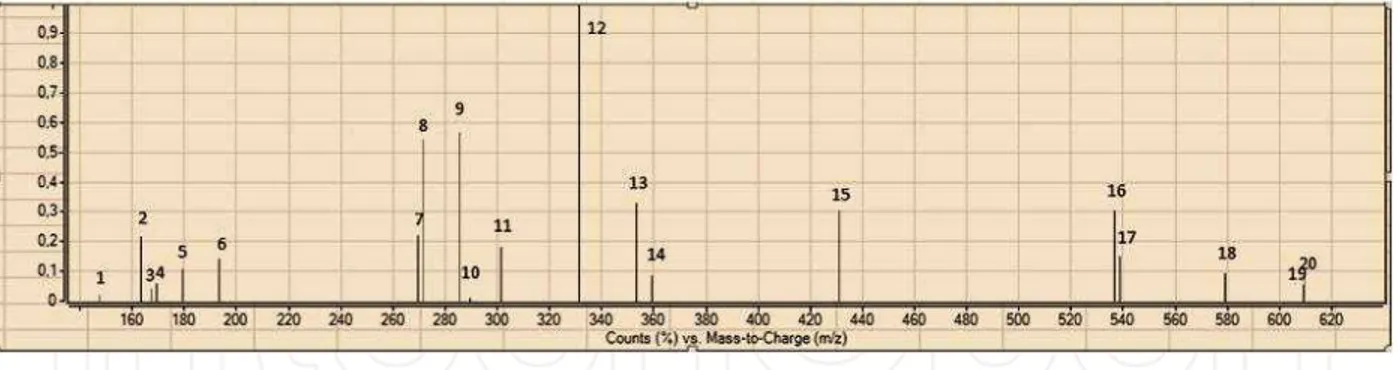
Fig. 1. ESI-MS Spectra of standard phenolics, 1; trans-cinnamic acid 2; p-coumaric acid 3; vanillic acid 4; gallic acid 5; caffeic acid 6; ferulic acid 7; apigenin 8; naringenin 9; luteolin 10; epicatechin 11; quercetin 12; carnosic acid 13; chlorogenic acid 14; rosmarinic acid 15;
apigenin 7-glucoside 16; amentoflavone 17; oleuropein 18; naringin 19; rutin hydrate 20; hesperidin
Fig. 2. ESI-TIC SIM chromatogram of standard phenolics, 1; trans-cinnamic acid 2; p-coumaric acid 3; vanillic acid 4; gallic acid 5; caffeic acid 6; ferulic acid 7; apigenin 8;
naringenin 9; luteolin 10; epicatechin 11; quercetin 12; carnosic acid 13; chlorogenic acid 14; rosmarinic acid 15; apigenin 7-glucoside 16; amentoflavone 17; oleuropein 18; naringin 19; rutin hydrate 20; hesperidin
A
B
D
E
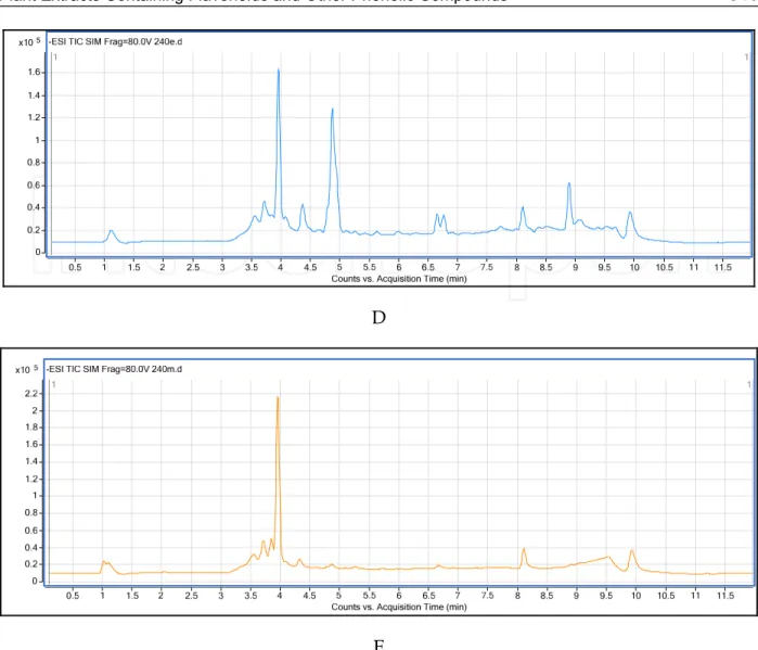
Fig. 3. ESI-TIC SIM chromatogram of O.sipyleum A) EA extract, B) ME extract; S. boissieri C) CL extract D) EA etract and E) ME extract. (Not all chromatograms are included).
The major phenolic compounds for T. syriacus were rutin hydrate and naringenin for CL extract; rosmarinic acid, apigenin naringenin, and vanillic acid for EA extract; rosmarinic acid, apigenin, luteolin, and oleropein for ME extract.
The major phenolic compounds of O. acutidens determined by LC-MS analyses were rutin hydrate for the CL extracts; rosmarinic acid and oleuropein for the EA extracts, rosmarinic acid; and vanillic acid for the ME extracts. The major phenolics of O. sipyleum were rutin hydrate for CL extracts; rosmarinic acid and vanillic acid for EA and ME extracts. The major phenolics of CL extracts of S. viridis were rosmarinic acid and rutin hydrate; for EA extracts, oleuropein followed by rosmarinic acid; for ME extracts, rosmarinic acid, chlorogenic acid and hesperidin.
The major phenolic compounds for S. microstegia were rutin hydrate for CL extracts; apigenin, luteolin, and rosmarinic acid for EA extract; and rosmarinic acid, apigenin and luteolin for ME extracts. In S. boissieri, the major phenolics for CL extracts were apigenin and naringenin; for EA extracts, rosmarinic acid, naringenin and hesperidin; for ME extracts, rosmarinic acid and hesperidin (Fig 3). The major phenolic compounds in the CL extracts of
An ti my co b a ct e ri a l Act ivi ty So me D if fe re n t L a mi a ce a e Pl a n t Ext ra ct s C o n ta in in g F la vo n o id s a n d O th e r P h e n o lic C o mp o u n d s
Pt: Plants; Exts: Extracts; CL: Chloroform; EA: Ethyl Acetat; ME: Methanol; 1; Rosmarinic Acid 2; Naringin 3; Quercet Rutin Hydrate 6; Caffeic Acid 7; Gallic Acid 8; Trans-cinnamic Acid 9; ρ-coumaric acid 10; Vanillic Acid 11; Ferulic Naringenin 13; Chlorogenic Acid 14; Luteolin 15; Apigenin 7-glucoside 16; Hesperidin 17; Oleuropein 18; Carnosic Amentoflavone 20; Apigenin
S. byzantina were rutin hydrate; in the EA extract, apigenin, luteolin and rosmarinic acid; and in the ME extract, rosmarinic acid, hesperidin and apigenin.
In S. cretica, the major phenolics in the CL extracts were trans-cinnamic acid and vanillic acid; oleuropein, vanillic acid and rosmarinic acid for EA extract; and chlorogenic acid and rosmarinic acid for ME extract. In S. smyrnaea, the major phenolics were rosmarinic acid and rutin hydrate for CL extracts; vanillic acid and chlorogenic acid for EA extract; chlorogenic acid and hesperidin for ME extracts.
The highest rutin hydrate contents were determined in O. sipyleum and S. viridis for CL extracts; T. cilicicus, S. viridis, and S. boissieri for EA extracts; S. cretica subsp. smyrnaea, S.
byzantina, and T. syriacus for ME extracts.
Gallic acid was determined only in methanol extracts of S. viridis. Carnosic acid was also found in CL extract of S. boissieri. Only the EA extracts of S. microstegia, S. byzantina, T.
cilicicus and T. striacus included the highest level of apigenin.
Trans-cinnamic acid was found in extracts of four plants (O. acutidens, S. byzantina, and S.
cretica subsp. smyrnaea). Quercetin and amentoflavone were not found. The highest level of chlorogenic acid was found in ME extracts of S. cretica subsp. smyrnaea, S. cretica, and S.
viridis. Luteolin occurred mostly in EA and ME extracts in S. microstegia, S. byzantina, and T.
cilicicus. The highest hesperidin level was found in S. boissieri ME extract and it follows S.
byzantina ME extracts; In addition, ME extracts of S. viridis and T. cilicicus also included high levels of hesperidin. The highest oleuropein content was determined in ME extracts of T.
cilicicus, followed by T. syriacus and S. boissieri. Within EA extracts, S. viridis and O. acutidens had the highest level of oleuropein.
3.2 Antimycobacterial activities, MICs & MBCs
The results were evaluated according to the literature. Extracts were tested against four mycobacteria strains (M. tuberculosis H37Ra, M. tuberculosis H37Rv, and two-positive M.
tuberculosis isolates) obtained from hospital patients, to determine the MIC and MBC using the micro dilution method (MABA) against reference strains.
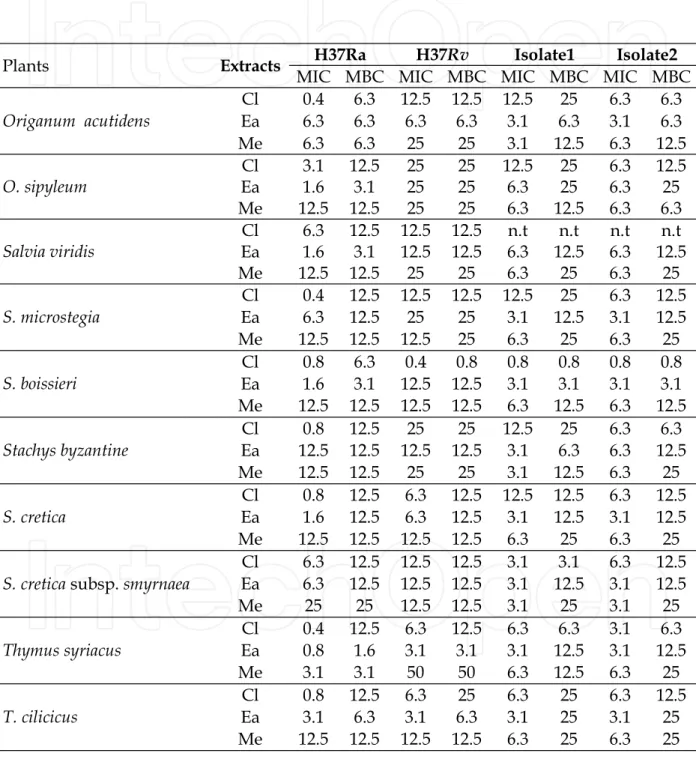
All plant extracts showed antimycobacterial activity (Table 4). Within all CL extracts, O.
acutidens, S. microstegia, and T. syriacus exhibited the lowest MIC value of 0.4 mg/mL against
M. tuberculosis H37 Ra. The lowest MBC value was 6.3 mg/mL for O. acutidens and S.
boissieri. The MBC value for the rest of species was 12.5 mg/mL.
The MIC value of CL extracts against M. tuberculosis H37 Rv was 0.4 mg/mL for S. boissieri, followed by S. cretica, T. syriacus, and T. cilicus at MIC 6.3 mg/mL. Although all CL extracts showed bactericidal activity against M. tuberculosis H37 Rv, the prominent MBC values are 0.8 mg/mL for S. boissieri and 3.1 mg/mL for T. syriacus. For TB-positive isolates1, the featured results were 0.8 mg/mL MIC and MBC for S. boissieri and 3.1 mg/mL MIC and MBC for S. cretica subsp. smyrnaea. S. boissieri was also effective at the concentration 0.8 mg/mL as MBC.
In the EA extracts, the most prominent efficacy was observed for T. syriacus at MIC 0.8 mg/mL; MBC 1.6 mg/mL for T. syriacus against M. tuberculosis H37 Ra. S. boissieri is also
effective at MIC and MBC 6.3 mg/mL against two TB-positive isolates; T. cilicus showed the same effect at MIC and MBC 3.1 on M. tuberculosis H37 Rv.
Of the ME extracts, the most effective against M. tuberculosis H37 Ra was T. syriacus (MIC and MBC 3.1 mg/mL). Stachys byzantine also showed considerable efficacy at MIC 3.1 mg/mL against TB-positive isolates1. Among the other extracts, MIC and MBC values ranged between 6.3-12.5 and 6.3-25 mg/mL, respectively (Table 4).
Plants Extracts H37Ra H37Rv Isolate1 Isolate2
MIC MBC MIC MBC MIC MBC MIC MBC
Origanum acutidens Cl 0.4 6.3 12.5 12.5 12.5 25 6.3 6.3 Ea 6.3 6.3 6.3 6.3 3.1 6.3 3.1 6.3 Me 6.3 6.3 25 25 3.1 12.5 6.3 12.5 O. sipyleum Cl 3.1 12.5 25 25 12.5 25 6.3 12.5 Ea 1.6 3.1 25 25 6.3 25 6.3 25 Me 12.5 12.5 25 25 6.3 12.5 6.3 6.3 Salvia viridis Cl 6.3 12.5 12.5 12.5 n.t n.t n.t n.t Ea 1.6 3.1 12.5 12.5 6.3 12.5 6.3 12.5 Me 12.5 12.5 25 25 6.3 25 6.3 25 S. microstegia Cl 0.4 12.5 12.5 12.5 12.5 25 6.3 12.5 Ea 6.3 12.5 25 25 3.1 12.5 3.1 12.5 Me 12.5 12.5 12.5 25 6.3 25 6.3 25 S. boissieri Cl 0.8 6.3 0.4 0.8 0.8 0.8 0.8 0.8 Ea 1.6 3.1 12.5 12.5 3.1 3.1 3.1 3.1 Me 12.5 12.5 12.5 12.5 6.3 12.5 6.3 12.5 Stachys byzantine Cl 0.8 12.5 25 25 12.5 25 6.3 6.3 Ea 12.5 12.5 12.5 12.5 3.1 6.3 6.3 12.5 Me 12.5 12.5 25 25 3.1 12.5 6.3 25 S. cretica Cl 0.8 12.5 6.3 12.5 12.5 12.5 6.3 12.5 Ea 1.6 12.5 6.3 12.5 3.1 12.5 3.1 12.5 Me 12.5 12.5 12.5 12.5 6.3 25 6.3 25
S. cretica subsp. smyrnaea
Cl 6.3 12.5 12.5 12.5 3.1 3.1 6.3 12.5 Ea 6.3 12.5 12.5 12.5 3.1 12.5 3.1 12.5 Me 25 25 12.5 12.5 3.1 25 3.1 25 Thymus syriacus Cl 0.4 12.5 6.3 12.5 6.3 6.3 3.1 6.3 Ea 0.8 1.6 3.1 3.1 3.1 12.5 3.1 12.5 Me 3.1 3.1 50 50 6.3 12.5 6.3 25 T. cilicicus Cl 0.8 12.5 6.3 25 6.3 25 6.3 12.5 Ea 3.1 6.3 3.1 6.3 3.1 25 3.1 25 Me 12.5 12.5 12.5 12.5 6.3 25 6.3 25
MIC:( mg/mL); MBC: ( mg/mL;). n.t: not tested.
Table 4. Antibacterial activity of extracts of the plants as MIC (mg/mL) and MBC
susceptibility test results against M. tuberculosis H37Ra (ATCC 25177) and M. tuberculosis H37Rv (ATCC 25618) obtained by MABA (Microplate Alamar blue assay) method.
4. Discussion
Lamiaceae plant extracts prepared by using different plant parts such as bark, stem, root, leaves, and fruits used in many biological activity studies. The extracts have been found to have antibacterial activity (Alma et al., 2003; Amanlou et al., 2004; Digrak et al., 2001; Bozin et al., 2006; Karaman et al., 2001), antifungal activity (Bouchra et al., 2003; Askun et al., 2008; Gulluce et al., 2003; Guynot et al., 2003; Souza et al., 2005), antimycobacterial activity (Ulubelen et al., 1997; Askun et al., 2009), antioxidant activity (Alma et al., 2003; Bozin et al., 2006; Mosaffa et al., 2006; Gulluce et al., 2003) and anti-inflammatory activity (Alcar´az et al., 1989; Jim´enez et al., 1986). Inhibitory effects of oregano components on some foodborne fungi were reported (Akgul & Kivanc, 1988). Askun et al. (2009) indicated that Origanum
minutiflorum and Thymbra spicata methanol extracts showed antimycobacterial activity against M. tuberculosis. T. spicata var. spicata showed greater antimycobacterial efficacy (at MIC 196 µg/ml) than O. minutiflorum (MIC 392 µg/ml). They stated that a high quantity of rosmarinic acid might be responsible for antimycobacterial activity.
Recently, investigations of plant extracts are attracting great attentions due to their antibacterial properties (Payne et al., 2007; Rukayadi et al., 2009; Guzman et al., 2010). Previous studies showed that some plant extracts were conciderably effective against M.
tuberculosis. Lall and Meyer (1999) reported that growth of M. tuberculosis is inhibited by acetone and water extracts of Cryptocarya latifolia, Euclea natalensis, Helichrysum melanacme,
Nidorella anomala and Thymus vulgaris. They screened these active acetone extracts against H37Rv and a TB strain that was resistant to the drugs isoniazid and rifampicin. They reported that, while some plants (Croton pseudopulchellus, Ekebergia capensis, Euclea natalensis,
Nidorella anomala and Polygala myrtifolia) exhibited MIC at 0.1 mg/mL against H37Rv, others (Chenopodium ambrosioides, Ekebergia capensis, Euclea natalensis, Helichrysum melanacme,
Nidorella anomala and Polygala myrtifolia) inhibited the resistant strain at the same MIC value. Many natural products have attracted much attention as potential antimycobacterial agents (Kinghorn, 2001; Gupta et al., 2010; Guzman et al., 2010). In recent years, there are pleny of researches on phenolics and their biological activities involved in the literature. Phenolic compounds obtained from plant extracts show great variety, with at least 8000 different structures (Bravo, 1998). Estevinho et al. (2008) showed that differences in the profiles of phenolic compounds are dependent of the flora predominance. Chun et al. (2005) reported that high phenolic and antioxidant activity was related to high antimicrobial activity against ulcer-associated H. pylori. Cinnamic acid is a naturally occurring phenolic compound that shows antimicrobial activity. Chen et al. (2011) showed that cis-cinnamic acid that was transformed from trans-cinnamic acid showed higher synergistic effect with INH or RIF against tuberculosis than trans-cinnamic acid.
Siedel & Taylor (2004) investigated plants, Pelargonium reniforme and P. sidoides (Geraniaceae) fractionation of n-hexane extracts against M. aurum, M. smegmatis, M.
fortuitum, M. abscessus and M. phlei. They reported that linoleic acid was the most potent compound (MIC of 2 mg/l) against M. aurum. Koysomboon et al. (2006) isolated flavonoids from the stems and roots of the mangrove plant Derris indica. They reported antimycobacterial activity at MIC values between 6.25 and 200 µg/mL, except in two of ten known compounds. Askun et al. (2009) indicated that Origanum minutiflorum and Thymbra
T. spicata var. spicata was more effective (MIC 196 µg/ml) than O. minutiflorum (MIC 392 µg/ml). They suggested that a high quantity of rosmarinic acid might be one of the responsible constituent for the observed antimycobacterial activity. Gordien et al. (2009) studied two terpenes, sesquiterpene and longifolene; and two diterpenes, totarol and trans-communic acid, obtained from the aerial parts and roots of Juniperus communis. They reported that totarol showed the highest activity against Mycobacterium tuberculosis H37Rv and that longifolene and totarol exhibited the most activity against rifampicin-resistant variants. These results supported the ethnomedicinal use of this species as a traditional anti-TB remedy. Kuete et al. (2010) investigated the antimycobacterial activity of five flavonoids (isobachalcone, kanzanol C, 4-hydroxylonchocarpin, stipulin, amentoflavone) and determined their effects on preventing the growth of mycobacteria with MIC < 10 µg/ml on
M. tuberculosis. In addition, isobachalcone and stipulin showed total inhibition of M.
tuberculosis strain H37Rv. Bernard et al. (1997) mentioned that rutin showed antibacterial activity on E. coli by inhibited topoisomerase IV-dependent decatenation activity and caused
E. coli topoisomerase IV which is essential for cell survival, dependent DNA cleavage (Bernard et al., 1997; Normark et al., 1969; Cushnie& Lamb., 2005). Huang et al. (2008) indicated that evidence that vanillic acid might be helpful to prevent of the development of the development of diabetic neuropathy by blocking the methylglyoxal-mediated glycation system.
Mandalari et al. (2007) also reported that, pair-wise combinations of eriodictyol, naringenin and hesperidin showed both synergistic and indifferent interactions that were dependent on the test indicator organism and their cell wall structure. Parekh and Chanda (2007) reported that the crude methanol extract of Woodfordia fruticosa contains certain constituents, such as tannins, with significant antibacterial properties, which enables the extract to overcome the Gram-negative cell wall barrier.
Kamatou et al. (2007) studied 16 South African Salvia species that are used in traditional medicine to treat microbial infection. They identified three species, S. verbenaca, S. radula and
S. dolomitica, which exhibited MIC value at 0.10 mg/mL and which also showed antibacterial activity. Green et al. (2010) reported on the activities of acetone extracts of four plants, while Berchemia discolor showed efficacy at MIC 12. 5µg/mL, on H37Ra and 10.5µg/mL on the clinical isolate; the others (Bridelia micrantha, Warbugia salutaris, and
Terminalia sericea) showed efficacy at 25µg/mL on both H37Ra and clinical isolate. The authors validated that these plants include mycobactericidal compounds that are effective against multidrug-resistant M. tuberculosis. Graham et al. (2003) presented an antimycobacterial evaluation of 216 species of Peruvian plants (in 63 families). Dichloromethane extracts from slightly more than half of the samples tested showed MIC value at 50 μg/ml concentration against M. tuberculosis. Billo et al. (2005) reported that methanolic extract of Amborella trichopoda fruits shows MIC value between 1 and 2.5 µg/ml, which was better than pyrazynamide and ethambutol in the same conditions.
Fabryet et al. (1998) reported that solvent extracts of plants with MIC values less than 8 mg/mL may be considered as antimicrobially effective. Gautam et al., (2007), shows that extracts of plant species from wide range of families and genera have exhibited significant in vitro antimycobacterial activities and this efficacy is interestingly compatible with the ethnomedicinal knowledge on plants.
Lechner et al. (2008) showed that myricetin was the most efficient intensifier of INH susceptibility in all tested strains by decreasing the MIC value of INH by as much as 64-fold; the second most effective compound was quercetin. Huang et al. (1980) tested two benzenoid compounds isolated from Ardisia japonica in-vivo on 201 patients infected with M.
tuberculosis (Okunade et al., 2004). They reported that both compounds showed over 80% efficacy.
5. Acknowledgments
The authors are grateful to TUBITAK. This research was supported by a grant from the Scientific and Technological Research Council of Turkey (TUBITAK), TBAG (Research Grant No. 104T336). In addition, we thank Ayhan Aysal for his assistances in LC-MS analyses.
6. Conclusion
In order to test the plant extracts, a potential drug resistant M. tuberculosis isolates was obtained from pulmonary tuberculosis hospital patients. The strains and isolates were then treated with plant extracts that are used for ethnopharmacological purposes. The level of the phenolic compounds and some flavonoids extracts were determined by liquid chromatography–mass spectrometry (LC-MS). The evaluation of results included the plants efficacy, their major phenolics, flavonoids and antimycobacterial activities. All plants extract showed antimycobacterial activity.
O. acutidens, S. microstegia, and T. syriacus were exhibited the lowest MIC value at 0.4 mg/mL against M. tuberculosis H37 Ra. S. boissieri and T. syriacus showed activity at MIC 0,4 mg/mL against M. tuberculosis H37 Rv. The prominent MIC and MBC values against M. tuberculosis H37 Rv were determined at 0,8 mg/mL for S. boissieri and 3,1 mg/mL for S. cretica subsp.
smyrnaea. S. boissieri and T. cilicicus were effective against two TB-positive isolates.
The present work provides a preliminary insight into the effects of phenolics against M.
tuberculosis. Plants of the Lamiaceae family have been shown to include new and effective constituents against Mycobacterium tuberculosis. Examination of these species, reported above, shows that rutin hydrate and vanillic acid were plentiful in all three extracts for these genera in Lamiaceae. All extracts of the Origanum species, Salvia, Satureja, Stachys and
Thymus genera were rich in rosmarinic acid. With the exception of S. viridis, these species did not contain gallic acid.
We suggest that phenolics and naturally occurring flavonoids (polyphenols) are mainly responsible for antimycobacterial, cytotoxicological and mutagenic activity against M.
tuberculosis. In some plants, (O. acutidens, O. sipyleum, S. microstegia, and Stachys byzantine) MIC and MBC values of CL extracts were in the same concentrations. These results might be due to several factors, such as a toxic effect caused by some compounds in the extracts. Liu et al. (2010) showed that a high concentration of cinnamic acid has toxic effects on soil bacteria. The other reason might be that the primary targets of the flavonoids have not been studied as widely in bacteria as in eukaryotes. While flavonoids affect enzyme systems such as prostaglandin, cyclooxygenase and lipoxygenase in eukaryotic cells, the bacteriocidal effect of the flavonoids might have caused the metabolic disorders on metalloenzymes by which their heavy metal atoms combine with flavonoids as ligand complexes in bacteria.
These strong complexes might disrupt the metabolism of organism (Havsteen, 2002). Flavonoids are also known to have mutagenic and antitumor activities (Hodec et al., 2002; Havsteen, 2002). Quercetin affects bacteria by inhibiting the catalytic activity of DNA topoisomerase I and II (Constantinou et al., 1995; Hodec et al., 2002). Quercetin was also reported by Xu et al. (2000) and Spedding et al. (1989) to have inhibitory effects on HIV1-protease and reverse transcriptase.
It is imperative to investigate the use of new, cheaper and efficient compounds to control
Mycobacteria tuberculosis. Recent studies have examined plants and the effectiveness of their different types of extracts on M. tuberculosis. Advanced research into the structure and activity relationships among naturally occurring flavonoids will yield greater understanding of their pharmacokinetics and effects on mycobacteria metabolism according to their structure. It is of great importance to determine the mechanisms of action of flavonoids on
M. tuberculosis .
7. References
Huang, PH., Chen, WS., Hu, Y. (1980). Studies on antituberculosis constituents from Ardisia
japonica. Yaoxue Tongbao 15, 39.
Okunade, AL., Elvin-Lewis, MPF. & Lewis, WH. (2004) Natural antimycobacterial metabolites: current status, Phytochemistry, Vol.65, No.8, (April 2004), pp. 1017-1032, ISSN 0031-9422.
Akgul, A., & Kivanc, M. (1988). Inhibitory effects of selected Turkish spices and oregano components on some foodborne fungi, International Journal of Food Microbiology, Vol.6, No.3, (May 1988), pp. 263-268, ISBN 0168-1605.
Alberto, MR., Farías, ME., & Manca de Nadra, MC. (2001). Effect of gallic acid and catechin on Lactobacillus hilgardii 5w growth and metabolism of organics compounds. Journal
of Agriculture and Food Chemistry, Vol.49, No.9, (September 2001), pp. 4359–4363, ISSN 0021-8561.
Alcar´az, MJ., Jim´enez, MJ., Valverde, S., Sanz, J., Rabanal, & RM., Villar, A. (1989). Anti-inflammatory compounds from Sideritis javalambrensis n-hexane extract. Journal of
Natural Products, Vol.52, No.5, (September-October 1989), pp. 1088– 1091, ISSN 0163-3864.
Alma, M., Mavi, A., Yildirim, A., Digrak, & M., Hirata, T. (2003). Screening chemical composition and in vitro antioxidant and antimicrobial activities of the essential oils from Origanum syriacum L. growing in Turkey, Biological & pharmaceutical
bulletin, Vol.26, No.12, pp. 1725-1729, ISSN 1347-5215.
Amanlou, M., Fazeli, M.R., Arvin, A., Amin, H.G. & Farsam, H. (2004). Antimicrobial activity of crude methanolic extract of Satureja khuzistanica, Fitoterapia, Vol.75, No.7-8, (December 2004), pp. 768-770, ISSN 0367-326X.
Antunes, ML., Aleixo-Dias, J., Antunes, AF., Pereira, MF., Raymundo, E., & Rodrigues, MF. (2000). Anti-tuberculosis drug resistance in Portugal. International Journal of
Tuberculosis and Lung Disease, Vol. 4, No.3, (March 2000), pp. 223—31, ISSN 1027-3719.
Askun, T., Tumen, G., Satil, F., & Kilic, T. (2008). Effects of Some Lamiaceae Species Methanol Extracts on Potential Mycotoxin Producer Fungi, Pharmaceutical Biology, Vol.46, No.10-11, pp. 688–694, ISSN 1744-5116.
Askun, T., Tumen, G., Satil, F., & Ates, M. (2009). In vitro activity of methanol extracts of plants used as spices against Mycobacterium tuberculosis and other bacteria. Food
Chemistry Vol. 116, No. 1 (September 2009), pp. 289–294, ISSN 0308-8146.
Barry III, CE., Lee, RE., Mdluli, K., Sampson, AE., Schroeder, BG., Slayden, RA., & Yuan, Y. (1998). Mycolic acids: structure, biosynthesis and physiological functions, Progress
in Lipid Research. Vol. 37, No. 2-3, (July-August 1998), pp. 143– 179, ISSN 0163-7827. Bernard, FX., Sable, S., Cameron, B., Provost, J, Desnottes, JF., Crouzet. J., & Blanche, F.
(1997). Glycosylated flavones as selective inhibitors of topoisomerase IV,
Antimicrobial Agents and Chemotherapy, Vol.41, No.5, (May 1997), pp. 992–998, ISSN 00664804.
Billo, M., Cabalion, P., Waikedre, J., Fourneau, C., Bouttier, S., Hocquemiller, R., & Fournet, A. (2005). Screening of some New Caledonian and Vanuatu medicinal plants for antimycobacterial activity, Journal of Ethnopharmacology, Vol.96, No.1-2, (January 2005), pp. 195-200. ISSN 0378-8741.
Bouchra, C., Achouri, M., Idrissi Hassani, L.M. & Hmamouchi, M. (2003) Chemical composition and antifungal activity of essential oils of seven Moroccan Labiatae against Botrytis cinerea Pers: Fr., Journal of Ethnopharmacology, Vol.89, No.1, (November 2003), pp. 165-169, ISSN 0378-8741.
Bozin, B., Mimica-Dukic, N., Simin, N., & Anackov, G. (2006). Characterization of the volatile composition of essential oils of some Lamiaceae spices and the antimicrobial and antioxidant activities of the entire oils. Journal of Agricultural and
Food Chemistry, Vol.54, No.5, (March 2006), pp. 1822-1828, ISSN 1579-4377.
Braibant, M., Gilot, P., & Content, J. (2000). The ATP binding cassette (ABC) transport systems of Mycobacterium tuberculosis, FEMS Microbiology Reviews, Vol.24, No.4, (Oct 2000), pp. 449-67, ISSN 0168-6445.
Bravo, L. (1998). Polyphenols: chemistry, dietary sources, metabolism and nutritional significance, Nutrition Reviews, Vol. 56, No. 11, (November 1998), pp. 317–333, ISSN 0029-6643.
Brennan, PJ., Nikaido, H. (1995) The envelope of mycobacteria, Annual Review of
Biochemistry, Vol.64, (July 1995), pp. 29–63, ISSN 15454509, 00664154.
Carter, AP., Clemons, WM., Brodersen, DE., Morgan-Warren, RJ., Wimberly, BT., & Ramakrishnan, V. (2000). Functional insights from the structure of the 30S ribosomal subunit and its interactions with antibiotics, Nature, Vol. 407, (September 2000), pp. 340-348, ISSN/ISBN 0028-0836.
Chan, SW., Li, S., Kwok, CY., Benzie, IFF., Szeto, YT., Guo, DJ., He, XP. & Yu, PHF. (2008) Antioxidant Activity of Chinese Medicinal Herbs, Pharmaceutical Biology, Vol.46, No.9, (September 2008), pp. 587-595, ISSN 1744-5116.
Chang, H.M., Yeung, H.W., Tso, W.-W., Koo, A., (1985). Advances in Chinese Medicinal
Materials Research, World Scientific, ISBN 9971966719, Singapore.
Chen, HJ., Shih, CK., Hsu, HY., & Chiang, W. (2010). Mast cell-dependent allergic responses are inhibited by ethanolic extract of adlay (Coix lachryma-jobi L. var. ma-yuen Stapf) testa, Journal of Agricultural and Food Chemistry, Vol.58, No. 4, (February 2010), pp. 2596–2601, ISSN 0021-8561.
Chen, YL., Huang, ST., Sun, FM., Chiang, YL., Chiang, CJ., Tsai CM., & Weng CJ. (2011). Transformation of cinnamic acid from trans- to cis-form raises a notable bactericidal and synergistic activity against multiple-drug resistant Mycobacterium
tuberculosis, European Journal of Pharmaceutical Sciences, Vol. 43, No.3, (Jun 2011), pp. 188-194, ISSN 0964-4679.
Chinnam, N., Dadi, PK., Sabri, SA., Ahmad, M., Kabir, MA., & Ahmad, Z. (2010). Dietary bioflavonoids inhibit Escherichia coli ATP synthase in a differential manner,
International Journal of Biological Macromolecules , Vol.46, No.5, (Jun 2010), pp. 478– 86, ISSN 0141-8130.
Chun, SS., Vattem, DA., Lin, YT., & Shetty, K. (2005). Phenolic antioxidants from clonal oregano (Origanum vulgare) with antimicrobial activity against Helicobacter pylori,
Process Biochemistry, Vol.40, No.2, (Feb 2005), pp. 809-816, ISSN 1359-5113.
Constantinou, A., Mehta, R., Runyan, C., Rao, K., Vaughan, A., Moon, R. (1995). Flavonoids as DNA topoisomerase antagonists and poisons: structure-activity relationships.
Journal of natural products, Vol.58, No.2, (Feb 1995), pp. 217-25, ISSN 0163-3864. Cushnie, T., & Lamb, AJ. (2006). Antimicrobial activity of flavonoids. International Journal of
Antimicrobial Agents , Vol. 26, No.5, (November 2005), pp.343-356, ISSN 0924-8579. Cushnie, TPT., & Lamb, AJ. (2005) Antimicrobial activity of flavonoids, International Journal
of Antimicrobial Agents, 26(5), (November 2005), pp. 343-356, ISSN 0924-8579.
Cushnie, TPT.,& Lamb, AJ. (2011). Recent advances in understanding the antibacterial properties of flavonoids. International Journal of Antimicrobial Agents, Vol.38, No.2, (August 2011), pp. 99-107, ISSN 0924-8579.
Davidson, PM., Sofos, JN.,& Brenem, AL. (Eds.Davidson, PM., John N . Sofos, JN., & Branen, LA.). (2005). Antimicrobials in Foods, CRC Press, ISBN 978-0-8247-4037-5 New York. De Rossi, E., Arrigo, P., Bellinzoni, M., Silva, PA., Martín, C., Aínsa, JA., Guglierame, P.,&
Riccardi, G. (2002). The multidrug transporters belonging to major facilitator superfamily in Mycobacterium tuberculosis, The Journal of Molecular Medicine, Vol.8, No.11, (November 2002), pp. 714-24, ISSN 1432-1440.
Digrak, M., Alma, MH., & Ilçim, A. (2001). Antibacterial and Antifungal Activities of Turkish Medicinal Plants, Pharmaceutical Biology, Vol.39, No.5, pp. 346-350, ISSN 1744-5116.
Estevinho, L., Pereira, AP., Moreira, L., Dias, LG., & Pereira, E. (2008). Antioxidant and antimicrobial effects of phenolic compounds extracts of Northeast Portugal honey,
Food and Chemical Toxicology, Vol.46, No.12, (December 2008), pp. 3774-3779, ISSN 0278-6915.
Fabry, W., Okemo, P.O. & Ansorg, R. (1998). Antibacterial activity of East African medicinal plants, Journal of Ethnopharmacology, Vol.60, No.1, (February 1998), pp. 79-84 ISSN 0378-8741.
Forbes, M., Kuck, NA., & Peets, EA. (1965). Effect of ethambutol on nucleic acid metabolism in Mycobacterium smegmatis and its reversal by polyamines and divalent cations. The
Journal of Bacteriology, Vol.89, No.5, (May 1965), pp. 1299-1305, ISSN 0021-9193. Gautam, R., Saklani, A. & Jachak, S.M. (2007) Indian medicinal plants as a source of
antimycobacterial agents, Journal of Ethnopharmacology, Vol.110, No.2, (March 2007), pp. 200-234, ISSN 0378-8741.
Gillespie, SH. (2002 ). Evolution of drug resistance in Mycobacterium tuberculosis: clinical and molecular perspective. Antimicrobial Agents and Chemotherapy, Vol.46, No.2, (Feb 2002), pp. 267-74, ISSN 1098-6596.
Gordien, AY., Gray, AI., Franzblau, SG. & Seidel, V. (2009). Antimycobacterial terpenoids from Juniperus communis L. (Cuppressaceae), Journal of Ethnopharmacology, Vol.126, No.3, (December 2009), pp. 500-505, ISSN 0378-8741.
Gradisar, H., Pristovsek, P., Plaper, A., & Jerala, R. (2007). Green tea catechins inhibit bacterial DNA gyrase by interaction with its ATP binding site, Journal of Medicinal
Graham, JG., Pendland, SL., Prause, JL., Danzinger, LH., Vigo, JS., Cabieses, F., & Farnsworth, NR. (2003). Antimycobacterial evaluation of Peruvian plants,
Phytomedicine, Vol.10, No.6-7, (July-August 2003), pp. 528-535, ISSN 0944-7113. Green, E., Samie, A., Obi, CL., Bessong, PO. & Ndip, RN. (2010). Inhibitory properties of
selected South African medicinal plants against Mycobacterium tuberculosis, Journal
of Ethnopharmacology, Vol.130, No.1, (July 2006), pp. 151-157, ISSN 0378-8741.
Gulluce, M., Sokmen, M., Daferera, D., Agar, G., Ozkan, H., Kartal, N., Polissiou, M., Sokmen, A., & Sahin, F. (2003). In vitro antibacterial, antifungal, and antioxidant activities of the essential oil and methanol extracts of herbal parts and callus cultures of Satureja hortensis L., Journal of Agricultural and Food Chemistry, Vol.51, No.14, pp. 3958-3965, ISSN 0021-8561.
Gupta, R., Thakur, B., Singh, P., Singh, H.B., Sharma, V.D., Katoch, V.M., & Chauhan, SV. (2010). Anti-tuberculosis activity of selected medicinal plants against multi-drug resistant Mycobacterium tuberculosis isolates. Indian Journal of Medical Research, Vol.131, (Jun 2010), pp. 809–813, ISSN 0019-5359.
Guynot, M., Ramos, AJ., Seto, L., Purroy, P., & Sanchis, V. (2003). Antifungal activity of volatile compounds generated by essential oils against fungi commonly causing deterioration of bakery products, Journal of General and Applied Microbiology, Vol.94, No.5, pp. 893-899, ISSN 0022-1260.
Guzman, JD., Gupta, A., Evangelopoulos, D., Basavannacharya, C., Pabon, LC., Plazas, EA., Muñoz, DR., Delgado, WA., Cuca, LE., Ribon, W., Gibbons, S., & Bhakta, S. (2010). Anti-tubercular screening of natural products from Colombian plants: 3-methoxynordomesticine, an inhibitor of MurE ligase of Mycobacterium tuberculosis,
Journal of Antimicrobial Chemotherapy, Vol.65, No.10, (October 2010), pp. 2101-7, ISSN 1460-2091.
Haraguchi, H., Tanimoto, K., Tamura, Y., Mizutani, K., & Kinoshita, T. (1998). Mode of antibacterial action of retrochalcones from Glycyrrhiza inflate, Phytochemistry, Vol.48, No. 2, ( May 1998), pp. 125–9, ISSN 0031-9422,
Harisinghani, MG., McLoud, TC., Shepard, JA., Ko, JP., Shroff, MM., & Mueller, PR. (2000). Tuberculosis from head to toe, Radiographics, Vol.20, No.2, (March-April 2000), pp. 449–70, ISSN 0271-5333.
Havsteen, B.H. (2002). The biochemistry and medical significance of the flavonoids,
Pharmacology & Therapeutics, Vol.96, No.2-3, (November 2002), pp. 67-202, ISSN 0163-7258.
Heym, B., & Cole, ST. (1997). Multidrug resistance in Mycobacterium tuberculosis. International
Journal of Antimicrobial Agents, Vol.8, No.1, (April-Jun 1998), pp. 61-70, ISSN 0924-8579.
Heym, B., Alzari, PM., Honore, N., & Cole, ST. (1995). Missense mutations in the catalase-peroxidase gene, katG, are associated with isoniazid resistance in Mycobacterium
tuberculosis. Molecular Microbiology, Vol.15, No.2, (January 1995), pp. 235–45, ISBN 0966438310.
Hodek, P., Trefil, P. & Stiborová, M. (2002) Flavonoids-potent and versatile biologically active compounds interacting with cytochromes P450, Chemico-Biological Interactions, Vol.139, No.1, (January 2002), pp. 1-21, ISBN 0009-2797.
Hosaka, T., Xu, J., & Ochi, K. (2006). Increased expression of ribosome recycling factor is responsible for the enhanced protein synthesis during the late growth phase in an antibiotic-overproducing Streptomyces coelicolor ribosomal rpsL mutant, Molecular
Huang, SM., Chuang, HC., Chi-HaoWu & Gow-Chin Yen (2008). Cytoprotective effects of phenolic acids on methylglyoxal-induced apoptosis in Neuro-2A cells, Molecular
Nutrition & Food Research, Vol. 52, No.8, (May 2008), pp. 940 – 949, ISSN 1613-4133. Hwang, SH., Kong, S-J., Park, J., & Kim, D-Y. (2010). Large intracranial tuberculomas
mimicking brain tumor in multidrug-resistant tuberculosis (MDR-TB). European
Journal of Radiology Extra, Vol.75, No.1, (July 2010), pp. 5-7, ISSN 0720-048X.
Ikigai, H., Nakae, T., Hara, Y., & Shimamura, T. (1993). Bactericidal catechins damage the lipid bilayer. Biochimica et Biophysica Acta, Vol.1147, No.1, (April 1993), pp. 132–6, ISSN 0005-2736.
Inderlied, CB., & Salfinger, M. (1995). Antimicrobial agents and susceptibility tests: Mycobacteria. In: Manual of Clinical Microbiology, Ed. Murray PR. pp. 1385–1404, ASM Press, ISBN 1-55581-371-2, Washington DC.
Jackson, M., Stadthagen, G., & Gicquel, B. (2007). Long-chain multiple methyl-branched fatty acid-containing lipids of Mycobacterium tuberculosis: Biosynthesis, transport, regulation and biological activities, Tuberculosis, Vol.87, No.2, (March 2007), pp. 78-86, ISSN 1472-9792.
Jäger, AK., & Saaby, L. (2011). Flavonoids and the CNS, Molecules, Vol.16, No.2, (February 2011), pp. 1471-1485, ISSN 1420-3049.
Jarlier, V., Nikaido, H. (1994). Mycobacterial cell wall: Structure and role in natural resistance to antibiotics, FEMS Microbiology Letters, Vol.123, No.1-2. (October 1994), pp.11-18, ISSN 1574-6968.
Jim´enez, MJ., Alcar´az, MJ., Ferrandis, ML., & Villar, A. (1986). Antiinflammatory activity of a flavone from Sideritis leucantha. Planta Medica, Vol.52,No.6, (December 1986), pp. 541–541, ISSN 0032-0943.
Kamatou, GPP., Van Vuuren, SF., Van Heerden, FR., Seaman, T., & Viljoen, AM. (2007). Antibacterial and antimycobacterial activities of South African Salvia species and isolated compounds from S. chamelaeagnea, South African Journal of Botany, Vol.73, No.4, (November 2007), pp. 552-557, ISSN 0254-6299.
Karaman, S., Digrak, M., Ravid, U., & Ilcim, A. (2001). Antibacterial and antifungal activity of the essential oils of Thymus revolutus Celak from Turkey, Journal of
Ethnopharmacology, Vol.76, No.2, (July 2001), pp. 183-186, ISSN 0378-8741.
Kilburn, JO., & Greenberg, J. (1977). Effect of ethambutol on the viable cell count in
Mycobacterium smegmatis, Antimicrobial Agents Chemotherapy, Vol.11, No. 3, (March 1997), pp. 534-540, ISSN 0066-4804.
Kinghorn, AD. (2001). Pharmacognosy in the 21st century. Journal of Pharmacy and
Pharmacology, Vol.53, No.2, (February 2010), pp. 135–148, ISSN 0022-3573.
Koysomboon, S., Van Altena, I., Kato, S., & Chantrapromma, K. (2006). Antimycobacterial flavonoids from Derris indica. Phytochemistry, Vol.67, No.10, (May 2006), pp. 1034-1040, ISSN 0031-9422.
Kozak, SF., Inderlied, CB., Hsu, HY., Heller, KB., & Sadun, AA. (1998). The Role of Copper on Ethambutol's Antimicrobial Action and Implications for Ethambutol-induced Optic Neuropathy, Diagnostic Microbiology and Infectious Disease, Vol.30, No.2, (February 1982), pp. 83-87, ISSN 0732-8893.
Kuete, V., Ngameni, B., Mbaveng, A.T., Ngadjui, B., Meyer, JJM., & Lall, N. (2010). Evaluation of flavonoids from Dorstenia barteri for their antimycobacterial, antigonorrheal and anti-reverse transcriptase activities, Acta Tropica, Vol.116, No.1, (October 2010), pp. 100-104, ISSN 0001-706X.
Kusuda, M., Inada, K., Ogawa, TO., Yoshida, T., Shiota, S., Tsuchiya, T., & Hatano T. (2006). Polyphenolic constituent structures of Zanthoxylum piperitum fruit and the antibacterial effects of its polymeric procyanidin on methicillin-resistant
Staphylococcus aureus. Bioscience, Biotechnology, and Biochemistry, Vol.70, (June 2006), No.6, pp. 1423–31, ISSN 0916-8451.
Lall, N., & Meyer JJM. (1999). In vitro inhibition of drug-resistant and drug-sensitive strains of Mycobacterium tuberculosis by ethnobotanically selected South African plants,
Journal of Ethnopharmacology, Vol.66, No.3, (September 1999), pp. 347-354, ISSN 0378-8741.
Lall, N., & Meyer JJM. (2001). Inhibition of drug-sensitive and drug-resistant strains of
Mycobacterium tuberculosis by diospyrin, isolated from Euclea natalensis, Journal of
Ethnopharmacology, Vol. 78, No.2-3, (December 2001), pp. 213-216, ISSN 0378-8741. Lechner, D., Gibbons, S., & Bucar, F. (2008). Modulation of isoniazid susceptibility by
flavonoids in Mycobacterium. Phytochemistry Letters, Vol.1, No.2, (August 2008), pp.71-75, ISBN 0521414210.
Lee, MY, Lin, HY., Cheng, F., Chiang, W., & Kuo, YH. (2008). Isolation and characterization of new lactam compounds that inhibit lung and colon cancer cells from adlay (Coix lachryma-jobi L. var. ma-yuen Stapf) bran. Food and Chemical Toxicology, Vol.46, No.6, (Jun 2008), pp. 1933–1939, ISSN 0278-6915.
Lee, RE., Brennan, PJ., & Besra, GS. (1996). Mycobacterium tuberculosis cell envelope, Current
Topics in Microbiology and Immunology, Vol.215, pp. 1–27, ISSN 0070-217X.
Liu, J., Wu, FZ. & Yang, Y. (2010). Effects of Cinnamic Acid on Bacterial Community Diversity in Rhizosphere Soil of Cucumber Seedlings Under Salt Stress, Agricultural
Sciences in China, Vol.9, No.2, (February 2010), pp. 266-274, ISSN 1671-2927.
Mabberley, DJ. (1997). The Plant Book: a Portable Dictionary of the Vascular Plants, Cambridge University Press, ISBN: 0521414210, Cambridge, UK.
Mandalari, G., Bennett, R N., Bisignano, G., Trombetta, D., Saija, A., Faulds, CB., Gasson, MJ., & Narbad, A. (2007). Antimicrobial activity of flavonoids extracted from bergamot (Citrus bergamia Risso) peel, a byproduct of the essential oil industry.
Journal of Applied Microbiology, Vol.103, No.6, (Dec 2007), pp. 2056–2064, ISSN 1364-5072.
Merza, MA., Farnia. P., Salih, AM., Masjedi, MR., & Velayati, AA. (2011). First insight into the drug resistance pattern of Mycobacterium tuberculosis in Dohuk, Iraq: Using spoligotyping and MIRU-VNTR to characterize multidrug resistant strains. Journal
of Infection and Public Health, Vol.4, No.1, (February 2011) pp. 41-47, ISSN 0749-3797.
Mohamad, S., Ibrahim ,P., & Sadikun, A. (2004) Susceptibility of Mycobacterium tuberculosis to isoniazid and its derivative, 1-isonicotinyl-2-nonanoyl hydrazine: investigation at cellular level. Tuberculosis (Edinb), Vol.84, No.1-2, pp. 56-62, ISSN 1472-9792. Mori, A., Nishino, C., Enoki, N., & Tawata, S. (1987). Antibacterial activity and mode of
action of plant flavonoids against Proteus vulgaris and Staphylococcus aureus,
Phytochemistry, Vol.26, (March 2001), pp. 2231–4, ISSN 0031-9422.
Morris, S., Bai, GH., Suffys, P., Portillo-Gomez, L., Fairchok, M., & Rouse, D. (1995). Molecular mechanisms of multiple drug resistance in clinical isolates of
Mycobacterium tuberculosis. American journal of infectious diseases, Vol.171, No.4, (April 1995), pp.54–60, ISSN1553-6203.
Mosaffa, F., Behravan, J., Karimi, G., & Iranshahi, M. (2006). Antigenotoxic effects of Satureja
hortensis L. on rat lymphocytes exposed to oxidative stress, Archives of pharmacal research, Vol.29, No.2, pp. 159-164, ISSN 0253-6269.
Muckart, DJ., & Bhagwanjee, S. (1997). American College of Chest Physicians/Society of Critical Care Medicine consensus conference definitions of the systemic inflammatory response syndrome and allied disorders in relation to critically injured patients, American Journal of Respiratory and Critical Care Medicine, Vol.25, No.11, (November 1997), pp. 1789–95, ISSN 1073-449X.
Naghibi, F., Mosaddegh, M., Motamed, SM,. & Ghorbani, A. (2005). Labiatae Family in folk Medicine in Iran:from Ethnobotany to Pharmacology, Iranian Journal of
Pharmaceutical Research, Vol.4, No. 2, (April 2005), pp. 63-79, ISSN 1735-0328.
Normark, S., Boman, HG., Matsson, E. (1969). Mutant of Escherichia coli with anomalous cell division and ability to decrease episomally and chromosomally mediated resistance to ampicillin and several other antibiotics, The Journal of Bacteriology, Vol.97, No.3, (March 1969), pp. 1334–1342, ISSN: 00219193.
O'Sullivan, DM., McHugh, TD., & Gillespie, SH. ( 2005). Analysis of rpoB and pncA mutations in the published literature: an insight into the role of oxidative stress in
Mycobacterium tuberculosis evolution?, The Journal of Antimicrobial Chemotherapy, Vol.55, No.5, (April 2005), pp. 674–9, ISSN 1460-2091.
Parekh, J., & Chanda, S. (2007). In vitro antibacterial activity of the crude methanol extract of
Woodfordia fruticosa Kurz, flower (Lythaceae). Brazilian Journal of Microbiology, Vol.38, No.2, (April 2007), pp. 204–207, ISSN 1517-8382.
Payne, DJ., Gwynn, MN., Holmes, DJ., & Pompliano, DL. (2007). Drugs for bad bugs: confronting the challenges of antibacterial discovery. Nature Reviews Drug
Discovery, Vol.6, (Jun 2007), pp. 29–40, ISSN : 1474-1776.
Perdigão, J., Macedo, R., Ribeiro, A., Brum, L., & Portugal, I. (2009). Genetic characterisation of the ethambutol resistance-determining region in Mycobacterium tuberculosis: prevalence and significance of embB306 mutations, International Journal of
Antimicrobial Agents, Vol.33, No.4, (April 2009), pp. 334-338, ISSN 0924-8579.
Pérez-Magariño, S., Revilla, I., González-SanJosé, M.L. & Beltrán, S. (1999) Various applications of liquid chromatography-mass spectrometry to the analysis of phenolic compounds, Journal of Chromatography A, Vol.847, No.1-2, (June 1999), pp. 75-81, ISSN 0021-9673.
Puupponen-Pimiä, R., Nohynek, L., Meier, C., Kähkönen, M., Heinonen, M., Hopia, A., & Oksman-Caldentey KM. (2001). Antimicrobial properties of phenolic compounds from berries. Journal of Applied Microbiology, Vol.90, No.4, (April 2001), pp. 494–507, ISSN 1364-5072.
Ramaswamy, S., & Musser, JM. (1998). Molecular genetic basis of antimicrobial agent resistance in Mycobacterium tuberculosis: 1998 update, Tubercle and Lung Disease, Vol.79, No1, pp. 3-29, ISSN 0962-8479.
Raviglione, MC., Snider, DE., & Kochi, A. (1995). Global epidemiology of tuberculosis.
Journal of the American Medical Association, Vol.273, No.3, pp. 220-226, ISSN 0098-7484.
Reguant, C., Bordons, A., Arola, L., & Roze´s, N. (2000). Influence of phenolic compounds on the physiology of Oenococcus oeni. Journal of Applied Microbiology, Vol.88, No.6, (Jun 2000), pp. 1065–1071, ISSN 1364-5072.