An Osteometric Study of Proximal and Distal Femur
Morphology
Proksimal ve Distal Femur Morfolojisinin Osteometrik Değerleri
Sema Özandaç1, Pınar Göker1, Ahmet Hilmi Yücel1, Memduha Gülhal Bozkır1
1Çukurova University Faculty of Medicine, Department of Anatomy, ADANA
Cukurova Medical Journal 2015;40(3):466-473.
ABSTRACT
Purpose: The current study was undertaken to determine the surgically relevant parameters of proximal and distal
femur from right and left femora in our population.
Methods: These measurements were taken from 400 adult femora (200 right, 200 left) from Department of Anatomy
laboratory of Çukurova University, Faculty of Medicine using an electronic digital caliper and goniometer. The mean values of the neck of the length (NL), maximum femoral length (FL), the length of femoral shaft (FSL), intertrochanteric apical axis length (ITAAL), maximum vertical diameter of the femoral head (VDH), neck shaft angle (NSA), superior-inferior femoral neck diameter (SID) and intercondylar notch witch (ICNW) were taken.
Results: The mean values of these measurements were found to be 22.2 ± 3.0 mm, 429.5 ± 35.0 mm, 306.5 ± 26.7
mm, 61.9 ± 5.7 mm, 44.2 ± 3.8 mm, 121.10 ± 4.00, 30.8 ± 3.1 and 2.4 ± 0.2 cm in the right side respectively. However, in
the left side same dimensions were 22.5 ± 4.4 mm, 431.3 ±26.2 mm, 299.5 ± 59.4 mm, 61.4 ± 5.1 mm, 44.2 ± 3.3 mm, 121.2 ± 4.00, 30.0 ± 2.9 and 2.3 ± 0.2 cm respectively.
Conclusion: The observations presented in the report have defined anatomic parameters that need to be taken into
consideration when surgical procedures are performed in hip and knee area for this population.
Key words: Femur morphometry, Proximal femur and Distal femur.
ÖZET
Amaç: Çalışmamızda, popülasyonumuzdaki sağ ve sol distal ve proksimal femurun cerrahi olarak ilgili ölçüm
değerlerinin belirlenmesi amaçlanmıştır.
Materyal ve Metod: Laboratuvarımızda bulunan 400 adet femurda milimetrik bir kumpas ve goniometre yardımıyla collum femoris ve femur’un maksimum uzunluğu ortalaması, femur şaftı uzunluğu ortalaması, linea intertrochanterica ile fovea capitis arasındaki uzaklık (intertrokanterik apikal aks uzunluğu) ortalaması, caput femoris’in maksimum vertikal uzunluk ortalaması, femur boyun şaft açısı ortalaması, collum femoris’in supero-inferior uzunluk ortalaması ve intercondylar mesafe uzunluğu ortalaması ölçülmüştür.
Bulgular: Ölçümlerimize ait değerler sırasıyla; sağda 22.2 ± 3.0 mm, 429.5 ± 35.0 mm, 306.5 ± 26.7 mm, 61.9 ± 5.7
mm, 44.2 ± 3.8 mm, 121.1 ± 4.00, 30.8 ± 3.1 ve 2.4 ± 0.2 cm bulunurken, aynı değerler sol tarafta 22.5 ± 4.4 mm, 431.3 ±26.2 mm, 299.5 ± 59.4 mm, 61.4 ± 5.1 mm, 44.2 ± 3.3 mm, 121.2 ± 4.00, 30.0 ± 2.9 ve 2.3 ± 0.2 cm olarak bulunmuştur.
Sonuç: Çalışmamızda elde edilen değerlerin popülasyonumuzda yapılacak olan kalça ve diz ile ilgili cerrahi girişimlerde
yardımcı olacağını düşünmekteyiz.
Anahtar kelimeler: Femur morfometrisi, proksimal femur ve distal femur.
INTRODUCTION
Antropometric measurements of long bones, provide important information including gender, race, age and stature of subjects and the lenght of the bone effects stature of subjects1. It has been thought that femur is the good predictor for stature1. The femur, bone of the thigh is the longest and most strongest of human body. It consists of a body (shaft) and two ends; proksimal and distal2,3,4. Moreover, knowledge of proximal femur anatomy is essential for hip joint. The femur morphometry is crucial for orthopedists, anatomists, anthropologists and forensic scientists. Femur morphometry could be used in treatment including osteotomy of proximal femur, hip and femur pathological situations1,5. Furthermore, neck shaft angle (NSA) effects the adaptation of the acetabulum to the femoral head, distribution forces and hip joint function5. The femoral neck is a flat surface of bone and It interconnect femoral head with femoral shaft6. It has been demonstrated that the measurements including femoral head diameter, bicondylar witdth are practical to use to sex identification7. Furthermore, proksimal femur geometry has been defined as an important parameter of hip fracture and longer neck lenght would be a indicator for hip fracture8,9.
NSA is used in diagnosis and treatment of the hip illness by orthopedists and radiologists. This angle decreases gradually. It is approximately 125 ±50 in normal adult population5,10. While this angle increases in congenital subluxation and dislocation of hip, poliomyelitis, cerebral palsy and idiopathic scoliosis. It decreases in some situations including congenital-posttraumatic coxa vara and perthes’ disease5. The neck length which is the good indicator of NSA, helps collo-diaphyseal angle correction, prosthesis design5,11. The measurements such as neck shaft angle and intertrochanteric apical axis length, maximum vertical diameter of the head affect each other and could be used in designing hip prostheses5.
Additionally, distal femur morphometry is substantial for total joint replacement and internal fixation material (12-16) and it can be used as the reference point for measuring patellar height17. Intercondylar notch width is important in injury of anterior cruciate ligament (ACL) and stenosis of the intercondylar notch in athletes3,18,19,20. As a result, femoral morphometric measurements have a great value in diagnosis and treatment of the congenital or post-traumatic deformity.
The current study aimed to document the normal values of a set of the neck length (NL), maximum femoral length (FL), the length of femoral shaft, intertrochanteric apical axis length (ITAAL), maximum vertical diameter of the femoral head (VDH), neck shaft angle (NSA), superior-inferior femoral neck diameter (SID) and intercondylar notch witch (ICNW) in our population.
MATERIALS and METHODS
The study was carried out from the 400 adult femora (200 right, 200 left). The data were collected from Department of Anatomy Laboratory of Çukurova University Faculty of Medicine in this study. The following 8 parameters were measured using an electronic digital caliper and goniometer. These measurements shown in Table 1 were as follows:
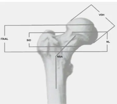
Femoral neck length (NL) (the distance between base of the femoral head and the intertrochanteric line at the junction of the front of the neck with the shaft) (Figure 1)
Maximum femoral length (FL) (measured the upper point of femur head and the lowest point of femoral condyle of femur) (Figure 2)
The length of femoral shaft (from lower point of the lesser trochanter to the upper end of the medial supracondylar ridge of femur) (Figure 2)
468 (the distance taken from fovea capitis to intertrochanteric line) (Figure 1)
Maximum vertical diameter of the femoral head (VDH) (the distance between the upper point of femoral head and the lowest point of femoral head) (Figure 1)
Collo-diaphysial or Neck shaft angle (NSA) (measured between the long axis of the femoral shaft with the axis of the femoral neck) (Figure 1)
Superior-inferior femoral neck diameter (SDI) (The distance taken from the narrowest part of
Intercondylar notch witch (ICNW) (The distance between ½ the antero-posterior diameter of the lateral surface of the medial condyle of femur and ½ the antero-posterior diameter of the medial surface of the lateral condyle of femur). (Figure 2)
The data were divided into two groups: right and left. The SPSS 10.0 program was used for statistical analysis of the measurement results. From these measurements, means, standard deviations (SD), and minimum and maximum (min.-max) values were calculated.
Table 1.Characteristics of proximal and distal femur from 400 (200 right, 200 left) adult femora from Cukurova University in Turkey
Measurements Minimum- Maksimum Mean SD
Femoral neck length (NL)
Right 17 mm-31 mm 22.2 mm 3.0 mm
Left 18 mm -42 mm 22.5 mm 4.4 mm
Maximum femoral length (Max.FL)
Right 361 mm-508 mm 429.5 mm 35.0 mm
Left 366 mm-485 mm 431.3 mm 26.2 mm
Length of femoral shaft (FSL)
Right 251 mm-367 mm 306.5 mm 26.7 mm
Left 240 mm-380 mm 299.5 mm 59.4 mm
Intertrochanteric apical axis length (ITAAL)
Right 47 mm-73 mm 61.9 mm 5.7 mm
Left 48 mm-77 mm 61.4 mm 5.1 mm
The maximum vertical lenght of head (VDH)
Right 35 mm- 51 mm 44.2 mm 3.8 mm
Left 35 mm-51 mm 44.2 mm 3.3 mm
Collo-diaphysial angle-Neck Shaft Angle (NSA)
Right 1170-1320 121.10 4.00
Left 1180-1320 122.20 4.00
Superior-inferior femoral neck diameter (SDI)
Right 24 mm-37 mm 30.8 mm 3.1 mm
Left 25 mm-38 mm 30.0 mm 2.9 mm
Intercondylar Notch Width (ICNW)
Right 2.0 cm-3.2 cm 2.4 cm 0.2 cm
Figure 1.-VDH- Maximum vertical diameter of the femoral head, NL-
Neck Length,SID-Superior-inferior femoral neck diameter, ITAAL-Intertrochanteric apical axis length, NSA-Neck Shaft Angle
Figure 2.-FL- Maksimum Femoral Length,
FSL- The length of the femoral Shaft, ICNW- Intercondylar notch width.
RESULT
The minimum-maximum, mean and SD values of the measurements from the right and left femora are shown in Table 1. From the 400 adult femora, neck of the length (NL), maximum femoral length (FL), the length of femoral shaft (FSL), intertrochanteric apical axis length (ITAAL), maximum vertical diameter of the femoral head (VDH), neck shaft angle (NSA), superior-inferior femoral neck diameter (SID) and intercondylar notch witch (ICNW) were evaluated. The mean values of neck of the length (NL), maximum femoral length (FL), the length of femoral shaft (FSL), intertrochanteric apical axis length (ITAAL), maximum vertical diameter of the femoral head (VDH), neck shaft angle (NSA), superior-inferior femoral neck diameter (SID) and intercondylar notch witch (ICNW) were found to be 22.2 ± 3.0 mm, 429.5 ±35.0 mm, 306.5 ± 26.7 mm, 61.9 ± 5.7 mm, 44.2 ± 3.8 mm, 121.1 ± 4.00
, 30.8 ± 3.1 mm and 2.4 ± 0.2 cm in the right side respectively.
22.5 ± 4.4 mm, 431.3 ±26.2 mm, 299.5 ± 59.4 mm, 61.4 ± 5.1 mm, 44.2 ± 3.3 mm, 121.2 ± 4.00
, 30.0 ± 2.9 mm and 2.3 ± 0.2 cm respectively in the left side respectively. According to our last measurement result, minimum-maximum corresponding values for right femora were found to be 17 31 mm, 361 508 mm, 251 mm-367 mm, 47 mm-73 mm, 35 mm- 51 mm, 1170 -1320, 24 mm-37 mm and 2.0 cm-3.2 cm in the right side respectively, and 18 mm-42 mm, 366 mm-485 mm, 240 mm-380 mm, 48 mm-77 mm, 35 mm- 51 mm, 1180-1320, 25 mm-38 mm and 1.7 cm-2.9 cm in left side respectively.
DISCUSSION
The femur is the longest and strongest bone of individual body. It consists of a body (shaft) and two ends; proksimal and distal2,3,21,22. Studies about long bones explain the data including race, gender, age and stature. Especially, race, hereditary, climate and nutritional status of population impress length of long bones and
470 other populations.
Proximal femur anatomy provide information about the hip joint mechanism5. Thus, proksimal femur geometry can be used in estimation of the hip fracture8,9. Moreover, loss of bone mineral density in proksimal femur increases inclination to fall. Many factors play role in hip fracture. Poor nutritional status, lower body weight, lean body mass, fat content and lifestyle including live by oneself, use of psychotropic drugs- alcohol and some diseases such as, senile dementia, neurologic disorders, hemiplegia are reasonss of hip fracture8,9. If impact stres increases and bone strength decreases, this fracture will occur easily. Furhermore, it has been shown that impact stres of proksimal femur leads to longer femoral neck length (NL) and smaller crosssectional area of the femoral neck8,9. It has been also determined that the racial differences in NL affect the differences in fracture risk incidence. Additionally, It was revealed that NL could be the important predictor of the NSA with FL and used for the NSA correction, prosthesis design. When only a fragment of the proksimal end of the femur is available, it can be useful in estimating maksimum FL too1,5,8,11. Maximum femoral length which depends on the stature is 27% of the stature. In a study consisting of Indians, the mean values of NL and FL were 28.6 mm and 434.9 mm in right side, and 28.1 mm and 435.3 mm in left side respectively5. Whereas, same values were 38.3 and 464.9 mm in males and 35.0 and 428.4 mm in females respectively in Scotland population23. According to Gujarat region, the mean value of the maksimum length of femur was 451.81 mm (right) and 453.35 mm (left) in males and 417.48 mm (right) and 420.44 mm (left) in females respectively24. The mean of the NL was found as 48.8 mm in chinese population8. Corresponding values were found to be 22.2 mm and 429.5 mm in the right side and 22.5 mm and 431.3 mm respectively in this study.
of the head and the mid-point of the intertrochanteric line, can be used for determining neck shaft angle and maksimum femoral length1,5. Also, it was determined that ITAAL and maksimum vertical diameter of head (VDH) might be useful parameters in estimating femoral length and sex identification. Furthermore, VDH measurement is taken from the upper point of head to inferior point of head1,22. Isaac et al reported that the mean values of ITAAL and VDH were 58.7 mm and 41.2 mm in right side respectively, and 57.1 mm and 41.2 mm in left side respectively5. Additionally, it was identified the mean value of VDH as 39.85 mm in males and 41.75 mm in females of South Indian population22. The mean values of same measurements were found to be 61.9 mm and 44.2 mm in the right side and 61.4 mm and 44.2 mm in the left side respectively for our population. Our results are different from Indians;having lower than Turkish group.
Hip disorders such as coxa vara or coxa valga affect femoral shaft angle (NSA). this angle changes as, she-he grows older. While the angle is aproximately 1500 in early infancy, it is 1400 in childhood, 1250 in adult life and 1200 in older5,10. When this value exceeds 1300,coxa valga is seen. Controversely, it is defined as coxa vara, when the angle is less than 1200 10,14,25. The NSA affects the adjusment of the femoral head in the acetabulum and forces dispersion and function hip joint. It is important for the surgeon during the internal fixation of fractured neck femur. Furthermore, it was estimated that femoral head diameters documented the variations of the bone in different populations5,11,14. According to Indian study, the mean value of NSA was 126.50 in right side and 126.90 in left side5. Additionally, same dimension was 122.640 for healthy females, and 125.53 mm for fractured females in Italian population9. However, this angle was reported as 126.650 in females and 136.700 in males in Nigerian region11. Moreover, in several papers from different
countries it has been reported the NSA value as 1260 in American population, 127.50 in Sweden, 122.40 in Belgium, in 132.1 in chinese population8,26,27,28,29. Owing to Finland, the same value was found to be 128.30 in healty group, and 133.70 in fractured groups30. However, this dimension was measured as 121.10 in right femur and 122.20 in left femur in our group. We found significant differences in the mean value of NSA of Indians, Nigerians, Americans, Sweeden, Chinese and Finland population with our population; having greater value than Turks. From this data, our results are more similar to Belgium and Italian healty group.
Superior inferior femoral neck diameter is practical to use for sex identification7,13. It was evaluated as 33.53 mm and 27.86 mm in American Indians, 33.91 mm and 28.92 mm in Mexican population and 35.09 mm and 30.85 mmin French population for males and females respectively13. Whereas, according to our data, corresponding value was found to be 30.8 mm in right side and 30.0 in left side.
Distal Femur anatomy is crucial determinant for total joint replacement design and internal fixation and patellar height measurement. It is associated with anterior cruciate ligament (ACL) rupture12,17. Moreover accurate knowledge of distal femur can be used for total knee prostheses design. Smaller intercondylar notch have been associated with smaller ACL width and this will increase the ACL rupture. It is also related with knee osteoarhritis3,12,18,20. Total knee arthroplasty is a reliable and cheap method for knee osteoarthritis treatment and it alters the mechanical loading of knee joint. The loss of bone in the distal anterior femur is also a risk factor for supracondylar fractures of the femur31,32 . It was found that the mean value of femur intercondylar width was 2.05 cm (right) and 2.05 cm (left) in Greek population12. Additionally, according to Nigerian population, the ICNW value was measured as 2.21 cm in left side and 2.27 cm in right side3. In this paper this value was 2.4 cm in
right side and 2.3 cm in left side. Our results are higher than other studies values.
In summary, when comparing the literature findings with this study, we observe that there are differences between Indians, Nigerians, Americans, Sweedens, Frenchs, Mexicans Finlands and our population about proksimal femur data. However there are similiarities between Belgiums, Italians and our population. We consider that these discrepancies could be a result of such factors like race, genetic variables, nutritional status, lifestyle, climate and socioeconomic status.
In conclusion, we think that precise knowledge of the femur anatomy is essential for safe and accurate surgical interventions. Especially, neck shaft angle is important for the surgeon during the internal fixation of fractured neck femur. Moreover, the differences of mean value of maksimum femoral length between populations could be a result of factors impressing bone morphology including heredity, diet, nutrition status, environment. Therefore, the observations presented in this study have defined anatomic parameters that need to be taken in consideration for performing safe surgical procedures such as total hip-knee replacement or prostheses design and guidelines for the use of devices in hip and knee. As a result, we believe that the data obtained in this study can provide crucial information for femur morphometry and may help the surgeon for having a succesful surgery and minimize the related problems.
REFERENCES
1. Prasad R, Vettivel S, Jeyaseelan L, Isaac B, Chandi G. Reconstruction of femur length from markers Its proksimal end. Clinical Anatomy. 1996;9:28-33. 2. Moore KL, Dalley AF. Clinically Oriented Anatomy.
4nd ed. Lippincott Williams&Wilkins, Canada. 1999;504-22.
3. Didia BC, Nwajagu GN, Dapper DV. Femoral Intercondylar Notch (ICN) width in Nigerians: Its relationship of femur length. West Afr J Med. 2002;21:265-68.
472 system: In Gray’s Anatomy. 38th ed., Churchill Livingstone. Newyork. 1995;678.
5. Isaac R, Vettivel S, Prasad R, Jeyaseelan L, Chandi G. Prediction of the femoral neck-shaft angle from the length of the femoral neck. Clinical anatomy. 1997;10:318-23.
6. Seidemann RM, Stojanowski CM, Doran GH. The use of the supero-inferior femoral neck diameter as a sex assessor. American Journal of physical anthropology. 1998;107:305-13.
7. Gill GW. Racial variation in the proximal and distal femur: heritability and forensic utility. J. Forensic Sci. 2001;46:791-9.
8. Yang RS, Wang SS, Liu TK. Proximal femoral dimension in elderly chinese women with hip fractures in taiwan. Osteoporosis International. 1999;10:109-13.
9. Gnudi S, Ripamonti C, Gualtieri G, Malavolta N. Geometry of proximal femur in the prediction of hip fracture in osteoporotic women. The British Journal of Radiology. 1999;72:729-33.
10. Byrne DP, Mulhall KJ, Baker JF. Anatomy & Biomechanics of the hip. The open sports medicine Journal. 2010;4,51-7.
11. Tahir A, Hassan AW, Umar IM. A study of the collodiaphyseal angle of the femur in the North- Eastern subregion of nigeria. Niger J. Med. 2001;10:34-6.
12. Terzidis I, Totlis T, Papathanasiou E, Sideridis A, Vlasis K, Natsis K. Gender and side to side differences of femoral condyles morphology: osteometric data from 360 caucasian dried femori. Hindavi Publishing Corporation anatomy research International. 2012;1-6.
13. Perret VA, Staccini P, Quatrehomme G. Reexamination of a measurement for sexual determination using the supero-inferior femoral neck diameter in a modern european population. J.Forensic Sci. 2003;48:517-21.
14. Asala SA. Mbajiorgu FE, Papandro BA. A comparative study of femoral head diameters and sex differentiation in Nigerians. Acta anatomy. 1998;162: 232-7.
Forensic Science International. 2002;127:114-8. 16. Asala SA. Sex determination from the head of the
femur of South african whites and blacks. Forensic science international. 2001;117:15-22.
17. Chareancholvanich K, Narkbunnam R. Novel method of measuring patellar height ratio using a distal femoral reference point. International Orthopaedics. 2012;36:749-53.
18. Good L, Odensten M, Gillquist J. Intercondylar notch measurements with special reference to anterior cruciate ligament surgery. Clinical Orthop. 1991;2:185-9.
19. Laprade RF, Burnett QM. Femoral intercondylar notch stenosis and correlation to anterior cruciate ligament injuries. A prospective study. Am J Sports Med. 1994;22:198-202.
20. Wada M, Tatsuo H, Baba H, Asamoto K, Nojyo A. Femoral intercondylar notch measurements in osteoarthritic knees. Rheumatology.1994;38:554-8. 21. Elbuken F, Baykara M, Ozturk C. Standardisation of
the neck shaft angle and measurement of age, gender and BMI related changes in the femoral neck using DXA. Singapore Med J. 2012;53: 587-90. 22. Nidugala H, Bhaskar B, Suresh S, Avadhani R.
Metric assessment of femur using discriminant function analysis in south indian population. Int J Anat Res. 2013;1: 29-32.
23. Duthie RA, Bruce MF, Hutchison JD. Changing proximal femoral geometry in north east scotland: an osteometric study. BMJ open respiratory research. 1998;316:1498.
24. Pandya AM, Singel TC, Akbari VJ, Dangar KP, Tank KC, Patel MP. Sexual dimorphism of maximum femoral length. National Journal of Medical Research. 2011;1; 67-70.
25. Ranade A, McCarthy JJ, Davidson RS. Acetabular changes in coxa vara. Clinical Orthop Relat Res.2008;466: 1688-91.
26. Michelotti J, Clark J. Femoral neck length and hip fracture risk. 1999;14:1714-9.
27. Boonen S, Koutri R, Dequeker J, Aerssens J, Lowet GJN, Verbeke G, Lesaffre E, Geusens P.
Measurement of femoral geometry in type I and type II osteoporosis: Differences in hip axis length consistent with heterogeneity in the pathogenesis of osteoporotic fractures. J Bone Miner Res. 1995;10:1908-12.
28. Faulkner KG, McClung M, Cummings SR. (1994) Automated evaluation of the hip axis length for predicting hip fracture. J Bone Miner Res. 1994;9:1065-70.
29. Karlsson KM, Sernbo I, Obrant KJ, Redlung JI, Johnell O. Femoral neck geometry and radiographic signs of osteoporosis as predictors of hip fracture. Bone.1996;18:327-30.
30. Partanen J, Jamsa T, Jalovaara P. Influence of the upper femur and pelvic geometry on the risk and type of hip fractures. 2001;16:1540-7.
31. Soininvaara TA, Miettinen HJA, Jurvelin JS, Suomalainen OT, Alhava EM, Kröger HPJ. Periprosthetic femoral bone loss after total knee arthroplasty: 1 year follow up study of 69 patients. The Knee. 2004;11;297-302.
32. Järvenpää J, Soininvaara T, Kettunen J, Miettinen H, Kröger H. Changes in bone mineral density of the distal femur after total knee arthroplasty: A 7 year DEXA follow up comparing results between obese and nonobese patients. The Knee. 2014;21:232-5. Yazışma Adresi / Address for Correspondence: Dr. Sema Özandaç
Cukurova University Faculty of Medicine Department of Anatomy
ADANA
E-mail : [email protected] Geliş tarihi/Received on : 22.01.2015 Kabul tarihi/Accepted on: 26.02.2015