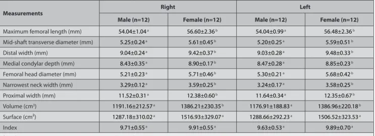
Morphometric evaluation of chinchillas (chinchilla lanigera) femur with different modelling techniques
Tam metin
Şekil


Benzer Belgeler
This study aims to evaluate the effectiveness of the program by comparing its implementation to that suggested by the literature, and, assisted by an assessment of teachers’ and
Bu ülkenin grafit üretimi, dünya grafit üretiminde önemli bir yer tutar.. Ülkedeki grafit yataklarının dağılımı
• HF/6-31G(d) is found as the best calculation level and it is taken into consideration in other calculations • Corrosion protection mechanism is predicted by using MEP maps, MEP
Bulgular: Araştırmamızda diyabetik nöropatili bireyler ile sağlıklı kontrol grubu karşılaştırıldığında MTHFR geni C677T ve A1298C polimorfizmlerinin genotip ve allel
1989 ve 1993 YIllan arasmda, Adalet Bakanhgl Adli TIp Kurumu Adana Grup Ba!jkanhgl'na Cumhuriyet Ba!jsavclhgl, Trafik ~ube Mlidlirlligli, Karakollar ve Iandarma Karakol
miidafaa ve hatta oldiirme amaCl ile kullamlmaktadlrlar. Bu incelemede Adli Tip Kurumu Bursa Grup Ba§kanhgmda otopsileri yapdan kesici-delici alet yaralanmasma bagh
The GLO-I isoenzyme typin g and ABO (H) anti gens detection could be performed on the same thread particular ly when the biological material submitted for
Akut intermitan porfiri ataðý sýrasýnda görülen hipertansiyon bazý vakalarda klinik tablonun tek bulgusu olup, diðer semptomlar daha sonra geliþebilir1-31. Hatta hipertansiyon,
