Summary
Mycoplasma synoviae (M. synoviae) infection is a cause of great economic loss in commercial egg layer hens. The aims of this study were to investigate the prevalence of M. synoviae and to compare the characteristics of M. synoviae infected and free flocks of commercial layers. A total of 400 tracheal swabs and 400 blood serum samples were collected from 20 different layer flocks. Random Amplified Polymorfic DNA (RAPD) analysis was used for the molecular typing of M. synoviae isolates to determine the source of infection. M. synoviae was isolated from 89 tracheal swabs collected from 5 out of 20 flocks. The genetic similarity between field strains ranged from 53% to 100% as determined by RAPD analysis. All M. synoviae strains separated into 2 main-clusters in UPGMA dendogram. These results revealed that the 5 infected flocks were contaminated from 2 different sources. The egg production of positive flocks was statistically lower (P<0.05) than that of the pathogen free flocks. Infection was more frequent in multi-age farms and on sites with several houses. The mortality of infected flocks was higher than uninfected flocks, but this difference was not statistically significant. The mean weight of eggs and the average live weight of hens were similar in free and infected flocks. In conclusion, M. synoviae infection significantly decreased egg production in layer hens and was more frequent in multiage farms.
Keywords: Mycoplasma synoviae, Layer, Epidemiology, RAPD
Ticari Yumurtacı Tavuklarda Mycoplasma synoviae
Enfeksiyonunun Moleküler Epidemiyolojisi
Özet
Mycoplasma synoviae (M. synoviae) yumurtacı tavuk sektöründe büyük ekonomik kayıplara neden olmaktadır. Bu çalışmada, ticari yumurtacı tavuk kümeslerinde M. synoviae’nın prevalansını araştırmak ve enfekte ve sağlıklı kümeslerin özelliklerini karşılaştırmak amaçlanmıştır. Toplam 400 trakeal sıvap ve 400 kan serum örneği 20 farklı kümesten toplandı. Enfeksiyonun kaynağını belirlemek için M. synoviae suşlarının moleküler tiplendirmesi Random Amplified Polymorfic DNA (RAPD) analizi ile yapıldı. Trakeal sıvap örneklerinin 89’undan M. synoviae izole edildi ve bu pozitif sıvap örnekleri 5 farklı kümesten toplanmıştı. Sahadan izole edilen suşlar arasındaki genetik benzerlik oranları RAPD analizi ile %53 ile %100 arasında bulundu. UPGMA dendogramında izolatlar 2 ayrı ana kümede yer aldılar ve bu sonuç gösterdi ki 5 farklı enfekte kümes 2 ayrı kaynaktan kontamine olmuştur. Enfekte kümeslerin yumurta verimi sağlıklı kümeslerden daha düşük (istatistiki olarak anlamlı) olarak bulundu. Enfeksiyon, farklı yaşlardaki tavuk kümeslerinin aynı bahçede olduğu çiftliklerde daha sık tespit edildi. Enfekte kümeslerin mortalite oranları sağlıklı kümeslere oranla daha yüksek (istatistiki olarak anlamsız) bulundu. Ortalama yumurta ağırlığı ve tavukların canlı ağırlığı ise enfekte ve sağlıklı sürülerde benzerdi. Sonuç olarak, M. synoviae enfeksiyonu yumurtacı tavuklarda yumurta verimini azaltmakta ve farklı yaşlardaki tavuk kümeslerinin aynı bahçede olduğu çiftliklerde daha sık görülmektedir.
Anahtar sözcükler: Mycoplasma synoviae, Yumurtacı tavuk, Epidemiyoloji, RAPD
Molecular Epidemiology of Mycoplasma synoviae
Infection in Commercial Layers
Zeki ARAS
1
Zafer SAYIN
21 2
Konya Provincial Directorate of Health, Kazımkarabekir cad, TR-42040 Selçuklu, Konya - TURKEY
Department of Microbiology, Faculty of Veterinary Medicine, Selçuk University, TR-42075 Campus, Konya - TURKEY
Makale Kodu (Article Code): KVFD-2013-9488
Mycoplasma synoviae (M. synoviae) mainly causes chronic subclinical upper respiratory disease, infectious synovitis, and airsacculitis in chickens and turkeys. It has
been acknowledged as the cause of great economic loss in commercial poultry industry due to poor growth and decreased egg production. The infection occurs world-
INTRODUCTION
İletişim (Correspondence)
+90 332 3511832wide in commercial egg layers, broilers, and turkeys. The disease may be transmitted either vertically through the eggs, or horizontally, often by direct contact between contaminated materials and susceptible animals [1].
The diagnosis of M. synoviae infection has traditionally been carried out by serological procedures and isolation of the causative organism [1,2]. The polymerase chain reaction (PCR) is an alternative to bacteriological isolation for diagnosis of avian mycoplasmosis. It is a rapid, highly sensitive, very specific and inexpensive technique to detect M. synoviae DNA [3-7]. M. synoviae PCR assays have been based on the 16S rRNA gene [3,4,6] and VlhA haemagglutinin gene [5,7,8].
Molecular typing of bacteria is an important tool for determining the source of an infection, for recognizing outbreaks, monitoring vaccination programs, and determining the relationships amongst strains isolated from neighboring flocks. Random amplified polymorphic DNA (RAPD) assay was first described in 1990 as a means of genotyping microorganisms [9] and has been employed for the molecular typing of M. synoviae isolates [10,11].
There are few studies that have attempted to characterise M. synoviae infected flocks and it has been reported that M. synoviae infection does not statistically effect egg production [11,12], egg quality [13] or the mortality [11] of the birds. However, there is no information concerning the effect of Turkish strains of M. synoviae on egg production, egg quality or mortality of hens.
The aims of this study were to investigate the prevalence of M. synoviae and to compare the characteristics of M. synoviae infected and free flocks in commercial egg layers. The characteristics determined were egg production, egg weight, live weights and mortality rates of the hens. Additionally, an RAPD based method was used to type the M. synoviae isolates to detect possible routes of contaminations.
MATERIAL and METHODS
Study SampleTracheal swabs and blood serum samples were collected from 20 different commercial egg laying flocks in the Konya region of Turkey during the later six months of 2010. As the farms of the main egg production companies in Turkey are located in the Konya region and contain more than 8 millions laying hens in 2010, it is an important city for poultry sector of Turkey. Twenty tracheal swabs were randomly collected from each flock and were transported in Frey’s medium to the laboratory for M. synoviae isolation. A flock was considered M. synoviae positive if at least one tracheal swab yielded M. synoviae. Additionally, 20 blood serum samples were collected from the same layer flocks for serological investigation. Sera were kept in aliquots
at -20°C until analysed. The study protocol was approved by Selcuk University Veterinary Faculty Ethical Committee (2009/49).
Egg production, egg weight, live weight of hens, mortality rate of flocks, and farm characteristics was investigated by questionnaire forms obtained from the farm veterinarians. Flocks were defined as hens of same age group (55-60 weeks) and same origin. All of the 20 flocks visited in this study belonged to different farms located in different parts of the region.
Culture and Identification
Specimens were cultured using the method described by Frey et al.[14]. All tracheal swabs were placed in 2 ml Frey’s broth for transport and used for mycoplasma culture. All broths were incubated at 37°C under an atmosphere with 10% CO2 and subcultured onto Frey’s agar until a color
change was observed. Isolates were identified according to a M. synoviae specific Multiplex PCR assay [4].
DNA Extraction
M. synoviae sample and reference (M. synoviae WVU 1853) strains were grown in Frey’s broth. DNA was extracted using the protocol provided in Promega Wizard Genomic DNA purification Kit (Cat No: A1120). DNA concentration was determined spectrophotometrically (Eppendorf, Model 6131, Germany) by absorbance readings at 260 and 280 nm. The samples were stored at -20°C until used as templates for amplification.
Identification of M. Synoviae by Multiplex PCR Assay
M. synoviae and M. gallisepticum specific primer pairs were used in PCR, as described by Wang et al.[4]. The sequence of forward primer was 5’- GAA GCA AAT AGT GAT ATC A -3’ and reverse primer was 5’- GTC GTC TCG AAG TTA ACA A -3’ for M. synoviae. The sequence of M. gallisepticum specific forward and reverse primer pairs were 5’-GGA TCC CAT CTC GAC CAG GAG AAA A-3’ and 5’-CTT TCA ATC AGT GAG TAA CTG ATG A-3’, respectively. PCR was performed as previously described [4].
RAPD Genotyping
Three M. synoviae clones per infected flock were analyzed by RAPD analysis. This analysis was performed as described by Geary et al.[15]. The 1254 primer (5’-CCG CAGCCAA-3’) coupled with the 1281 primer (5’-AACGC GCAAC-3’) were used in assay and obtained from IDT Technologies, USA.
Computer Analysis of RAPD Patterns
All fragments were compared on the same gel. Each band was treated on gel and was scored as 1 (when present) or 0 (when absent). These data matrices were recorded to the software program of PubMLST data analysis (http://
pubmlst.org/), and then obtained Unweighted Pair Group Method with Arithmetic Means (UPGMA) dendogram. Additionally, the genetic diversity for the bacteria was calculated according to Nei and Li [16]. Similarity index between pairs of strains were calculated with the following formula: F=2NAB/NA+NB, where NA and NB are the number of fragments in strains A and B, respectively, and NAB is the number of fragments in strains A that match fragments in strain B.
Hunter and Gaston’s Index
The discriminatory power of RAPD assay was evaluated by the Gaston and Hunter’s index D [17] which is calculated according to the following equation:
Where N is the total number of strains, s is the total number of types described, and nj is the number of strains belonging to the j-th type. An index higher than 0.90 indicates that the typing system is discriminates and can be used as epidemiological typing system.
The reproducibility of the PCR products was tested by interassay analysis of five isolates randomly chosen. The same five isolates were tested for five consecutive days during the interassay analysis.
Rapid Serum Agglutination Test (RSAT)
M. synoviae RSAT antigen was provided from Pendik Veterinary Control and Research Institute, Turkey. RSAT was performed according to procedures described by Kleven and Ferguson-Noel [1]. Briefly, an equal volume (30 μl) of the antigen and serum was mixed on a clean glass slide and shaken gently. Formation of agglutination was accepted as a positive result.
Statistical Test
Chi-square test was used to compare the distributions of M. synoviae infected or free flocks according to flock
characteristics. A significance level of 5% was used. The statistical analyses were performed using SPSS software version 12.
RESULTS
M. synoviae was isolated from 89 tracheal swabs which were collected from 5 (25%) out of 20 flocks. Three hundred and eleven of the tracheal swabs from remaning 15 (75%) flocks were bacteriologically negative. A total of 89 M. synoviae strains gave 207 bp M. synoviae specific bands but M. gallisepticum was not detected by Multiplex PCR.
Of the 400 sera examinated in this study, 90 (22.5%) were found to be positive for M. synoviae antibodies by RSAT. These 90 positive sera were collected from the 5 flocks that were bacteriologically positive. The 5 M. synoviae positive flocks were examined for other viral and bacterial diseases, but no disease was detected in these flocks.
The mean size of flocks was 26.375 hens. Three out of 20 flocks were kept in a one-house farm, 6 flocks in two-house farms, 3 flocks in three-two-house farms and 8 flocks in more than 3 house farms. Ten of 20 farms harboured hens of multiple ages. The five infected flocks belonged to five different farms which harboured hens of multiple ages.
The mortalities of M. synoviae positive or negative flocks were compared and while the mortality of the infected flocks was higher (2.80%) than that of the uninfected flocks (2.62%), this difference was not statistically significant.
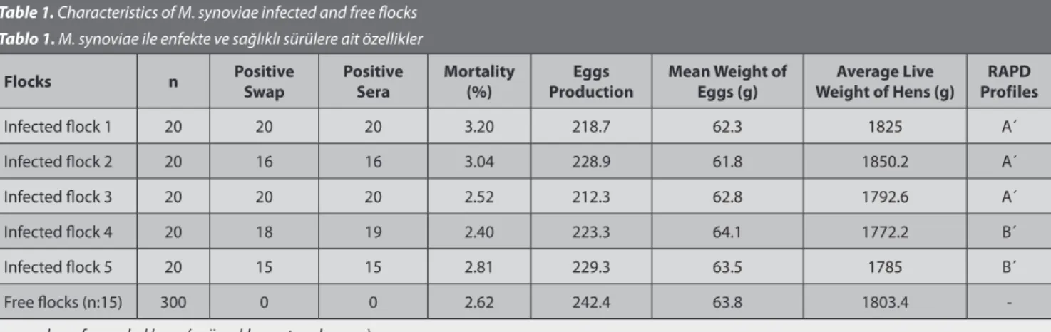
The number of eggs per hen produced by the positive flocks (222.5) was statistically lower than the number produced in free flocks (242.4, P<0.05). The age of the infected flocks was 56-58 weeks. The mean weight of eggs and the average live weight of the hens were similar in the free and infected flocks at 63.8/62.9 g and 1803.4/1805 g, respectively (Table 1). These differences were not statistically significant.
For each positive flock, three different M. synoviae
Table 1. Characteristics of M. synoviae infected and free flocks Tablo 1. M. synoviae ile enfekte ve sağlıklı sürülere ait özellikler
Flocks n Positive Swap Positive Sera Mortality(%) ProductionEggs Mean Weight of Eggs (g) Weight of Hens (g)Average Live ProfilesRAPD
Infected flock 1 20 20 20 3.20 218.7 62.3 1825 A´
Infected flock 2 20 16 16 3.04 228.9 61.8 1850.2 A´
Infected flock 3 20 20 20 2.52 212.3 62.8 1792.6 A´
Infected flock 4 20 18 19 2.40 223.3 64.1 1772.2 B´
Infected flock 5 20 15 15 2.81 229.3 63.5 1785 B´
Free flocks (n:15) 300 0 0 2.62 242.4 63.8 1803.4
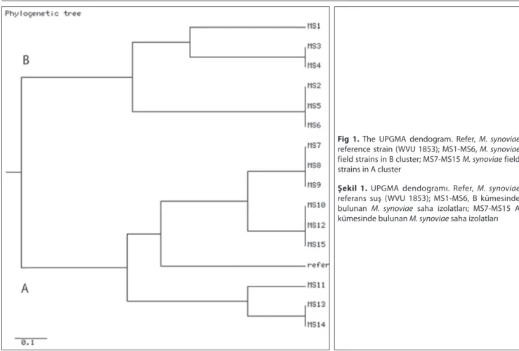
clones could be typed by RAPD and typeability of assay was found 100%. The reproducibility and discriminatory power index (Gaston and Hunter’s index) for RAPD were 100% and 0.90, respectively. Using the RAPD assay 7 different patterns were obtained from the 15 M. synoviae clones. The genetic similarity between strains was calculated to range from 53% to 100%, according to Nei and Li [16]. The UPGMA dendogram is shown in Fig. 1 and all M. synoviae clones were placed in 2 main separate clusters (A and B profiles). The reference strain (M. synoviae WVU 1853) and 9 of the field isolates made up the A´ cluster while the other 6 field clones formed cluster B´. The source of M. synoviae field isolates which were separated in cluster A´ and B´ are shown on Table 1.
DISCUSSION
In the present study M. synoviae and specific antibodies were detected by culture and serological examination, respectively, in 5 (25%) of the 20 flocks examined. However, this is in sharp contrast to the incidence of M. synoviae infection in commercial flocks in Australia, France and Brazil [6,11,13] where the reported infection rates are 69%, 68% and 72.7%, respectively.
Serological tests are widely used to identify infected flocks, but cross-reaction with M. gallisepticum and other nonspecific reactions may occur. Reactors are confirmed as
positive by isolation and identification of the organism [1]. In this study, 90 of 400 serum samples were found to be M. synoviae positive by RSAT, which was also confirmed by culture. This result indicates that RSAT can be used for the diagnosis of subclinical M. synoviae infection. Indeed RSAT is recommended as a screening test for poultry flocks by the Turkish Ministry of Agriculture.
In this study, the characteristics of M. synoviae infected and free flocks of commercial egg layers were compared. The characteristics examined were egg production, egg weight, live weight of the hens and their mortality rate. Few studies have investigated the effect of M. synoviae infection on egg laying and found no significant association between egg production and naturally infection with M. synoviae [11,12,18]. In contrast, we found that the number of eggs per hen was statistically lower in M. synoviae infected flocks. The mean weight of eggs and the mean live weight of hens were found similar in free and infected flocks. These differences were also not statistically significant.
The mortality rate of M. synoviae positive or negative flocks was compared and it was found that although the mortality of the infected flocks was marginally higher than that of the uninfected flocks the difference was not statistically significant. Similar results have been observed by other researchers [11,12]. Mohammed et al.[12] reported that mortality can be as high as 10% following infection
Fig 1. The UPGMA dendogram. Refer, M. synoviae reference strain (WVU 1853); MS1-MS6, M. synoviae field strains in B cluster; MS7-MS15 M. synoviae field strains in A cluster
Şekil 1. UPGMA dendogramı. Refer, M. synoviae referans suş (WVU 1853); MS1-MS6, B kümesinde bulunan M. synoviae saha izolatları; MS7-MS15 A kümesinde bulunan M. synoviae saha izolatları
with M. synoviae. In the present work, the mortality rate of infected flocks was 2.80%.
Hagan et al.[19] reported that M. synoviae infection was more frequent on sites where the hens were kept in several houses. In the present study, all of infected flocks were found in farms those the birds were kept in 5 to 6 house supporting that this may be a risk factor for M. synoviae infection. It has also been reported that infection was more frequent in multiage farms [11]. In our study, all of the infected flocks contained hens with varied widely in age.
The diversity of M. synoviae field strains was investigated by RAPD genotyping method to determine if a there was genetic links between the isolates obtained from the different egg laying flocks. The reproducibility, typeability, and discriminatory power index for the RAPD assay was 100%, 100% and 0.90, respectively. All the M. synoviae strains analysed in this study could be put into 2 separate clusters comprising 8 sub-groups (Fig. 1). The genetic similarity between strains was found to vary from 53% to 100%. Nine (60%) out of 15 M. synoviae clones from 3 different infected flocks belonged to the same cluster (Fig. 1, A´). This group also included the reference strain (M. synoviae WVU 1853). The 6 remaning clones belonged to the other cluster (Fig. 1, B´). These results supported that the 5 infected flocks were contaminated from 2 different sources.
In conclusion, M. synoviae infection significantly decreased egg production in layer hens and it was more frequent in multiage farms and on sites with several houses. RAPD profiles showed that infected flocks were contaminated from 2 different sources. This genotyping method appears valuable tool for epidemiological studies on various M. synoviae isolates.
REFERENCES
1. Kleven SH, Ferguson-Noel N: Mycoplasma synoviae infection. In, Saif YM, Fadly AM, Glisson JR, McDougald LR, Nolan LK, Swayne DE (Eds): Diseases of Poultry. 846-851, Blackwell Publishing, 2008.
2. Ewing L, Cookson KC, Phillips RA, Turner KR, Kleven SH: Experimental infection and transmissibility of Mycoplasma synoviae with delayed serological response in chickens. Avian Dis, 42, 230-238, 1998. 3. Lauerman LH, Hoerr FJ, Sharpton AR, Shah SM, Van Santen VL: Development and application of a polymerase chain reaction assay for
Mycoplasma synoviae. Avian Dis, 37, 829-34, 1993.
4. Wang H, Fadl AA, Khan MI: Multiplex PCR for avian pathogenic mycoplasmas. Mol Cell Probes, 11, 211-216, 1997.
5. Ben Abdelmoumen Mardassi B, Ben Mohamed R, Gueriri I, Boughattas S, Mlik B: Duplex PCR to differentiate between Mycoplasma
synoviae and Mycoplasma gallisepticum on the basis of conserved
species-specific sequences of their hemagglutinin genes. J Clin Microbiol, 43, 948-958, 2005.
6. Buim MR, Mettifogo E, Timenetsky J, Kleven SK, Ferreira AJP: Epidemiological survey on Mycoplasma gallisepticum and M. synoviae by multiplex PCR in commercial poultry. Pesq Vet Bras, 29 (7): 552-556, 2009. 7. Hammond PP, Ramırez AS, Morrow CJ, Bradbury JM: Development and evaluation of an improved diagnostic PCR for Mycoplasma synoviae using primers located in the haemagglutinin encoding gene vlhA and its value for strain typing. Vet Microbiol, 136, 61-68, 2009.
8. Jeffery N, Gasser RB, Steer PA, Noormohammadi AH: Classification of Mycoplasma synoviae strains using single-strand conformation polymorphism and high-resolution melting-curve analysis of the vlhA gene single-copy region. Microbiology, 153, 2679-2688, 2007.
9. Olive DM, Bean P: Principles and applications of methods for DNA-based typing of microbial organisms. J Clin Microbiol, June, 1661-1669, 1999.
10. Marois C, Dufour-Gesbert F, Kempf I: Comparison of pulsed-field gel electrophoresis with random amplified polymorphic DNA for typing of Mycoplasma synoviae. Vet Microbiol, 79, 1-9, 2001.
11. Dufour-Gesbert F, Dheilly A, Marois C, Kempf I: Epidemiological study on Mycoplasma synoviae infection in layers. Vet Microbiol, 114, 148-154, 2006.
12. Mohammed HO, Carpenter TE, Yamamoto R: Economic impact of
Mycoplasma gallisepticum and M. synoviae in commercial layer flocks. Avian Dis, 31, 477-482, 1987.
13. Gole VC, Chousalkar KK, Roberts JR: Prevalence of antibodies to
Mycoplasma synoviae in laying hens and possible effects on egg shell
quality. Prev Vet Med, 106 (1): 75-78, 2012.
14. Frey ML, Hanson RP, Anderson DP: A medium for the isolation of avian mycoplasmas. Am J Vet Res, 29, 2163-2171, 1968.
15. Geary SJ, Forsyth MH, Aboul Saoud S,Wang G, Berg DE, Berg CM:
Mycoplasma gallisepticum strain differentiation by arbitrary primer PCR
(RAPD) fingerprinting. Mol Cell Probes, 8, 311-316, 1994.
16. Nei M, Li WH: Mathematical modeling for studying genetic variation in terms of restriction endonucleases. Proc Natl Acd Sci, 76, 5269-5273, 1979. 17. Hunter P, Gaston M: Numerical index of the discriminatory ability of typing systems: An application of Simpson’s index of diversity. J Clin
Microbiol, 27, 2156-2160, 1988.
18. Branton SL, Lott BD, May JD, Maslin WR, Pharr GT, Brown JE, Boykin DL: The effects of F strain Mycoplasma gallisepticum, Mycoplasma
synoviae, and the dual infection in commercial layer hens over a 44-
week laying cycle when challenged before beginning of lay. II. Egg size distribution. Avian Dis, 43, 326-330, 1999.
19. Hagan JC, Morgan KL, Bradbury JM: Epidemiology of Mycoplasma
synoviae in layers. In, Congress of the Society for Veterinary Epidemiology