https://doi.org/10.1007/s00296-019-04480-9
OBSERVATIONAL RESEARCH
Enthesitis and its relationship with disease activity, functional status,
and quality of life in psoriatic arthritis: a multi‑center study
Ismihan Sunar
1· Sebnem Ataman
1· Kemal Nas
2· Erkan Kilic
3· Betul Sargin
4· Sevtap Acer Kasman
5·
Hakan Alkan
6· Nilay Sahin
7· Gizem Cengiz
8,9· Nihan Cuzdan
10· Ilknur Albayrak Gezer
11·
Dilek Keskin
12· Cevriye Mülkoğlu
13· Hatice Resorlu
14· Ajda Bal
15· Mehmet Tuncay Duruöz
5·
Okan Küçükakkaş
16· Ozan Volkan Yurdakul
16· Meltem Alkan Melikoglu
17· Yıldıray Aydın
2·
F. Figen Ayhan
13,18· Hatice Bodur
19· Mustafa Calis
8· Erhan Capkın
20· Gul Devrimsel
21· Kevser Gok
22·
Sami Hizmetli
23· Ayhan Kamanlı
2· Yaşar Keskin
16· Hilal Kocabas
24· Oznur Kutluk
25· Nesrin Şen
26·
Omer Faruk Şendur
27· Ibrahim Tekeoğlu
2· Sena Tolu
29· Murat Toprak
28· Tiraje Tuncer
25Received: 22 August 2019 / Accepted: 19 November 2019 / Published online: 26 November 2019 © Springer-Verlag GmbH Germany, part of Springer Nature 2019
Abstract
Psoriatic arthritis (PsA) is an inflammatory arthritis with distinct phenotypic subtypes. Enthesitis is assigned as a hallmark
of the disease, given its significant relations to disease activity and quality of life. Our objective is to evaluate the prevalence
of enthesitis and its association with some clinical parameters, particularly quality of life, using data from a national registry.
Patients with PsA meeting ClASsification criteria for Psoriatic Arthritis (CASPAR) were enrolled by means of a multi-centre
Turkish League Against Rheumatism (TLAR) Network Project. The following information was recorded in web-based case
report forms: demographic, clinical and radiographic data; physical examination findings, including tender and swollen joint
counts (TJC and SJC); nail and skin involvement; Disease Activity Score-28 for Rheumatoid Arthritis with Erythrocyte
Sedimentation Rate (DAS 28-ESR); Bath Ankylosing Spondylitis Disease Activity Index (BASDAI); Maastricht
Ankylos-ing Spondylitis Enthesitis Score (MASES); Psoriasis Area Severity Index (PASI); Bath AnkylosAnkylos-ing Spondylitis Radiology
Index for the spine (BASRI-s); Health Assessment Questionnaire (HAQ); Bath Ankylosing Spondylitis Functional Index
(BASFI); Health Assessment Questionnaire for the spondyloarthropathies (HAQ-s); Psoriatic arthritis quality of Life scale
(PsAQoL); Short Form 36 (SF-36); Hospital Anxiety Depression Scale (HADS); Functional Assessment of Chronic Illness
Therapy-Fatigue (FACIT-F); and Fibromyalgia Rapid Screening Tool (FiRST) scores. The patients were divided into two
groups, namely with and without enthesitis, based on the triple Likert-type physician-reported statement of ‘active enthesitis’,
‘history of enthesitis’ or ‘none’ in the case report forms. Patients with active enthesitis were compared to others in terms of
these clinical parameters. A total of 1130 patients were enrolled in this observational study. Of these patients, 251 (22.2%)
had active enthesitis according to the clinical assessment. TJC, HAQ-s, BASDAI, FiRST and PsAQoL were significantly
higher whereas the SF-36 scores were lower in patients with enthesitis (p < 0.05). Chronic back pain, dactylitis, and
teno-synovitis were more frequent in the enthesopathy group (59.4%/39%, 13.1%/6.5% and 24.7%/3.4%, respectively). Significant
positive correlations between the MASES score and the TJC, HAQ, DAS 28-ESR, BASDAI, FiRST and PsAQoL scores,
Rheumatology
INTERNATIONALThe preliminary results of the article were presented as poster presentation at the GRAPPA 2019 Annual meeting Meeting on July 11–13. The abstracts presented in that event were published in the October issue of the Journal of Psoriasis and Psoriatic Arthritis (JPPA). The full bibliographic information is https ://journ als.sagep ub.com/doi/full/10.1177/24755 30319 87350 2. However, that work is almost different from this submitted version. Because we had divided patients into two categories as ones with active enthesitis and history of enthesitis, and ones without active enthesitis and any history of enthesitis. According to precious advice of senior authors given in that meeting, we have performed another classification in the current manuscript. We assigned patients with active enthesitis as enthesitis group, and ones without active enthesitis and with/without history of enthesitis were the ‘no enthesitis’ group. Therefore, all analysis results have changed completely. This paper exhibits the data of a wide group of Turkish patients with PsA from almost all regions of the country. A total of 1130 patients were divided into two groups as ones with and without enthesitis and compared in terms of some clinical characteristics. This cross-sectional study suggests that enthesitis has relations to disease activity, functional status, and quality of life.
and a negative correlation with the SF-36 score were found. When linear regression analysis was performed, the SF-36
MCS and PCS scores decreased by − 9.740 and − 11.795 units, and the FiRST scores increased by 1.223 units in patients
with enthesitis. Enthesitis is an important involvement of PsA with significant relations to quality of life determined with
PsAQoL and SF-36 scores. Our study found higher frequency of dactylitis and chronic back pain, and worse quality of life
determined with SF-36 and PsAQoL scores in patients with enthesitis.
Keywords
Enthesitis · Psoriatic arthritis · Enthesopathy · Registry · Disease activity · Quality of life
Introductıon
Psoriatic arthritis (PsA) is an inflammatory arthritis that
develops in up to 30% of patients with psoriasis [
1
]. It is
characterized by a broad spectrum of clinical conditions,
including axial skeletal involvement, enthesitis, dactylitis,
uveitis and arthritis. Among those, enthesitis is assigned
to be the hallmark of PsA [
2
,
3
]. Recently, the Group for
Research and Assessment of Psoriasis and Psoriatic
Arthri-tis (GRAPPA) advised that six clinical domains of PsA
should be taken into consideration in the management of
the disease. These domains are enthesitis, peripheral
arthri-tis, dactyliarthri-tis, axial disease, skin disease and nail disease
[
4
]. Moreover, Outcome Measures in Rheumatoid
Arthri-tis Clinical Trials (OMERACT) revised the PsA core set to
include musculoskeletal disease activity (peripheral
arthri-tis, dactyliarthri-tis, enthesitis and axial symptoms), skin disease
activity (skin psoriasis and nail dystrophy), fatigue, patient
global evaluation, physical function, pain, health-related
quality of life (HRQoL) and systemic inflammation. The
non-mandatory items were economic costs, emotional
well-being, participation and structural damage [
5
]. Enthesitis,
which is defined as the inflammation of the junction where
the tendon, ligament or joint capsule inserts into the bone,
may be the primary pathological process underlying
spon-dyloarthritis (SpA)-associated skeletal inflammation [
6
].
Enthesopathy can be a consequence of several clinical
con-ditions including metabolic syndrome, mechanical injuries
and degeneration, and rheumatologic conditions including
SpA, in which enthesitis most commonly occurs at
fibro-cartilaginous attachments [
7
]. Although enthesitis affects
35–50% of patients with PsA, it can be challenging for the
clinician to identify enthesitis in patients with PsA [
2
,
8
,
9
].
Enthesopathy may either be asymptomatic, interpreted as
a mechanical injury or mistaken as central
hypersensitiza-tion [
9
]. In most cases, enthesitis needs to be distinguished
from fibromyalgia. Patients’ genetic and
socio-environ-mental factors may influence the pattern and severity of the
disease. Therefore, correct analysis of data from different
epidemiologies may contribute to improving comprehension
of the disease, identification of its course and prognosis, as
well as facilitating phenotype definition [
10
,
11
]. Real-life
data are of great importance to enhance the clinical
under-standing of physicians.
In the current study, we determine the prevalence of
enthesitis and related clinical factors, particularly
qual-ity of life, in a observational multi-centre cohort of
Turk-ish patients with PsA. According to an analysis of the
PSUMMIT 1 and PSUMMIT 2 trials on patients with PsA,
improvement in enthesitis was reported to be related to
improvements in physical function and HRQoL [
12
].
There-fore, the primary endpoint of this study was to determine
the clinical differences between patients with and without
enthesitis in terms of disease activity and HRQoL. The
sec-ondary end points involved assessing whether these groups
differed as regards to skin and nail changes and dactylitis.
Methods
Patients
Patients with PsA meeting the CASPAR (ClASsification
cri-teria for Psoriatic ARthritis) [
13
] were enrolled by means
of the multicenter Turkish League Against Rheumatism
(TLAR)-Network in 2018. This multi-center, independent
project involved 1130 patients from 25 university or public
hospitals across Turkey. TLAR-Network is a collaboration
platform to conduct scientific studies in rheumatology by
supporting researchers from the proposal of a scientific
pro-ject to all processes from data collection to control of data,
analysis, and creation of publication. The study was
con-ducted in accordance with the Helsinki Declaration. Ethics
committee approval was obtained from the Sakarya
Univer-sity Ethics Committee on 25.01.2018 and with the number
of 42 and all centers obtained written consents from patients.
The inclusion criteria were being over 18 years old, meeting
the CASPAR and accepting to participate in the study [
13
,
14
]. The exclusion criteria were pregnancy, lactation and
coexistent malignancy or other connective tissue diseases
[
15
].
Main outcome variable
Demographic, clinical and radiographic data including age,
gender, body mass index (BMI), smoking status, physical
examination findings such as presence of enthesitis and sites,
dactylitis, chronic back pain, tender and swollen joint counts
(TJC, SJC) over 53 joints including the distal and
proxi-mal interphalangeal joints of the hands,
metacarpophalan-geal and metatarsophalanmetacarpophalan-geal joints, temporomandibular,
manubriosternal, sternoclavicular, wrist, elbow, shoulder,
knee, and ankle joints were analyzed. All assessments were
performed by rheumatologists or physical medicine and
rehabilitation specialists (17 rheumatologists and 20
physi-cal medicine and rehabilitation specialists) taking care of
rheumatic patients routinely in each center. Erythrocyte
sedimentation rate (ESR), Disease Activity Score 28-ESR,
(DAS 28-ESR) [
16
], the Bath Ankylosing Spondylitis
Dis-ease Activity Index (BASDAI) [
17
], the Bath Ankylosing
Spondylitis Functional Index (BASFI) [
18
], the Maastricht
Ankylosing Spondylitis Enthesitis Score (MASES) [
19
],
Health assessment Questionnaire (HAQ) [
20
] and the Health
Assessment Questionnaire for the spondyloarthropathies
(HAQ-s) scores [
21
], nail and skin findings and Psoriasis
Area Severity Index (PASI) [
22
], the Bath Ankylosing
Spon-dylitis Radiology Index-spine (BASRI-s) [
23
], Psoriatic
arthritis Quality of Life scale (PsAQoL) [
24
], Short form 36
(SF-36) [
25
], Hospital Anxiety Depression Scale (HADS)
[
26
], Functional Assessment of Chronic Illness
Therapy-Fatigue (FACIT-F) [
27
], and Fibromyalgia Rapid Screening
Tool (FiRST) [
28
] scores of patients were recorded in
elec-tronic case report forms (CRFs). The MASES was evaluated
on 13 sites (1st costochondral joint left/right, 7th
costochon-dral joint left/right, posterior superior iliac spine left/right,
anterior superior iliac spine left/right, 5th Lumbar spinous
process, and proximal insertion of Achilles tendon left/right)
dichotomously as “no pain, 0 point” or “painful, 1 point” and
summed up to a maximum score of 13 [
19
]. For the
statisti-cal analysis, the patients were divided into two groups as
ones with and without active enthesitis. For this purpose,
clinicians’ triple Likert-type assessment in CRFs marked as
‘active enthesitis’ or ‘enthesitis history’ or ‘no enthesitis’
was used. The main outcome variables were determined as
quality of life determined by PsAQoL and SF-36.
Statistical analysis
Statistical analyses were performed on SPSS v11.5
pack-age program. Categorical variables were summarized using
percentages, and continuous variables were given by mean,
median, interquartile range and standard deviation. X
2test
was used for comparison of the categorical data. Whether
data distributed normally were anayzed with
Kolmogo-rov–Smirnov test and histograms. Statistical comparisons
between subgroups were evaluated using t tests for
con-tinuous variables with normal distribution. When the
dis-tribution of continuous data was not normal,
Mann–Whit-ney U test was used. Correlations between variables were
investigated using Spearman’s coefficient. We performed
linear regression analysis to investigate whether enthesitis
is an independent predictor of disease activity and quality of
life. Confidence intervals were calculated for 95%. p < 0.05
was considered significant.
Results
This multicenter observational study included 1130 patients
with PsA (724 female, 406 male). The mean age of patients
was 46.9 ± 12.2 years. Enthesitis, tenosynovitis, dactylitis,
chronic back pain, fingernail and toenail involvement, and
current skin lesions were positive in 251 (22.2%), 92 (8.1%),
90 (8.0%), 492 (43.5%), 610 (54.0%), 556 (49.2%), and 840
(74.3%) patients at enrolment, respectively. Of the 1130
patients, 577 patients (51.1%) did not have active enthesitis
or history of enthesitis. Of the remaining 553 patients with
active or past enthesitis, 251 (45.4%) had active enthesitis
at the time of enrolment and 302 (54.6%) had enthesitis in
the past.
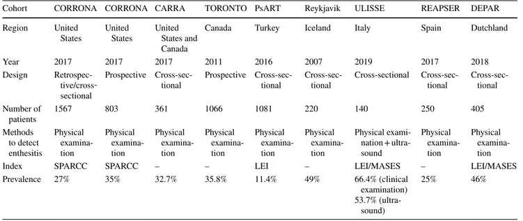
When 251 (22.2%) patients with enthesopathy and 879
patients (77.8%) without enthesopathy were compared,
patients with enthesitis had more frequent tenosynovitis,
dactylitis, and chronic back pain (24.7%/3.4%, 13.1%/6.5%,
59.4%/39.0%, respectively). TJC, HAQ-s, BASDAI, FiRST,
and PsAQoL were significantly higher whereas SF-36 scores
were lower in patients with enthesitis (p < 0.05 for all). The
comparison of clinical parameters of patients with and
with-out enthesitis is given in Table
1
.
Moreover, significant but weak positive correlations
between the MASES score and TJC, HAQ, DAS 28-ESR,
BASDAI, FiRST, and PsAQoL scores, and negative
cor-relations with SF-36 scores were found (p < 0.005 for all).
The correlations between MASES and other indices are
pre-sented in Table
2
.
The most common enthesitis site was Achilles insertion
(39.6%) followed by lumbar 5th spinous process (36.5%) and
1st costochondral sites (27.6%). The distribution of enthesitis
sites according to the MASES is given in Table
3
.
According to the linear regression analysis, BASDAI and
FiRST scores of patients with enthesitis increased by 0.857
and 1.223 units compared to the patients without enthesitis
(R
2= 0.033, F = 31.438, R
2= 0.054, F = 64.214, respectively,
p < 0.001). Also, PsAQoL score of patients with enthesitis
increased by 2.461 units and SF-36 MCS and SF-36 PCS
scores decreased by − 9.740 and − 11.795 units compared
to the patients without enthesitis (R
2= 0.026, F = 30.589,
R
2= 0.035, F = 40.346, R
2= 0.044, F = 51.500, respectively,
Table 1 Comparison of two groups with and without enthesitis regarding physical examination and clinical evaluation
Statistically significant p values are given in bold (p < 0.05)
TJC tender joint count, SJC swollen joint count, FiRST Fibromyalgia Rapid Screening Tool, MASES Maastricht Ankylosing Spondylitis Enthesitis Score, HAQ-s Health Assessment Questionnaire for the spondyloarthropa-thies, PASI Psoriasis Area Severity Index, BASRI-s The Bath Ankylosing Spondylitis Radiology Index, PsAQoL Psoriatic Arthritis Quality of Life, ESR erythrocyte sedimentation rate, BASDAI Bath Ankylosing Spondylitis Disease Activity Index, BASFI Bath Ankylosing Spondylitis Functional Index, SF-36 Short form 36, HADS Hospital Anxiety Depression Scale, FACIT-F Functional Assessment of Chronic Illness Therapy-Fatigue
Clinical parameter Enthesitis positive
n:251 (22.2%) Enthesitis negativen:879 (77.8%) p
BMI (mean ± SD) 29.12 ± 4.72 28.70 ± 5.10 0.226
Duration of PsA (years) (median IQR) 3 (6) 4 (8.5) 0.001
Diagnostic delay (years) (median IQR) 1.2 (4) 1 (4) 0.036
TJC (median IQR) 6 (10) 4 (7) 0.001 SJC (median IQR) 2 (3) 2 (3) 0.585 Active smoker, n (%) 77 (30.7) 214 (24.3) 0.126 Dactylitis, n (%) 33 (13.1) 57 (6.5) 0.001 Tenosynovitis, n (%) 62 (24.7) 30 (3.4) < 0.001 Fingernail involvement, n (%) 141 (56.2) 469 (53.4) 0.429 Fingernail onycholysis 24 (9.6) 100 (11.4 0.417
Fingernail transverse lines 100 (39.8) 386 (43.9)) 0.250
Fingernail hemorrhaige 2 (0.8) 9 (1.0) 1.000
Fingernail hyperkeratosis 20 (8.0) 74 (8.4) 0.820
Fingernail oil-drop discoloration 14 (5.6) 27 (3.1) 0.061
Fingernail pitting 72 (28.7) 187 (21.3) 0.014
Toenail involvement, n (%) 127 (50.6) 429 (48.8) 0.616
Toenail transverse lines 84 (33.5) 278 (31.6) 0.582
Toenail pitting 22 (8.8) 66 (7.5) 0.512
Toenail hyperkeratosis 27 (10.8) 143 (16.3) 0.031
Toenail hemorrhaige 2 (0.8) 5 (0.6) 0.655
Overall skin involvement, n (%) 206 (82.1) 634 (72.1) 0.006
Auricula 63 (25.1) 157 (17.9) 0.011
Scalp 131 (52.2) 403 (45.8) 0.076
Umbilical 55 (21.9) 163 (18.5) 0.233
Gluteal cleft 25 (10) 55 (6.3) 0.044
Extensor area 176 (70.1) 603 (68.6) 0.647
Chronic back pain, n (%) 149 (59.4) 343 (39.0) < 0.001
MASES score (median IQR) 4 (4) 0 (3) < 0.001
FiRST score (median IQR) 4 (3) 2 (4) < 0.001
BASRI-s (median IQR) 2 (4) 2 (4) 0.451
PASI score (median IQR) 2.1 (3.9) 1.5 (3) < 0.001
HAQ score (median IQR) 0.4 (0.65) 0.25 (0.65) < 0.001
HAQ-s score (median IQR) 0.67 (0.83) 0.33 (1) < 0.001
DAS 28-ESR score (mean ± SD) 3.63 ± 1.16 3.33 ± 1.23 0.001
ESR (mm/h) (mean ± SD) 21.23 ± 14.86 20.97 ± 15.57 0.809
PsAQoL score (median IQR) 8 (10) 4 (10) < 0.001
BASFI score (median IQR) 3.4 (3.2) 3.1 (3.0) 0.108
BASDAI score (median IQR) 4.95 (2.9) 3.9 (2.9) < 0.001
FACIT-F (median IQR) 22 (17) 18 (15) < 0.001
SF-36 MCS (median IQR) 46.25 (31.75) 62.63 (37.81) < 0.001
SF-36 PCS (median IQR) 45 (35.75) 61.25 (38.75) < 0.001
HADS depression (median IQR) 7 (6) 6 (6) 0.004
Dıscussıon
We analyzed the prevalence of enthesitis and its association
with clinical factors, particularly HRQoL in patients with
PsA, all of which seem rather compatible with the literature.
We found that approximately half of the patients with PsA
experienced enthesitis either at enrolment or in the past, and
that patients with active entheseal involvement had higher
rates of tenosynovitis, dactylitis and chronic back pain as
well as higher TJC, FiRST and PsAQoL scores and lower
SF-36 scores. Enthesitis may be considered as a sign of
increased disease burden due to its association with several
clinical aspects, and a major determinant of disease activity.
Enthesitis is often not considered as the primary outcome
measure in studies of peripheral SpA and PsA although it is
assumed to be a key pathology for these disorders [
6
]. The
main limitations for reporting enthesitis in daily practice are
absence of overt clinical inflammatory signs such as
objec-tive swelling or increase in acute phase reactants. Enthesitis
is frequently assessed via clinical examination and rarely
radiography as it may be inconclusive [
29
]. Several
enthesi-tis assessment tools including the Leeds Enthesienthesi-tis Index
(LEI), Mander Enthesitis Index (MEI) [
30
],
Spondyloar-thritis Research Consortium of Canada (SPARCC), and the
MASES with some variations in reliability, validity, and
sen-sitivity are commonly used in practice [
30
]. In the present
study, we used the MASES, and an imaging modality was
not employed due to multi-center design.
There are several PsA cohorts worldwide. In Corrona
Psoriatic Arthritis/Spondyloarthritis Registry from the
United States, both cross-sectional and prospective
analy-ses were conducted on enthesitis [
14
,
31
]. In a similar way,
our patients with enthesitis had significantly higher rates of
chronic back pain than patients without enthesitis. In a
pro-spective longitudinal cohort study, conducted between 2008
and 2014, the prevalence of enthesitis was reported to be
35%. Similar to our data, the Achilles tendon was the most
common site of involvement. They reported that enthesitis
was associated with more active disease as determined based
on joint count and the presence of tenosynovitis and
dacty-litis, which is similar to our results.[
8
]. In the multi-centre
cohort of the GRACE Project, 49% of the PsA patients
had enthesitis with a median MASES score of 1.1. [
32
].
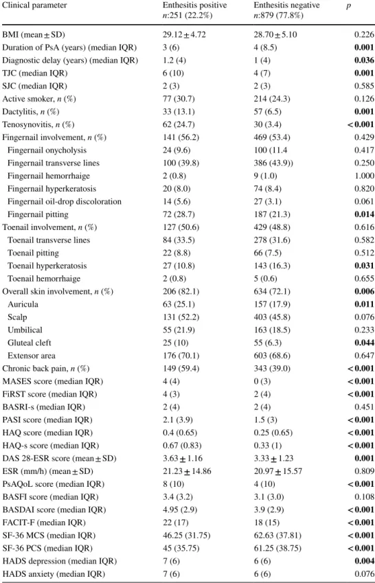
Table
5
presents information on the prevalences of
enthesi-tis in these two studies as well as other studies including
the Toronto PsA cohort study [
10
], Dutch southwest Early
Psoriatic Arthritis cohoRt study (DEPAR) [
33
],
Recent-Onset PsA Registry of the Spanish Society of
Rheumatol-ogy (REAPSER) [
34
], Reykjavik cohort of the Iceland [
35
],
the PsART study from our country [
11
], and juvenile PsA
cohort CARRA [
36
] study. The different rates may
poten-tially be attributed to the patients’ disease onset profile, the
differences in the enthesitis indices used, ethnic differences,
and variations in number of patients included in the studies.
Table 2 Correlations between MASES and other clinical indices
BMI Body mass index, TJC tender joint count, SJC swollen joint count, HAQ-s Health Assessment Questionnaire for the spondyloar-thropathies, FiRST Fibromyalgia Rapid Screening Tool, MASES Maastrich Ankylosing Spondylitis Enthesitis Score, PASI Psoriasis Area Severity Index BASRI-s The Bath Ankylosing Spondylitis Radi-ology Index, PsAQoL Psoriatic Arthritis Quality of Life, DAS 28-ESR Disease Activity Score28, ESR erythrocyte sedimentation rate, BAS-DAI Bath Ankylosing Spondylitis Disease Activity Index, BASFI Bath Ankylosing Spondylitis Functional Index, SF 36 Short form 36, HADS Hospital Anxiety Depression Scale, FACIT-F Functional Assessment of Chronic Illness Therapy-Fatigue
p < 0.05 statistical significance; rs Spearman’s correlation coefficient
MASES 95% CI rs p Lower Upper BMI 0.082 0.013 0.018 0.146 TJC 0.341 < 0.001 0.272 0.406 SJC 0.123 0.018 0.022 0.221 FiRST score 0.334 < 0.001 0.275 0.390 BASRI-s score − 0.039 0.241 − 0.104 0.026 PASI score 0.192 < 0.001 0.129 0.254 HAQ score 0.201 < 0.001 0.138 0.262 HAQ-s score 0.194 < 0.001 0.131 0.255 DAS28-ESR score 0.275 < 0.001 0.212 0.335 PsAQoL score 0.276 < 0.001 0.215 0.335 BASDAI score 0.290 < 0.001 0.224 0.354 BASFI score 0.160 < 0.001 0.081 0.237 ESR (mm/h) 0.030 0.371 − 0.035 0.094 FACIT-F 0.233 < 0.001 0.171 0.293 SF-36 MCS − 0.290 < 0.001 − 0.348 − 0.230 SF-36 PCS − 0.269 < 0.001 − 0.328 − 0.208 HADS anxiety 0.173 < 0.001 0.110 0.235 HADS Depression 0.175 < 0.001 0.112 0.237
Table 3 Distribution of affected entesitis sites in MASES
Enthesitis site n %
1st costo-condral right (n = 692) 188 27.4
1st costo-condral left (n = 685) 191 27.6
7th costo-condral right (n = 651) 132 20.3
7th costo-condral left (n = 654) 136 20.8
Anterior superior iliac spine, right (n = 657) 147 22.4 Anterior superior iliac spine, left (n = 668) 157 23.5 Posterior superior iliac spine, right (n = 636) 138 21.7 Posterior superior iliac spine, left (n = 642) 150 23.4
Right iliac crest (n = 666) 154 23.1
Left iliac crest (n = 659) 159 24.1
Lumbar 5th spinous process (n = 718) 262 36.5
Achilles tendon, right (n = 740) 276 37.3
Higher FiRST scores among patients with enthesitis, the
positive correlation between the FiRST and MASES index
scores and the linear regression analysis results, indicating
that enthesitis is an independent predictor for an increase
in the FiRST scores, are other striking results of our study.
Although FiRST is considered as a screening tool rather than
as a diagnostic tool, it may provide with substantial data
regarding widespread pain [
28
]. It is well known that central
sensitisation syndromes are more frequent in patients with
several types of rheumatic diseases, including SpA, PsA,
and RA and it is detected in 10–40% of all cases [
37
].
There-fore, the importance of distinguishing between fibromyalgia,
which is the prototype of central sensitisation syndromes
[
38
], and polyenthesitis in SpA patients has been addressed
in prior studies [
9
]. In a study to identify the clinical features
used in the differential diagnosis of PsA and fibromyalgia,
Table 4 Linear regression analysis of enthesitis and clinical parameters df Estimate (B) 95% CI p value Lower Upper BASDAI Constant 4.144 3.993 4.295 < 0.0001 Presence of enthesititis 1 0.857 0.557 1.156 < 0.0001 PsAQoL Constant 6.292 5.881 6.704 < 0.0001 Presence of enthesititis 1 2.461 1.588 3.334 < 0.0001 HAQ score Constant 0.400 0.370 0.431 < 0.0001 Presence of enthesititis 1 0.115 0.050 0.180 < 0.0001 HAQ-s Constant 0.614 0.570 0.658 < 0.0001 Presence of enthesititis 1 0.192 0.098 0.285 < 0.0001 SF-36 MCS Constant 57.714 56.306 59.143 < 0.0001 Presence of enthesititis 1 − 9.740 − 12.749 − 6.732 < 0.0001 SF-36 PCS Constant 59.045 57.524 60.565 < 0.0001 Presence of enthesititis 1 − 11.795 − 15.019 − 8.570 < 0.0001 FiRST score Constant 2.189 2.048 2.330 < 0.0001 Presence of enthesititis 1 1.223 0.923 1.522 < 0.0001
Table 5 Comparison of the PsA cohorts/studies evaluating enthesitis
Cohort CORRONA CORRONA CARRA TORONTO PsART Reykjavik ULISSE REAPSER DEPAR
Region United
States United States United States and Canada
Canada Turkey Iceland Italy Spain Dutchland
Year 2017 2017 2017 2011 2016 2007 2019 2017 2018
Design Retrospec-tive/cross‐ sectional
Prospective
Cross-sec-tional Prospective Cross-sec-tional Cross-sec-tional Cross-sectional Cross-sec-tional Cross-sec-tional Number of patients 1567 803 361 1066 1081 220 140 250 405 Methods to detect enthesitis Physical examina-tion Physical examina-tion Physical examina-tion Physical examina-tion Physical examina-tion Physical examina-tion Physical exami-nation + ultra-sound Physical examina-tion Physical examina-tion
Index SPARCC SPARCC – – LEI – LEI/MASES – LEI/MASES
Prevalence 27% 35% 32.7% 35.8% 11.4% 49% 66.4% (clinical
examination) 53.7%
(ultra-sound)
the presence of ≥ 6 fibromyalgia-associated symptoms
and ≥ 8 tender points were reported to be the best
predic-tors of fibromyalgia [
39
]. However, many of the entheseal
points used in the enthesitis assessment tools are near the
joints and tender points for fibromyalgia, contributing to the
possibility of misclassification. Ultrasound (US) is alleged
to be more sensitive than clinical examination in enthesitis
[
40
]. However, it is challenging for clinicians to incorporate
US into their daily practice in PsA due to the time needed
to examine multiple enthesopathy sites [
41
]. Fibromyalgia
should be considered when dealing with PsA patients with
higher enthesopathy scores upon evaluation. Unfortunately,
our study did not involve questioning the patients about
somatic symptoms or examining their tender points to
dif-ferentiate fibromyalgia based on the available classification
criteria [
42
,
43
].
Another hallmark of PsA, dactylitis with specificity
approaching 95% in SpA [
44
] was present in 8.0% of our
patients. In an international multi-center psoriasis and PsA
trial, the frequency of active dactylitis was found to be
3.4–12.8% [
45
]. In our study, dactylitis was also found to
be more frequent in the enthesopathy subgroup. Although
accepted evidence suggests that dactylitis is primarily related
to flexor tenosynovitis [
46
], a recent study using
high-res-olution MRI to explore dactylitis in PsA demonstrated that
enthesitis was common in PsA dactylitis. Furthermore, the
authors concluded that ’digital polyenthesitis’, related to the
flexor tendon pulleys and fibrous sheaths, provides a
pos-sible explanation for its association with flexor
tenosyno-vitis [
47
]. Our results regarding significantly higher rates
of dactylitis and tenosynovitis in patients with enthesitis
are in accordance with that study’s findings. An association
between extensor tendon enthesopathy, distal
interphalan-geal (DIP) joint involvement, and nail pathology has been
widely acknowledged [
48
]. Some authors proposed the term
‘nail-enthesitis theory’ to refer to the increased prevalence of
extensor tendon enthesitis in digits with involved nails [
49
].
In line with these data, our patients with enthesitis had more
frequent fingernail pitting.
Among patients with PsA, arthritis is known to impair
HRQoL. However, the association between HRQoL and
enthesitis was not thoroughly evaluated. In a recent study
on DEPAR PsA cohort, SF-36 was used to assess HRQoL,
and enthesitis was evaluated using LEI and/or MASES
scores. Given that higher SF-36 scores represent a better
state of HRQoL, based on clinical examination, patients with
enthesitis were reported to have significantly lower scores
on all the SF-36 domains than patients without enthesitis
[
33
]. Furthermore, in our study, the patients with enthesitis
had higher PsAQoL and lower SF-36 scores. Although the
association between enthesitis and HRQoL has been poorly
investigated in PsA, some studies were performed in SpA.
In a study on 1505 Brazilian patients with SpA, of whom
18.4% had PsA, it was reported that 53.8% of the patients
with PsA had enthesitis, and the SpA patients with enthesitis
at clinical examination had a lower HRQoL [
50
]. The higher
PsAQoL and lower SF-36 scores we observed in patients
with enthesopathy are in correspondence with previous
stud-ies [
12
].
One of the strengths of our study is that it reports on
data of a wide group of patients with PsA from almost all
regions of Turkey. The population is highly representative
and selection bias is unlikely since the patients were enrolled
consecutively. However, our study has some limitations as
well. It did not use other disease activity indices
involv-ing laboratory data such as ASDAS (Ankylosinvolv-ing
Spondyli-tis Disease Activity Index) [
51
] instead of the BASDAI to
determine the severity of axial disease. DAPSA (Disease
Activity Index for Psoriatic Arthritis) [
52
] would be more
appropriate than DAS 28 for peripheral arthritis because it
counts more joints prone to psoriatic involvement. One of
the major limitations of the study is that it used the MASES
to assess enthesitis. The MASES does not score some of the
main enthesitis sites in PsA, such as plantar fascia insertions
into the calcaneum, medial femoral condyles, and lateral
epi-condyles of the humerus. The LEI has been used in several
PsA trials and it was developed and validated specifically
for PsA. Therefore, if LEI, MEI or SPARCC had been used
in our study, more reliable results could have been obtained.
Another limitation is that our description of enthesitis solely
depends on clinical symptoms and signs; it is not based on
objective evidence exhibited by imaging because enthesitis
lacks apparent inflammatory characteristics, such as swelling
and erythema. It was already emphasized that it is not good
to be too reliant on clinical examination of enthesitis as a
marker of underlying disease, except for the Achilles tendon
insertion [
53
]. The relatively low prevalence of enthesitis
determined in our study may have been due to this
limita-tion. Another limitation is the missing data, which is not a
challenge for HAQ, HAQ-s, FiRST, PASI, PsAQoL,
BASRI-s, and ESR. However, missing data may be a concern for the
BASDAI, MASES, and BASFI scores around 20%,
prob-ably due to not having been performed on patients that only
have peripheral involvement. Finally, this study presents an
observational analysis of the relationship between
enthesi-tis and disease activity, function, and HRQoL. It did not
analyze the changes in these parameters over time or after
treatments. In the light of our findings, as announced by the
GRAPPA and the OMERACT, we consider that enthesitis
should be incorporated to daily practice as well as evaluation
for arthritis, spondylitis, and skin. For this purpose,
com-posite indices such as Comcom-posite Psoriatic Disease Activity
Index (CPDAI) or modified CPDAI (mCPDAI) [
54
], and
The Psoriatic Arthritis Disease Activity Score (PASDAS)
[
55
] which involve assessment for enthesitis should be
used in determination of overall psoriatic disease activity.
Furthermore, patients with PsA and polyenthesitis should
also be overviewed to exclude coexisting fibromyalgia and
chronic widespread pain conditions.
Conclusion
Enthesitis is among the most important clinical PsA
phe-notypes displaying significant associations with HRQoL,
shown by lower PsAQoL and SF-36 scores in this study.
All patients with PsA, particularly those with dactylitis,
chronic back pain, and tenosynovitis should be examined
for enthesitis. Fibromyalgia should be distinguished from
polyenthesitis in patients with PsA.
Acknowledgements We acknowledge dr Nazmiye Kurşun for
statis-tical consulting and Scribendi Editing Services for external editing.
Author contributions All authors have substantial contributions to the conception or design of the work, drafting or revising it critically for important intellectual content, have approved the final version to be published, and in agreement to be accountable for all aspects of the work in ensuring that questions related to the accuracy or integrity of any part of the work are appropriately investigated and resolved. All co-authors are fully responsible for all aspects of the study and the final manuscript in line with the IJME 4 criteria.
Compliance with ethical standards
Conflict of interest Authors declare no conflicts of interest or financial
support.
Ethical approval Ethics committee approval was obtained from the
Sakarya University Ethics Committee on 25.01.2018 and with the num-ber of 42 and conducted in accordance with the Helsinki Declaration.
References
1. Zachariae H (2003) Prevalence of joint disease in patients with psoriasis: implications for therapy. Am J Clin Derma-tol 4(7):441–447. https ://doi.org/10.2165/00128 071-20030 4070-00001
2. Kaeley GS, Eder L, Aydin SZ, Gutierrez M, Bakewell C (2018) Enthesitis: a hallmark of psoriatic arthritis. Semin Arthri-tis Rheum 48(1):35–43. https ://doi.org/10.1016/j.semar thrit .2017.12.008
3. Ritchlin CT, Colbert RA, Gladman DD (2017) Psoriatic arthri-tis. N Engl J Med 376(10):957–970. https ://doi.org/10.1056/ NEJMr a1505 557
4. Coates LC, Kavanaugh A, Mease PJ, Soriano ER, Laura Acosta-Felquer M, Armstrong AW, Bautista-Molano W, Boehncke W-H, Campbell W, Cauli A, Espinoza LR, FitzGerald O, Gladman DD, Gottlieb A, Helliwell PS, Husni ME, Love TJ, Lubrano E, McHugh N, Nash P, Ogdie A, Orbai A-M, Parkin-son A, O’Sullivan D, Rosen CF, Schwartzman S, Siegel EL, Toloza S, Tuong W, Ritchlin CT (2016) Group for research and assessment of psoriasis and psoriatic arthritis 2015 treatment
recommendations for psoriatic arthritis. Arthritis Rheumatol 68(5):1060–1071. https ://doi.org/10.1002/art.39573
5. Orbai AM, de Wit M, Mease PJ, Callis Duffin K, Elmamoun M, Tillett W, Campbell W, FitzGerald O, Gladman DD, Goel N, Gossec L, Hoejgaard P, Leung YY, Lindsay C, Strand V, van der Heijde DM, Shea B, Christensen R, Coates L, Eder L, McHugh N, Kalyoncu U, Steinkoenig I, Ogdie A (2017) Updating the psoriatic arthritis (PsA) core domain set: a report from the PsA workshop at OMERACT 2016. J Rheumatol 44(10):1522–1528.
https ://doi.org/10.3899/jrheu m.16090 4
6. Watad A, Cuthbert RJ, Amital H, McGonagle D (2018) Enthesitis: much more than focal insertion point inflammation. Curr Rheumatol Rep 20(7):41. https ://doi.org/10.1007/s1192 6-018-0751-3
7. Sudoł-Szopińska I, Kwiatkowska B, Prochorec-Sobieszek M, Maśliński W (2015) Enthesopathies and enthesitis. Part 1. Etiopathogenesis. J Ultrasonogr 15(60):72–84. https ://doi. org/10.15557 /JoU.2015.0006
8. Polachek A, Li S, Chandran V, Gladman DD (2017) Clinical enthesitis in a prospective longitudinal psoriatic arthritis cohort: incidence, prevalence, characteristics, and outcome. Arthritis Care Res 69(11):1685–1691. https ://doi.org/10.1002/acr.23174
9. Sakkas LI, Alexiou I, Simopoulou T, Vlychou M (2013) Enthesi-tis in psoriatic arthriEnthesi-tis. Semin ArthriEnthesi-tis Rheum 43(3):325–334.
https ://doi.org/10.1016/j.semar thrit .2013.04.005
10. Gladman DD, Chandran V (2010) Observational cohort studies: lessons learnt from the University of Toronto Psoriatic Arthritis Program. Rheumatology 50(1):25–31. https ://doi.org/10.1093/ rheum atolo gy/keq26 2
11. Kalyoncu U, Bayindir Ö, Ferhat Öksüz M, Doğru A, Kimyon G, Tarhan EF, Erden A, Yavuz Ş, Can M, Çetin GY, Kılıç L, Küçükşahin O, Omma A, Ozisler C, Solmaz D, Bozkirli EDE, Akyol L, Pehlevan SM, Gunal EK, Arslan F, Yılmazer B, Ata-kan N, Aydın SZ, Psoriatic Arthritis Registry of Turkey Study Group (2016) The Psoriatic Arthritis Registry of Turkey: results of a multicentre registry on 1081 patients. Rheumatology 56(2):279–286. https ://doi.org/10.1093/rheum atolo gy/kew37 5
12. McInnes IB, Puig L, Gottlieb AB, Ritchlin CT, Song M, You Y, Kafka S, Morgan GJ, Rahman P, Kavanaugh A (2019) Association between enthesitis and health-related quality of life in psoriatic arthritis in biologic-naive patients from 2 phase III ustekinumab trials. J Rheumatol. https ://doi.org/10.3899/jrheu m.18079 2
13. Taylor W, Gladman D, Helliwell P, Marchesoni A, Mease P, Mielants H (2006) Classification criteria for psoriatic arthritis: development of new criteria from a large international study. Arthritis Rheum 54(8):2665–2673. https ://doi.org/10.1002/ art.21972
14. Mease PJ, Karki C, Palmer JB, Etzel CJ, Kavanaugh A, Ritchlin CT, Malley W, Herrera V, Tran M, Greenberg JD (2017) Clinical characteristics, disease activity, and patient-reported outcomes in psoriatic arthritis patients with dactylitis or enthesitis: results from the Corrona Psoriatic Arthritis/Spondyloarthritis Registry. Arthri-tis Care Res 69(11):1692–1699. https ://doi.org/10.1002/acr.23249
15. TRASD-NETWORK Scientific Studies Collaboration Platform.
https ://trasd -netwo rk.org/proje ct_detai l.php?id=2. Accessed 01 Oct 2019
16. Prevoo ML, van ’t Hof MA, Kuper HH, van Leeuwen MA, van de Putte LB, van Riel PL (1995) Modified disease activity scores that include twenty-eight-joint counts. Development and validation in a prospective longitudinal study of patients with rheumatoid arthritis. Arthritis Rheum 38(1):44–48
17. Garrett S, Jenkinson T, Kennedy LG, Whitelock H, Gaisford P, Calin A (1994) A new approach to defining disease status in ankylosing spondylitis: the Bath Ankylosing Spondylitis Disease Activity Index. J Rheumatol 21(12):2286–2291
18. Calin A, Garrett S, Whitelock H, Kennedy LG, O’Hea J, Mal-lorie P, Jenkinson T (1994) A new approach to defining func-tional ability in ankylosing spondylitis: the development of the Bath Ankylosing Spondylitis Functional Index. J Rheumatol 21(12):2281–2285
19. Heuft-Dorenbosch L, Spoorenberg A, van Tubergen A, Landewé R, van ver Tempel H, Mielants H, Dougados M, van der Heijde D (2003) Assessment of enthesitis in ankylosing spondylitis. Ann Rheum Dis 62(2):127–132. https ://doi.org/10.1136/ard.62.2.127
20. Fries JF, Spitz PW, Young DY (1982) The dimensions of health outcomes: the health assessment questionnaire, disability and pain scales. J Rheumatol 9(5):789–793
21. Daltroy LH, Larson MG, Roberts NW, Liang MH (1990) A modi-fication of the Health Assessment Questionnaire for the spondy-loarthropathies. J Rheumatol 17(7):946–950
22. Fredriksson T, Pettersson U (1978) Severe psoriasis—oral therapy with a new retinoid. Dermatology 157(4):238–244. https ://doi. org/10.1159/00025 0839
23. Lubrano E, Marchesoni A, Olivieri I, D’Angelo S, Spadaro A, Par-sons WJ, Cauli A, Salvarani C, Mathieu A, Zaccara E, Ferrara N, Helliwell PS (2009) The radiological assessment of axial involve-ment in psoriatic arthritis: a validation study of the BASRI total and the modified SASSS scoring methods. Clin Exp Rheumatol 27(6):977–980
24. McKenna SP, Doward LC, Whalley D, Tennant A, Emery P, Veale DJ (2004) Development of the PsAQoL: a quality of life instru-ment specific to psoriatic arthritis. Ann Rheum Dis 63(2):162– 169. https ://doi.org/10.1136/ard.2003.00629 6
25. Ware JE Jr., Sherbourne CD (1992) The MOS 36-item short-form health survey (SF-36). I. Conceptual framework and item selec-tion. Med Care 30(6):473–483
26. Zigmond AS, Snaith RP (1983) The hospital anxiety and depres-sion scale. Acta Ppsychiatrica Scandinavica 67(6):361–370 27. Webster K, Cella D, Yost K (2003) The functional assessment of
chronic illness therapy (FACIT) measurement system: properties, applications, and interpretation. Health Qual Life Outcomes 1:79.
https ://doi.org/10.1186/1477-7525-1-79
28. Perrot S, Bouhassira D, Fermanian J (2010) Development and validation of the fibromyalgia rapid screening tool (FiRST). Pain 150(2):250–256. https ://doi.org/10.1016/j.pain.2010.03.034
29. Weckbach S, Schewe S, Michaely HJ, Steffinger D, Reiser MF, Glaser C (2011) Whole-body MR imaging in psoriatic arthritis: additional value for therapeutic decision making. Eur J Radiol 77(1):149–155. https ://doi.org/10.1016/j.ejrad .2009.06.020
30. Mease PJ (2011) Measures of psoriatic arthritis: Tender and Swol-len Joint Assessment, Psoriasis Area and Severity Index (PASI), Nail Psoriasis Severity Index (NAPSI), Modified Nail Psoriasis Severity Index (mNAPSI), Mander/Newcastle Enthesitis Index (MEI), Leeds Enthesitis Index (LEI), Spondyloarthritis Research Consortium of Canada (SPARCC), Maastricht Ankylosing Spon-dylitis Enthesis Score (MASES), Leeds Dactylitis Index (LDI), Patient Global for Psoriatic Arthritis, Dermatology Life Qual-ity Index (DLQI), Psoriatic Arthritis QualQual-ity of Life (PsAQOL), Functional Assessment of Chronic Illness Therapy-Fatigue (FACIT-F), Psoriatic Arthritis Response Criteria (PsARC), Pso-riatic Arthritis Joint Activity Index (PsAJAI), Disease Activity in Psoriatic Arthritis (DAPSA), and Composite Psoriatic Disease Activity Index (CPDAI). Arthritis Care Res (Hoboken) 63(Suppl 11):S64–85. https ://doi.org/10.1002/acr.20577
31. Mease PJ, Palmer JB, Liu M, Kavanaugh A, Pandurengan R, Ritchlin CT, Karki C, Greenberg JD (2018) Influence of axial involvement on clinical characteristics of psoriatic arthritis: analy-sis from the corrona psoriatic arthritis/spondyloarthritis registry. J Rheumatol 45(10):1389–1396. https ://doi.org/10.3899/jrheu m.17109 4
32. Helliwell PS, FitzGerald O, Fransen J, Gladman DD, Kreuger GG, Callis-Duffin K, McHugh N, Mease PJ, Strand V, Waxman R, Azevedo VF, Beltran Ostos A, Carneiro S, Cauli A, Espinoza LR, Flynn JA, Hassan N, Healy P, Kerzberg EM, Lee YJ, Lubrano E, Marchesoni A, Marzo-Ortega H, Porru G, Moreta EG, Nash P, Raffayova H, Ranza R, Raychaudhuri SP, Roussou E, Scarpa R, Song YW, Soriano ER, Tak PP, Ujfalussy I, de Vlam K, Walsh JA (2013) The development of candidate composite disease activity and responder indices for psoriatic arthritis (GRACE project). Ann Rheum Dis 72(6):986–991. https ://doi.org/10.1136/annrh eumdi s-2012-20134 1
33. Wervers K, Luime JJ, Tchetverikov I, Gerards AH, Kok MR, Appels CWY, van der Graaff WL, van Groenendael JHLM, Korswagen L-A, Veris-van Dieren JJ, Hazes JMW, Vis M (2018) Influence of disease manifestations on health-related quality of life in early psoriatic arthritis. J Rheumatol 45(11):1526–1531.
https ://doi.org/10.3899/jrheu m.17140 6
34. Rubén Queiro AL, Montilla-Morales CA, Galindez-Agirregoikoa E, Bethencourt JJ, Seoane D (2017) Spondyloarthropathies and psoriatic arthritis—clinical aspects and treatment poster III: out-comes, outcome measures, and comorbidities. In: Paper presented at the 2017 ACR/ARHP annual meeting, September 18, 2017 35. Love TJ, Gudbjornsson B, Gudjonsson JE, Valdimarsson H (2007)
Psoriatic arthritis in Reykjavik, Iceland: prevalence, demograph-ics, and disease course. J Rheumatol 34(10):2082–2088 36. Zisman D, Gladman DD, Stoll ML, Strand V, Lavi I, Hsu JJ,
Mellins ED (2017) The juvenile psoriatic arthritis cohort in the CARRA Registry: clinical characteristics, classification, and out-comes. J Rheumatol. https ://doi.org/10.3899/jrheu m.16071 7
37. Goldenberg DL Overview of chronic widespread (centralized) pain in the rheumatic diseases. https ://www.uptod ate.com/conte nts/overv iew-of-chron ic-wides pread -centr alize d-pain-in-the-rheum atic-disea ses/print . Accessed 05 Mar 2019
38. Boomershine CS (2015) Fibromyalgia: the prototypical central sensitivity syndrome. Curr Rheumatol Rev 11(2):131–145 39. Marchesoni A, Atzeni F, Spadaro A, Lubrano E, Provenzano G,
Caulı A, Olivieri I, Melchıorre D, Salvaranı C, Scarpa R, Sarzı-Puttını P, Montepaone M, Porru G, D’angelo S, Catanoso M, Costa L, Manara M, Varısco V, Rotunno L, de Lucıa O, de Marco G (2012) Identification of the clinical features distinguishing pso-riatic arthritis and fibromyalgia. J Rheumatol 39(4):849–855. https ://doi.org/10.3899/jrheu m.11089 3
40. Balint PV, Kane D, Wilson H, McInnes IB, Sturrock RD (2002) Ultrasonography of entheseal insertions in the lower limb in spondyloarthropathy. Ann Rheum Dis 61(10):905–910. https :// doi.org/10.1136/ard.61.10.905
41. Kristensen S, Christensen JH, Schmidt EB, Olesen JL, Johansen MB, Arvesen KB, Schlemmer A (2016) Assessment of enthesitis in patients with psoriatic arthritis using clinical examination and ultrasound. Muscles Ligaments Tendons J 6(2):241–247. https :// doi.org/10.11138 /mltj/2016.6.2.241
42. Wolfe F, Clauw DJ, Fitzcharles MA, Goldenberg DL, Hauser W, Katz RL, Mease PJ, Russell AS, Russell IJ, Walitt B (2016) 2016 Revisions to the 2010/2011 fibromyalgia diagnostic criteria. Semin Arthritis Rheum 46(3):319–329. https ://doi.org/10.1016/j. semar thrit .2016.08.012
43. Wolfe F, Smythe HA, Yunus MB, Bennett RM, Bombardier C, Goldenberg DL, Tugwell P, Campbell SM, Abeles M, Clark P et al (1990) The American College of Rheumatology 1990 criteria for the classification of fibromyalgia. Report of the multicenter criteria committee. Arthritis Rheum 33(2):160–172. https ://doi. org/10.1002/art.17803 30203
44. Dougados M, Linden SVD, Juhlin R, Huitfeldt B, Amor B, Calin A, Cats A, Dijkmans B, Olivieri I, Pasero G, Veys E, Zeidler H (1991) The European spondylarthropathy study group preliminary
criteria for the classification of spondylarthropathy. Arthritis Rheum 34(10):1218–1227. https ://doi.org/10.1002/art.17803 41003
45. Chandran V, Gottlieb A, Cook RJ, Duffin KC, Garg A, Helliwell P, Kavanaugh A, Krueger GG, Langley RG, Lynde C, McHugh N, Mease P, Olivieri I, Rahman P, Rosen CF, Salvarani C, Thaci D, Toloza SMA, Wong MYW, Zhou QM, Gladman DD (2009) International multicenter psoriasis and psoriatic arthritis reliabil-ity trial for the assessment of skin, joints, nails, and dactylitis. Arthritis Care Res 61(9):1235–1242. https ://doi.org/10.1002/ art.24562
46. Olivieri I, Barozzi L, Favaro L, Pierro A, de Matteis M, Borghi C, Padula A, Ferri S, Pavlica P (1996) Dactylitis in patients with seronegative spondylarthropathy. Assessment by ultrasonography and magnetic resonance imaging. Arthritis Rheum 39(9):1524– 1528. https ://doi.org/10.1002/art.17803 90912
47. Tan AL, Fukuba E, Halliday NA, Tanner SF, Emery P, McG-onagle D (2015) High-resolution MRI assessment of dactylitis in psoriatic arthritis shows flexor tendon pulley and sheath-related enthesitis. Ann Rheum Dis 74(1):185–189. https ://doi. org/10.1136/annrh eumdi s-2014-20583 9
48. Zabotti A, Idolazzi L, Batticciotto A, De Lucia O, Scire CA, Tinazzi I, Iagnocco A (2017) Enthesitis of the hands in psori-atic arthritis: an ultrasonographic perspective. Med Ultrasonogr 19(4):438–443. https ://doi.org/10.11152 /mu-1172
49. Acosta-Felquer ML, Ruta S, Rosa J, Marin J, Ferreyra-Garrot L, Galimberti ML, Galimberti R, Garcia-Monaco R, Soriano ER (2017) Ultrasound entheseal abnormalities at the distal inter-phalangeal joints and clinical nail involvement in patients with psoriasis and psoriatic arthritis, supporting the nail-enthesitis theory. Semin Arthritis Rheum 47(3):338–342. https ://doi. org/10.1016/j.semar thrit .2017.05.002
50. Carneiro S, Bortoluzzo A, Goncalves C, Silva JA, Ximenes AC, Bertolo M, Ribeiro SL, Keiserman M, Skare T, Menin R, Azevedo
V, Vieira W, Albuquerque E, Bianchi W, Bonfiglioli R, Campan-holo C, Carvalho HM, Costa I, Duarte A, Kohem C, Leite N, Lima SA, Meirelles ES, Pereira IA, Pinheiro MM, Polito E, Resende GG, Rocha FA, Santiago MB, Sauma Mde F, Valim V, Sampaio-Barros PD (2013) Effect of enthesitis on 1505 Brazilian patients with spondyloarthritis. J Rheumatol 40(10):1719–1725. https :// doi.org/10.3899/jrheu m.12114 5
51. Lukas C, Landewé R, Sieper J, Dougados M, Davis J, Braun J, van der Linden S, van der Heijde D (2009) Development of an ASAS-endorsed disease activity score (ASDAS) in patients with ankylosing spondylitis. Ann Rheum Dis 68(1):18–24. https ://doi. org/10.1136/ard.2008.09487 0
52. Schoels MM, Aletaha D, Alasti F, Smolen JS (2016) Disease activity in psoriatic arthritis (PsA): defining remission and treat-ment success using the DAPSA score. Ann Rheum Dis 75(5):811– 818. https ://doi.org/10.1136/annrh eumdi s-2015-20750 7
53. Helliwell PS (2019) Assessment of enthesitis in psoriatic arthri-tis. J Rheumatol 46(8):869–870. https ://doi.org/10.3899/jrheu m.18138 0
54. Mumtaz A, Gallagher P, Kirby B, Waxman R, Coates LC, Veale JD, Helliwell P, FitzGerald O (2011) Development of a preliminary composite disease activity index in psoriatic arthri-tis. Ann Rheum Dis 70(2):272–277. https ://doi.org/10.1136/ ard.2010.12937 9
55. Coates LC, FitzGerald O, Mease PJ, Gladman DD, Strand V, Goel N, Campbell I, Krueger G, McHugh NJ, Helliwell PS (2014) Development of a disease activity and responder index for psori-atic arthritis–report of the Psoripsori-atic Arthritis Module at OMER-ACT 11. J Rheumatol 41(4):782–791. https ://doi.org/10.3899/ jrheu m.13125 0
Publisher’s Note Springer Nature remains neutral with regard to jurisdictional claims in published maps and institutional affiliations.
Affiliations
Ismihan Sunar
1· Sebnem Ataman
1· Kemal Nas
2· Erkan Kilic
3· Betul Sargin
4· Sevtap Acer Kasman
5·
Hakan Alkan
6· Nilay Sahin
7· Gizem Cengiz
8,9· Nihan Cuzdan
10· Ilknur Albayrak Gezer
11·
Dilek Keskin
12· Cevriye Mülkoğlu
13· Hatice Resorlu
14· Ajda Bal
15· Mehmet Tuncay Duruöz
5·
Okan Küçükakkaş
16· Ozan Volkan Yurdakul
16· Meltem Alkan Melikoglu
17· Yıldıray Aydın
2·
F. Figen Ayhan
13,18· Hatice Bodur
19· Mustafa Calis
8· Erhan Capkın
20· Gul Devrimsel
21· Kevser Gok
22·
Sami Hizmetli
23· Ayhan Kamanlı
2· Yaşar Keskin
16· Hilal Kocabas
24· Oznur Kutluk
25· Nesrin Şen
26·
Omer Faruk Şendur
27· Ibrahim Tekeoğlu
2· Sena Tolu
29· Murat Toprak
28· Tiraje Tuncer
25* Ismihan Sunar [email protected] Sebnem Ataman [email protected] Kemal Nas [email protected] Erkan Kilic [email protected] Betul Sargin [email protected] Sevtap Acer Kasman [email protected] Hakan Alkan [email protected] Nilay Sahin [email protected] Gizem Cengiz [email protected] Nihan Cuzdan [email protected] Ilknur Albayrak Gezer [email protected] Dilek Keskin [email protected] Cevriye Mülkoğlu [email protected] Hatice Resorlu [email protected]
Ajda Bal
[email protected] Mehmet Tuncay Duruöz [email protected] Okan Küçükakkaş [email protected] Ozan Volkan Yurdakul [email protected] Meltem Alkan Melikoglu [email protected] Yıldıray Aydın [email protected] F. Figen Ayhan [email protected] Hatice Bodur [email protected] Mustafa Calis [email protected] Erhan Capkın [email protected] Gul Devrimsel [email protected] Kevser Gok [email protected] Sami Hizmetli [email protected] Ayhan Kamanlı [email protected] Yaşar Keskin [email protected] Hilal Kocabas [email protected] Oznur Kutluk [email protected] Nesrin Şen [email protected] Omer Faruk Şendur [email protected] Ibrahim Tekeoğlu [email protected] Sena Tolu [email protected] Murat Toprak [email protected] Tiraje Tuncer [email protected]
1 Division of Rheumatology, Department of Physical
Medicine and Rehabilitation, Ankara University School of Medicine, Hacettepe, Talatpaşa Blv No:82, Altındağ, 06230 Ankara, Turkey
2 Division of Rheumatology and Immunology, Department
of Physical Medicine and Rehabilitation, Sakarya University School of Medicine, Sakarya, Turkey
3 Rheumatology Clinic, Afyonkarahisar State Hospital,
Afyonkarahisar, Turkey
4 Division of Rheumatology, Department of Physical Medicine
and Rehabilitation, Adnan Menderes University School of Medicine, Aydın, Turkey
5 Division of Rheumatology, Department of Physical Medicine
and Rehabilitation, Marmara University School of Medicine, Istanbul, Turkey
6 Department of Physical Medicine and Rehabilitation,
Pamukkale University School of Medicine, Denizli, Turkey
7 Department of Physical Medicine and Rehabilitation,
Balıkesir University School of Medicine, Balıkesir, Turkey
8 Division of Rheumatology, Department of Physical Medicine
and Rehabilitation, Erciyes University School of Medicine, Kayseri, Turkey
9 Rheumatology Clinic, Van Training and Research Hospital,
Van, Turkey
10 Rheumatology Clinic, Şanlıurfa Training and Research
Hospital, Şanlıurfa, Turkey
11 Department of Physical Medicine and Rehabilitation, Selçuk
University School of Medicine, Konya, Turkey
12 Department of Physical Medicine and Rehabilitation,
Kırıkkale University School of Medicine, Kirikkale, Turkey
13 Department of Physical Medicine and Rehabilitation, Ankara
Training and Research Hospital, Ankara, Turkey
14 Department of Physical Medicine and Rehabilitation,
Çanakkale Onsekiz Mart University School of Medicine, Çanakkale, Turkey
15 Department of Physical Medicine and Rehabilitation, Dışkapı
Yıldırım Beyazıt Training and Research Hospital, Ankara, Turkey
16 Department of Physical Medicine and Rehabilitation,
Bezmiâlem Foundation University, Istanbul, Turkey
17 Division of Rheumatology, Department of Physical Medicine
and Rehabilitation, Atatürk University School of Medicine, Erzurum, Turkey
18 Department of Physical Theraphy and Rehabilitation, Uşak
University, High School of Health Sciences, Uşak, Turkey
19 Department of Physical Medicine and Rehabilitation,
Yıldırım Beyazıt University School of Medicine, Ankara, Turkey
20 Department of Physical Medicine and Rehabilitation,
Karadeniz Technical University School of Medicine, Trabzon, Turkey
21 Department of Physical Medicine and Rehabilitation, Recep
Tayyip Erdoğan University School of Medicine, Rize, Turkey
22 Rheumatology Clinic, Ankara Numune Training
and Research Hospital, Ankara, Turkey
23 Division of Rheumatology, Department of Physical
Medicine and Rehabilitation, Cumhuriyet University School of Medicine, Sivas, Turkey
24 Division of Rheumatology, Department of Physical Medicine
and Rehabilitation, Necmettin Erbakan University Meram School of Medicine, Konya, Turkey
25 Division of Rheumatology, Department of Physical Medicine
and Rehabilitation, Akdeniz University School of Medicine, Antalya, Turkey
26 Rheumatology Clinic, Kartal Dr. Lutfi Kirdar Training
and Research Hospital, Istanbul, Turkey
27 Department of Physical Medicine and Rehabilitation, Adnan
Menderes University School of Medicine, Aydın, Turkey
28 Department of Physical Medicine and Rehabilitation,
Yuzuncu Yıl University School of Medicine, Van, Turkey
29 Department of Physical Medicine and Rehabilitation,
![Table 5 presents information on the prevalences of enthesi- enthesi-tis in these two studies as well as other studies including the Toronto PsA cohort study [10], Dutch southwest Early Psoriatic Arthritis cohoRt study (DEPAR) [33], Recent-Onset PsA Regi](https://thumb-eu.123doks.com/thumbv2/9libnet/4971525.100622/5.892.461.814.105.382/presents-information-prevalences-including-toronto-southwest-psoriatic-arthritis.webp)