Hong Kong J. Dermatol. Venereol. (2018) 26, 133-137
Case Report
Localised loose skin on the back; acquired localised cutis
laxa
M Aksoy and R Anadolu
Cutis laxa (CL); is a rare disease characterised clinically by loose puffy skin and histologically by dermal elastic tissue loss. Both forms of hereditary or acquired CL may be generalised or localised. Acquired CL, which is less common than congenital CL, carries the risk of systemic involvement. The localised form of acquired CL is not common, and it is often preceded by an inflammatory phase. We present a 34 year-old male patient with a localised CL who developed loosening of the skin overlying the right scapula without a preceding inflammatory phase.
34
Keywords: Keywords: Keywords: Keywords:
Keywords: Acquired, Cutis laxa, Elastic tissue, Localised, Rare disease
Harran University Faculty of Medicine, Department Harran University Faculty of Medicine, Department Harran University Faculty of Medicine, Department Harran University Faculty of Medicine, Department Harran University Faculty of Medicine, Department of Dermatology and V
of Dermatology and V of Dermatology and V of Dermatology and V
of Dermatology and Venereal Diseases, enereal Diseases, enereal Diseases, enereal Diseases, anlenereal Diseases, anlanlanlanllllllurfa/urfa/urfa/urfa/urfa/
TTTTTurkurkurkurkurkeyeyeyeyey
M Aksoy, MD, MSc
Dermatology Clinic, Ank Dermatology Clinic, Ank Dermatology Clinic, Ank Dermatology Clinic, Ank
Dermatology Clinic, Ankara/Tara/Tara/Tara/Turkara/Turkurkurkeyurkeyeyeyey
R Anadolu, MD, MSc
Correspondence to: Dr. M Aksoy
Harran University Faculty of Medicine, Department of Dermatology and Venereal Diseases, anllllllurfa/Turkey
Introduction
Introduction
Introduction
Introduction
Introduction
Cutis laxa (CL) is a rare connective tissue disease resulting from defects in the elastic fibre network, and can affect many organs, particularly the skin.1
While clinically characterised by loose puffy skin, histopathological loss of elastic tissue in the papillary and reticular dermis is seen.1,2 In general,
the incidence and prevalence are uncertain.3 Two
forms, congenital and acquired, have been described.4 Both forms can be generalised or
localised. Acquired cutis laxa (ACL) occurs most frequently in the second and third decade of life, although it can be seen at any age.5 ACL can
be in two forms, the generalised insidious form (type 1) and the localised form (type 2) which is associated with inflammation.6 The localised form
of ACL is not common, and is often preceded by an inflammatory stage.2 In this article, we present
a case of localised of ACL that developed without any preceding inflammatory stage.
Case report
Case report
Case report
Case report
Case report
A 34-year-old male patient was admitted to our polyclinic with complaints of loosening of the skin overlying the lower part of the right scapula for unknown duration. No colour change in that area was noted by the patient before the laxity began, as the laxity was on his back. He first became aware of this when his spouse informed him of loose skin on his back one week earlier. There was no itching, pain or burning in the affected area. There was no history of inflammatory dermatoses, topical or systemic drug use, insect bites or eczema and there was no significant past medical, drug or family history. There was also no history of fever, weight loss, photosensitivity, sun exposure, smoking or alcohol habit. He had not received any treatment for his back lesions.
On examination, the skin overlying the lower part of the left scapula was normal (Figure 1), although there was increased skin laxity with a size of 10x10 cm over the lower part of the right scapula. There were no lesions associated with the increased laxity. The skin was soft and thin on palpation, and sparse terminal hairs were present in the affected area (Figure 2). There were no abnormalities of the mucosal membranes, hair or nails.
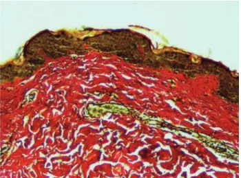
Histopathology revealed mild hyperkeratosis, increased pigmentation in the basal layer, and fibrosis in the dermis (Figure 3). A total loss of dermal elastic fibres was seen with Verhoeff-van Gieson stain (Figure 4).
Complete blood count, biochemistry, electrolytes, kidney, liver and thyroid function tests, coagulation
Figure 1. Figure 1. Figure 1. Figure 1.
Figure 1. Midline of the upper back. Figure 2.Figure 2.Figure 2.Figure 2.Figure 2. Increase in skin laxity over the lower part of
the right scapula.
Figure 3. Figure 3. Figure 3. Figure 3.
Figure 3. Mild hyperkeratosis, increased pigmentation
in the basal layer, and fibrosis in the dermis (H&E, x10).
Figure 4. Figure 4. Figure 4. Figure 4.
Figure 4. Verhoeff-van Gieson stain. Total loss of
factors, complement levels, RF, ANA and anti-ds DNA levels, complete urine analysis and viral marker levels were normal.
There was neither pathological involvement of the cardiovascular, pulmonary, haematological, gastrointestinal systems, nor involvement of the ocular region or connective tissues.
When these findings were taken together, the diagnosis of idiopathic ACL was made and the patient was referred to the Plastic and Reconstructive Surgery outpatient clinic for cosmetic intervention.
Discussion
Discussion
Discussion
Discussion
Discussion
CL is a rare connective tissue disease that is due to a deficiency of elastic fibres. It is inherited in a dominant, recessive, and X-linked recessive inheritance or it may be acquired.3 Acquired forms
are less common and the aetiopathogenesis is not fully understood.7 However, several hypotheses
have been suggested, including immunologically-mediated mechanisms, decreased production of tropoelastin, copper vacancy, abnormal copper metabolism, dysfunction of elastase inhibitors, and hyper-elastase activity.2
Although ACL can be seen at any age, it is most commonly seen in the second and third decade of life. This disease, which affects both sexes equally, also has no race predeliction.5 ACL is
clinically seen in two forms, the generalised insidious form (type 1) and the localised form (type 2) associated with inflammation.6 The lesion of
our patient was localised over the lower part of the right scapula. The localised form of ACL is uncommon, but there is an inflammatory phase that frequently precedes the erythematous
plaques.2 Our patient did not report any
inflammatory lesions in the affected area. Localised ACL on sun-exposed areas such as the neck, shoulder, face, and periorbital area, which were progressive despite surgical procedures have
been reported.7 It is well-known that ultraviolet
rays induce extracellular matrix degradation and skin ageing. The skin lesion of the patient was on the back which was not often exposed to the sun.3
Localised ACL, unlike generalised ACL, has a risk of systemic involvement.5 In addition to changes
on the skin, there is usually systemic involvement predominantly of the lungs (emphysema). However, cardiovascular (aortic aneurysm, mitral valve prolapse), gastrointestinal (hiatus and inguinal hernias) and genitourinary anomalies can
also be present.1 Complete blood count,
biochemistry, electrolytes, kidney, liver and thyroid function tests, thyroid autoantibody levels, coagulation factors, serum copper level, immunoglobulin and complement levels, RF, ANA and anti-ds DNA levels, complete urine analysis and viral marker levels of our patient were normal. No systemic involvement was found after multidiscliplinary assessment by the haematology, oncology, cardiology, gastroenterology, rheumatology, chest and eye departments. ACL may develop following inflammatory dermatoses s u c h a s S w e e t ' s s y n d r o m e , d e r m a t i t i s herpetiformis, systemic lupus erythematosus and erythema multiforme, urticaria attacks and angioedema. Some cases may be preceded by penicillin hypersensitivity, and some may be associated with coeliac disease, nephrotic syndrome, isoniazid therapy, cutaneous lymphoma, melanoma, heavy chain deposition diseases, multiple myeloma and monoclonal gammopathy.8 There was no history of drug use
or dermatosis in our patient.
The differential diagnosis of ACL includes rare degenerative elastic tissue diseases such as
anetoderma and granulomatous loose skin.5
Anetoderma is a rare benign dermatosis caused by a loss of mid-dermal elastic tissue resulting in well-circumscribed areas of pouch-like herniations of flaccid skin.9 Lesions are usually on the neck, upper
body, and proximal extremities. Anetoderma can be seen in both adults and children. There are
two forms: primary and secondary. While the primary anetoderma occurs on the normal skin, the secondary anetoderma occurs after an inflammatory dermatosis. The secondary form is observed mostly in women between 20-40 years. Secondary forms may be accompanied by systemic diseases.5 Mid-dermal elastolysis is
usually symmetrically located on the neck, trunk and upper limbs. In 50% of cases, erythematous papules and plaques, persisting retinal erythema and telangiectasia are observed. Mid-dermal elastolysis is most common in young and middle-aged (30-50 years) women. The lesions are common in sun-exposed skin. There is no extracutaneous involvement but may accompany autoimmune diseases.5 Granulomatous slack skin
is characterised by lax skin and wrinkles, mainly in the flexures with pruritus and erythema, usually occuring in males in their third or fourth decade. Granulomatous mycosis fungoides may be preceded by erythematous scaly patches or macules and may co-exist with classical mycosis fungoides lesions. In 50% of cases, there is co-existence with Hodgkin disease. Histologically, granulomatous mycosis fungoides can be distinguised from granulomatous slack skin.10
Diagnosis is usually clinical and histopathological examination is necessary. Making a definite diagnosis of localised ACL is sometimes difficult because elastic fibres cannot be routinely fixed and stained on histological sections. Therefore, clinical presentation, histopathological findings of affected tissue, laboratory results and sometimes electron microscopy are required.3
Histopathological findings include shrunken elastic fibre residues and significantly reduced elastic fibres. Collagen is usually normal.11 Verhoeff-van
Gieson stain is a simple method for examining elastic fibres. Histopathology of our patient revealed mild hyperkeratosis, increased pigmentation in the basal bed, and fibrosis in the dermis. The presence of total dermal elastic fiber loss was seen with Verhoeff-van Gieson stain.
Treatment of CL targets mostly symptomatic relief.3
Plastic and reconstructive surgical interventions constitute an important part of the treatment.1,9
Recurrence after reconstructive surgery is an important problem, and detection and treatment of underlying diseases can prevent the recurrence.5
In conclusion, localised ACL can be difficult to diagnose as it is rare and the clinic presentation is often late. It is important to diagnose the disease early in order to provide surgical care to prevent future malformations and to provide psychosocial support. ACL patients should also be evaluated in terms of visceral involvement and regular cardiac monitoring should be provided to prevent a possible lethal aortic rupture.1
Declaration of interest
Declaration of interest
Declaration of interest
Declaration of interest
Declaration of interest
There is no scientific or medical committee membership or membership, consultancy, expertise, working status, shareholding, etc. in the work of authors and/or family members in relation to this work, which might be a potential conflict of interest.
The cost of the study was covered by the researchers and no financial aid was received from any person or institution.
R
R
R
R
References
eferences
eferences
eferences
eferences
1. Kim DP, Klein PA. Acquired cutis laxa in a 55-year-old female with multiple myeloma and serologic evidence of systemic lupus erythematosus. Dermatol Online J 2011;7:8.
2. Riveros CJ, Gavilan MF, França LF, Sotto MN, Takahashi MD. Acquired localized cutis laxa confined to the face: case report and review of the literature. nt J Dermatol 2004;43:931-5.
3. Gveric T, Bari M, Bulat V, Situm M, Pusic J, Huljev D, et al. Clinical presentation of a patient with localized acquired cutis laxa of abdomen: a case report. Dermatol Res Pract 2010: 402093. doi:10.1155/2010/402093 4. Marti N, Monteagudo C, Revert A, Reig I, Gamez L,
Jorda E. Acral localized acquired cutis laxa. Int J Dermatol 2013;52:983-6.
5. Öztürk Durmaz E. Rarely encountered degenerative elastic tissue disorders. Turk J Dermatol 2013;7: 121-9.
6. Gupta A, Helm TN. Acquired cutis laxa associated with multiple myeloma. Cutis 2002;69:114-8. 7. Filippopoulos T, Paula JS, Torun N, Hatton MP,
Pasquale LR, Grosskreutz CL. Periorbital changes associated with topical bimatoprost. Ophthalmic Plastic Reconstr Surg 2008;24:302-7.
8. Greenbaum SS, Krull EA, Rubin MG, Lee R. Localized acquired cutis laxa in one of identical twins. Int J Dermatol 1989;28:402-6.
9. Kineston DP, Xia Y, Turiansky GW. Anetoderma: a case report and review of the literature. Cutis 2008; 81:501-6.
10. Shah A, Safaya A. Granulomatous slack skin disease: a review, in comparison with mycosis fungoides. J Eur Acad Dermatol Venereol 2012;26:1472-8. 11. Johnson BL, Honig Paul. Congenital diseases. In:
Elder D, Elenitsas R, Johnson B Jr, Murphy GF, eds. Lever's histopathology of the skin, 9th edit. Philadelphia: Lippincott-Raven, 2005;164-5. 12. Kazlouskaya V, Malhotra S, Lambe J, Idriss MH,
Elston D, Andres C. The utility of elastic Verhoeff-Van Gieson staining in dermatopathology. J Cutan Pathol 2013;40:211-25.