LCMSMS Analysis of 8-OHdG and Measuring Metallothionein Level for
Evaluation of Protective Role of Geraniol in Lead Acetate Administered Rats
Aysel ALKAN UÇKUN1,*, Ertan YOLOĞLU2, Miraç UÇKUN3, Ahmet ÖZKAYA4
1Department of Environmental Engineering, Faculty of Engineering, Adıyaman University, Adıyaman,
Turkey
[email protected], ORCID: 0000-0002-8957-7476
2Department of Mathematics and Science Education, Faculty of Education, Adıyaman University,
Adıyaman, Turkey
[email protected], ORCID: 0000-0002-9730-9471
3Department of Food Engineering, Faculty of Engineering, Adıyaman University, Adıyaman, Turkey
[email protected], ORCID ID: 0000-0002-9018-8515
4Department of Chemistry, Faculty of Science-Literature, Adıyaman University, Adıyaman, Turkey
[email protected], ORCID ID: 0000-0002-0173-3084
Received: 03.03.2020 Accepted: 21.05.2020 Published: 25.06.2020
Abstract
In this study, DNA damage and metallothionein levels were used as biomarkers to evaluate the protective potential of geraniol, a monoterpen against lead stress, in rats. Hepatic 8-hydroxy-2-deoxyguanosine (8-OHdG) level used as a marker of oxidative DNA damage was measured by LCMSMS. Experimental groups were formed in four ways: control, lead acetate, geraniol and lead acetate + geraniol. Seven animals were used in each group. Geraniol and lead acetate were administered to rats for 30 days. In geraniol administered rats, 8-OHdG and metallothionein levels decreased significantly compared to lead acetate administered rats. The highest DNA damage and metallothionein levels were observed in lead acetate administered rats. According to the results of this study, it can be suggested that geraniol protects cells against lead-caused damage by reducing ROS production. In addition, studies on the measurement of 8-OHdG by LCMSMS in
the literature are limited. Therefore, it is thought that the presented study will contribute to the evaluation of the applicability of this method in the literature.
Keywords: 8-hydroxy-2-deoxyguanosine; Metallothionein; Geraniol; Lead; LCMSMS. Kurşun Asetat Uygulanmış Sıçanlarda Geraniolün Koruyucu Rolünün Değerlendirilmesi
için Metallothionein Düzeyinin Ölçülmesi ve 8-OHdG’nin LCMSMS Analizi Öz
Bu çalışmada, sıçanlarda kurşun stresine karşı bir monoterpen olan geraniolün koruyucu potansiyelini değerlendirmek için DNA hasarı ve metallotiyonein seviyeleri biyobelirteç olarak kullanıldı. Oksidatif DNA hasarının bir belirteci olarak kullanılan hepatik 8-hidroksi-2-deoksiguanozin (8-OHdG) seviyesi LCMSMS ile ölçüldü. Her grupta yedi hayvan kullanıldı. Geraniol ve kurşun asetat 30 gün boyunca uygulandı. Geraniol uygulanan sıçanlarda, 8-OHdG ve metallotiyonein düzeyleri, kurşun asetat uygulananlara kıyasla önemli derecede düşüş göstermiştir. En yüksek DNA hasarı ve metallotiyonein seviyesi, kurşun asetat uygulanan sıçanlarda gözlenmiştir. Bu çalışmanın sonuçlarına göre, geraniolün hücreleri kurşunun yol açtığı hasara karşı ROS üretimini azaltmak suretiyle koruduğu ileri sürülebilir. Ayrıca, literatürde 8-OHdG’nin LCMSMS ile ölçümü konusunda çalışmalar sınırlı sayıdadır. Bu nedenle, sunulan çalışmanın bu yöntemin literatürde uygulanabilirliğinin değerlendirilmesine katkıda bulunacağı düşünülmektedir.
Anahtar Kelimeler: 8-hidroksi-2-deoksiguanozin; Metallotiyonein; Geraniol; Kurşun; LCMSMS.
1. Introduction
Environmental or occupational exposures to numerous chemicals can occur during various periods of human life. Metals such as mercury, cadmium, lead etc. could pose to serious health problems [1, 2]. Lead causes to oxidative stress by increasing free radicals and reducing antioxidant sources [3]. Additionaly lead displaces zinc in many proteins and may cause damage to DNA [4]. Many scientists have suggested that various antioxidant treatments can prevent oxidative stress caused by lead [5]. Herbs and spices have been used as alternative and traditional medicines for many years. Terpenoid compounds found in essential oils of plants such as rose, citronella and coriander have been reported to be beneficial to human health [6]. Geraniol (3,7-dimethylocta-trans-2,6-dien-1-ol) is an acyclic monoterpene alcohol with the chemical formula C10H18O. Geraniol was isolated from palmarosa oil and has many biochemical, pharmacological
antitumor effect against many cancer types and has also been shown by many authors to have antimicrobial activity [8, 9].
Metallothioneins (MTs) are cysteine rich intracellular proteins with low molecular weight that bind to metals such as zinc and copper to provide intracellular metal homeostasis [10, 11]. MTs are responsible for maintaining transition metal ion homeostasis and redox stabilization by protecting the cell against DNA damage and apoptosis [12]. Reduced expression of MT has been observed in liver [13], colon [14] and prostate [15] cancer. The protective role of MT in these cancer types has not been fully explained, but when considering the antioxidant effect and protective properties against DNA damage, this reduction may increase sensitivity to toxin-induced damage [16].
Reactive oxygen species are produced as a result of exposure to environmental factors affecting human health such as xenobiotics, radiation and during normal cellular metabolic functions [17, 18].Among the cellular biomolecules, DNA is the most affected by reactive oxygen species [19, 20].An increase in the 8-OHdG level occurs after oxidative DNA damage in the cells [21, 22].For this, 8-OHdG is an important biomarker used in the monitoring of cancer and other diseases [23]. Purpose of measuring 8-OHdG level in this study was to determine the level of DNA damage caused by reactive oxygen species in rats treated with lead and the protective effect of geraniol against this damage.
Many analytical methods such as including enzyme-linked immunosorbent assay (ELISA), high-performance liquid chromatography with electrochemical detection (HPLC-ECD), gas chromatography-mass spectrometry (GCMS) and liquid chromatography-tandem mass spectrometry (LCMSMS) have been developed to measure 8-OHdG. Recently, due to its specificity, reproducibility, accuracy and structural characterization, there is a great interest in LCMSMS [24].For these reasons, we preferred to measure the level of 8-OHdG by LCMSMS to estimate lead acetate-induced DNA damage in our study. In the literature, the study of 8-OHdG analysis with LCMSMS is very limited. Therefore, our study will also contribute to the 8-OHdG analysis by LCMSMS.
The objective of this study was to determine whether geraniol had a protective effect against lead damage by changing 8-OHdG and MT levels in rat liver.
2. Materials and Methods
2.1. Animals and experimental procedure
In this study, twenty-eight Wistar albino rats with an average weight of 330 ± 10 g were used. Animals were grown under a standard light / dark cycle with regular temperature and humidity. For the study of animal experiments, approval was obtained from the Ethics Committee of Fırat University, Elazığ, Turkey (Document No: 146/2011-11). Experimental groups were formed in four ways: control, Pb acetate, geraniol and Pb acetate + geraniol. Seven animals were used in each group. Geraniol was administered to animals at 1 day intervals for 30 days by gavage at a dose of 50 mg / kg by dissolving in corn oil.Pb acetate (dissolved in acetic acid) was administered to the animals by adding 500 ppm of Pb acetate to each liter of drinking water daily for 30 days. Only the solvents used to dissolve geraniol and Pb acetate were administered to the animals in the control group. At the end of 30 days, the animals were sacrificed and the liver tissues were removed and stored at -20 for biochemical analysis. The amount of Pb in liver was determined by inductively coupled plasma mass spectrometry (ICP-MS, Perkin Elmer, MA, USA) in Adıyaman University Central Research Laboratory by Ozkaya et al. [25].
2.2. Quantification of 8-hydroxy-2-deoxyguanosine in rat livers by LCMSMS
DNeasy tissue kit (Qiagen) was used for DNA isolation from the control, Pb acetate, geraniol and Pb acetate + geraniol exposed liver of rats. DNA isolation was performed according to the protocol of kit. The level of 8-OHdG in the liver was measured by liquid chromotgraphy tandem mass spectrometry (LCMSMS, Shimadzu Quadropole 8040) in Adıyaman University Central Research Laboratory. Information on LCMSMS analysis conditions is shown in Table 1 and Table 2, respectively. The Inertsil ODS-4 (2.1 mm I.D. × 50 mm L., 3 μm) column was used. Mobil phase A (5 mM ammonium formate in 100% water) and mobil phase B (5 mM ammonium formate in 100% methanol) were used at a flow rate of 0.2 mL min-1. The retention time of
8-OHdG was around 5.16 minutes. The curve was linear (r2 = 0.9998). The calibration curve range
prepared from the standards for the calculation of 8-OHdG concentrations was in the range of 1-10 µg/L. The samples were spiked over the samples so that the resulting values corresponded to the calibration curve values, and the total concentration of standart was given to the LCMSMS as 5 µg/L. The standard of 8-OHdG were gotten from Sigma-Aldrich with ≥ 98% purity. This method quantifies the molecular level of DNA damage by measuring the level of 8-OHdG directly. One milliliter purified DNA extract was imported to a vial. Quantitative readings were made in three replicates.
Table 1: LCMSMS conditions
Instrument Shimadzu Prominence LC-20A/XR Shimadzu
LCMS-8040
Mobil phase A 5 mM ammonium formate in 100% water
Mobil phase B 5 mM ammonium formate in 100% methanol
Column Inertsil ODS-4 (2.1 mm I.D. × 50 mm L., 3 μm)
Column oven temperature 40 °C
Flow rate 0.2 mL/min
Interface current 4.5 kV
Spraying gas flow rate 3 mL/min
Drying gas flow rate 15 mL/min
DL temperature 250 °C
HB temperature 450 °C
Table 2: Gradient mode of LCMSMS
2.3. Quantification of metallothionein in rat livers by microplate reader
To measure the MT level, the method was used developed by Viarengo et al. [26] which was modified for the microplate reader. The livers were homogenized for one minute at 2000 rpm on ice in 20 mM Tris HCl buffer (pH 8.6) containing 0.5 M sucrose, 0.5 M PMSF and 0.01% b-mercaptoethanol. The homogenates were centrifuged at 15000 g for 30 minutes and the supernatants obtained from this assay were used for analysis. To each 1 ml supernatant was added 1.05 ml of cold (-20 ºC) absolute ethanol and 80 µL of chloroform, and these mixtures were centrifuged for 10 min at 6000 x g at 4 ºC. The mixtures were allowed to incubate for one hour at -20 ° C and then centrifuged for 10 minutes at 6000 x g at 4 °C. Supernatants discharged after centrifugation, the pellets were washed with homogenizing buffer containing 87% ethanol and 1% chloroform and centrifuged at 6000 x g for 10 min at 4 ºC. Finally, the pellets were resuspended in 300 µL 5 mM Tris-HCl buffer (pH 7.0) containing 1 mM EDTA, and 4.2 mL 0.43mM. DTNB buffer containing 0.2 M Na-phosphate (pH 8.0) was added to each sample and then these mixtures were incubated at room temperature for 30 minutes in the dark and centrifuged at 3000 x g for 5 minutes. For the calculation of the MT concentration using GSH as a reference, samples were read at 412 nm absorbance.
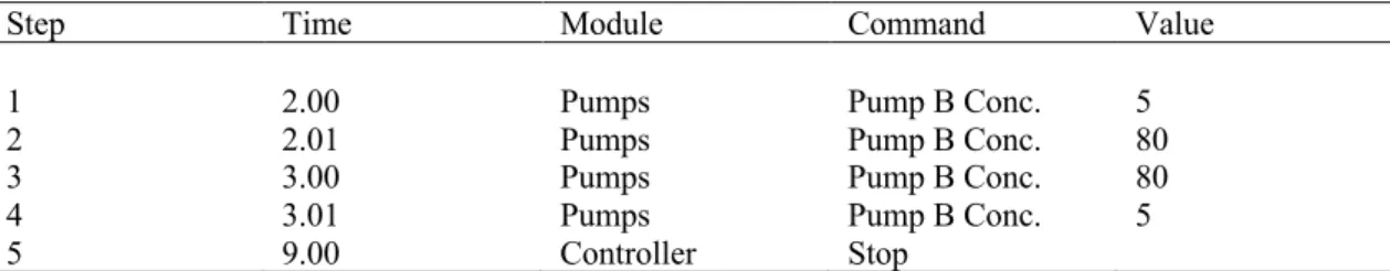
Step Time Module Command Value
1 2.00 Pumps Pump B Conc. 5
2 2.01 Pumps Pump B Conc. 80
3 3.00 Pumps Pump B Conc. 80
4 3.01 Pumps Pump B Conc. 5
2.4. Statistical analysis
In the statistical analysis of the data, computer software package SPSS 22 was used. Data normality was determined using Shapiro-Wilk test (p < 0.05). Kruskal Wallis test was used to determine the comparison of biochemical data between groups. Bonferroni Mann Whitney-U test was used to determine whether there was a significant difference within the groups. The statistical significance level was based on p < 0.05.
3. Results and Discussion
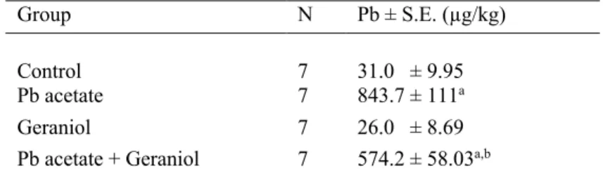
The levels of Pb determined by ICPMS in rats [25] were shown in Table 3. The concentration of Pb was found significantly higher in Pb acetate (843.7 ± 111 µg/kg) group than the geraniol group (26.0 ± 8.69 µg/kg) (p < 0.05). Likewise, the amount of Pb was significantly higher in Pb acetate group than Pb acetate + geraniol group (574.2 ± 58.03 µg/kg) (p < 0.05). Pb exposure is known to cause an increase in reactive oxygen species and a decrease in cellular antioxidant capacity. Also it is known that an imbalance in the prooxidant / antioxidant ratio causes damage to membranes, DNA and proteins in tissue and cellular components [27].
Table 3: Pb concentrations in the liver of rats
Group N Pb ± S.E. (µg/kg)
Control 7 31.0 ± 9.95
Pb acetate 7 843.7 ± 111a
Geraniol 7 26.0 ± 8.69
Pb acetate + Geraniol 7 574.2 ± 58.03a,b a : p < 0.05 compared with the control group
b : p < 0.05 compared with the Pb acetate group
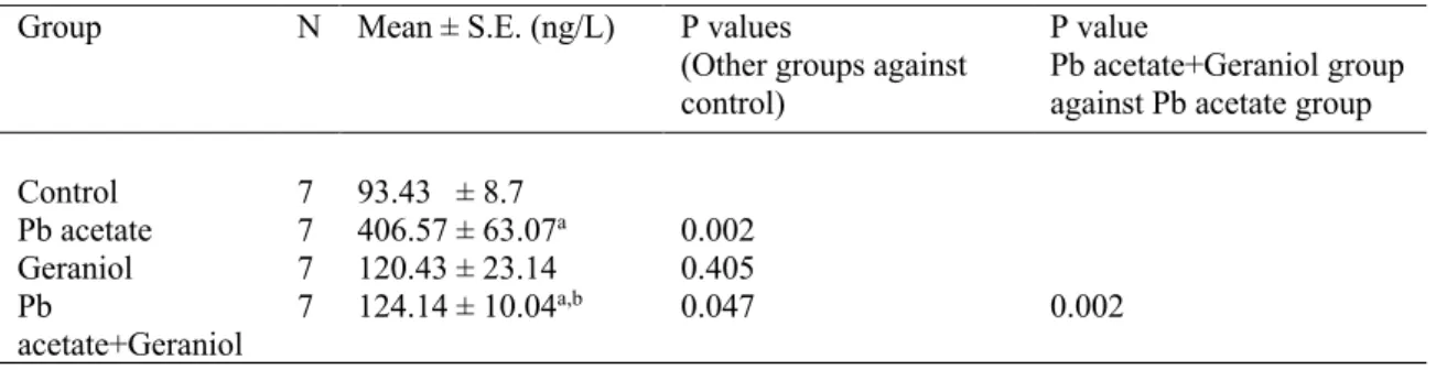
The concentrations of 8-OHdG determined by LCMSMS were presented in Table 4. The recovery value was calculated over seven samples and was found in the range of 90% - 105 %. The % relative standard deviation (RSD) value was between 1.23 and 27.64. Measurement of 8-OHdG level in the liver is an integral indicator of many parameters such as free radical production, antioxidant defense system and cellular redox potential [28].The mean concentration of 8-OHdG in DNA of liver from Pb acetate applied group was in the range of 181 ± 16.09 – 664 ± 79.22 ng L-1. A significant increase was observed in 8-OHdG level in the Pb acetate and Pb
acetate+geraniol groups, compared to the control group (406.57 ± 63.07; 124.14 ± 10.04; 93.43 ± 8.7 ng/L respectively), however there was no significant difference between the control and geraniol group. In short, we can say that there was significant DNA damage in the group where Pb is applied alone, compared to the other groups, whereas there was little or no damage in the geraniol-treated groups. Oxidative stress increases ROS production, which can cause oxidative
damage to DNA and proteins [29].Since 8-OHdG is a product of oxidative DNA damage induced by ROS, its level indirectly reflects the level of ROS in the body [30]. In our study, elevation of the 8-OHdG level in Pb acetate-treated rats may be attributed to oxidative stress resulting in an increase in the amount of free radicals because of metabolic activation between the blood and membrane. Significant declines in the level of 8-OHdG in geraniol administered rats can be interpreted as the reduction of reactive oxygen species by geraniol. At the same time, geraniol may have reduced hepatic DNA damage which caused by Pb acetate. Similarly, Bolin et al. [31] found that 8-OHdG levels increased significantly in rats treated with Pb compared to the control group. Xu et al. [32] exposed rats to different doses of Pb acetate for 4 weeks and as a result they observed an increase in DNA damage along with ROS production. In a study conducted by Liu et al. [33] the liver of rats exposed to 500 mg/L Pb acetate for 75 days by drinking water showed an increase in the 8-OHdG level in parallel with ROS production, and it was suggested that DNA was a target in ROS-induced liver injury, furthermore, the increase of 8-OHdG induced by Pb was effectively suppressed by puerarin. In a study investigating the linkage of Pb-induced DNA damage to oxidative stress, Wang et al. [34] observed that an increase in ROS production in the mitochondria of mice exposed to 0.2% Pb acetate for 42 days was associated with apoptosis induced by caspase 3 activation and 8-OHdG lesion. Tiwari and Kakkar [35] reported that geraniol has antioxidant potential by reducing lipid peroxidation and inhibiting ROS production. It has been reported by Ong et al. [36] that geraniol plays a protective role against liver cancer by inhibiting cell proliferation and DNA damage. Ozkaya et al. [25] observed that 8-OHdG immunoreactivity in rats did not differ significantly between the control and geraniol treated groups, but there was a significant difference between the Pb acetate and Pb acetate + geraniol treated groups. In our study, there was no significant difference between the control and geraniol group in terms of 8-OHdG level.
There are limited studies about the measurement of 8-OHdG level in LCMSMS, which is a marker of DNA damage, so our use of this method will have contributed to the literature. Chen et al. [37] developed a stable isotope dilution by using an automated on-line SPE LCMSMS method to simultaneously measure 8-OHdG and cotinine in human urine. Guo et al. [24] have developed a sensitive, specific and applicable method to detect 8-OHdG in human urine in UPLC-MSMS. They observed higher levels of 8-OHdG in patients with colorectal cancer and tumor metastasis compared to the control group and patients without tumor metastasis respectively. Li et al. [38] determined the levels of 8-OHdG in the livers and kidneys of cetaceans through isotope-dilution LCMSMS and based on the results of the study, it has been suggested that measurement of 8-OHdG by LCMSMS is possible and can be associated with environmental pollutants.
Table 4: Levels of 8-OHdG in the liver of rats
Group N Mean ± S.E. (ng/L) P values
(Other groups against control)
P value
Pb acetate+Geraniol group against Pb acetate group
Control 7 93.43 ± 8.7 Pb acetate 7 406.57 ± 63.07a 0.002 Geraniol 7 120.43 ± 23.14 0.405 Pb acetate+Geraniol 7 124.14 ± 10.04a,b 0.047 0.002
a : p < 0.05 compared with the control group b : p < 0.05 compared with the Pb acetate group
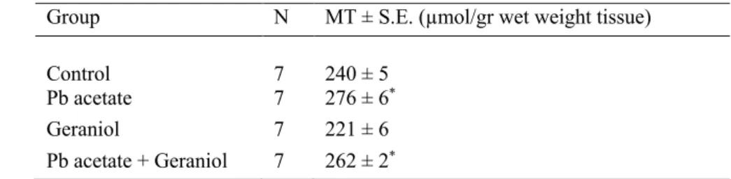
Table 5 shows the changes in MT levels between groups. As can be seen in this table, the highest MT level was observed in the Pb acetate group but there was no significant difference between Pb acetate and Pb acetate + geraniol groups (276 ± 6 and 262 ± 2 µmol/gr wet weight tissue; respectively) (p > 0.05). The lowest MT level was detected in the geraniol treated group (221 ± 6 µmol/gr wet weight tissue). There was no significant difference between the geraniol treated and control groups in MT levels (p > 0.05). Metallothionein is a protein that is slightly induced by lead but more induced by zinc and cadmium. The lead is better bonded to a pre-induced MT by zinc and cadmium so that a lead-thionine complex is formed. In our study, MT levels were higher in Pb acetate group than geraniol treated and control groups. There are many studies showing increases in MT expression due to the protective mechanism of MT against Pb administration. It was observed that MT synthesis was increased in liver, kidney and other tissues of rats exposed to sublethal doses of Pb [39, 40]. MT gene expression was increased by more than 3-fold in cultured human mononuclear blood cells due to Pb exposure [41].MT expression was significantly increased due to the intra cerebral administration of Pb [42].Similarly, Ikebuchi et al.[40] observed that when they applied sublethal dose of Pb acetate intraperitoneally to the rats, there was an increase in the synthesis of lead metallothionein and zinc metallothionein. Chidinma et al. [43] observed increased MT expression in mouse livers exposed to different concentrations of Pb. In addition, several studies have shown that geraniol has an antioxidant effect by preventing oxidative stress. Ozkaya and his colleagues [25] suggested that hepatic MDA level, which is an important indicator of lipid peroxidation, was lower in the group where geraniol was administered alone than the group in which Pb acetate was applied alone, and this may be caused by the antioxidant effect of geraniol. Wang et al. [44] stated that at a dose of 250 mg kg-1 geraniol can
reduce oxidative stress by reducing lipid peroxidation and affecting the activity of GSH and other antioxidant enzymes. Prasad and Muralidhara [45] have also suggested that geraniol can be a promising antioxidant by reducing oxidative stress.
Table 5: The MT levels in the liver of rats
Group N MT ± S.E. (µmol/gr wet weight tissue)
Control 7 240 ± 5
Pb acetate 7 276 ± 6*
Geraniol 7 221 ± 6
Pb acetate + Geraniol 7 262 ± 2* * : p < 0.05 compared with the control group
In conclusion, it can be said that Pb causes oxidative and DNA damage in the liver of rats, and geraniol may reduce these damages by inhibiting ROS production. 8-OHdG and MT are also useful biomarkers reflecting lead toxicity in rats. In addition, it can be said that exogenous supplementation of geraniol may play an advantageous role in antioxidant defense of cells to prevent Pb toxicity.
References
[1] Sinicropi, M.S., Amantea, D., Caruso, A., Saturnino, C., Chemical and biological properties of toxic metals and use of chelating agents for the pharmacological treatment of metal poisoning, Archives of Toxicology, 84, 501-520, 2010.
[2] Carocci, A., Rovito, N., Sinicropi, M.S., Genchi, G., Mercury toxicity and neurodegenerative effects, Reviews of Environmental Contamination and Toxicology, 229, 1-18, 2014.
[3] Silbergeld, E.K., Waalkes, M., Rice, J.M., Lead as a carcinogen, experimental evidence and mechanisms of action, American Journal of Industrial Medicine, 38, 316-323, 2000.
[4] Adonaylo, V.N., Oteiza, P.I., Pb2+ promotes lipid peroxidation and alteration in
membrane physical properties, Toxicology, 132, 19-32, 1999.
[5] Garcia, M.T.A., Gonzalez, E.L.M., Toxic effects of perinatal lead exposure on the brain of rats, involvement of oxidative stress and the beneficial role of antioxidants, Food Chemistry and Toxicology, 46, 2089-2095, 2008.
[6] Jayachandran, M., Chandrasekaran, B., Namasivayam, N., Geraniol attenuates fibrosis and exerts anti-inflammatory effects on diet induced atherogenesis by NF-κB signaling pathway, European Journal of Pharmacology, 762, 102-111, 2015.
[7] Barnard, D.R., Xue, R., Laboratory evaluation of mosquito repellents against Aedes albopictus, Culex nigripalpus, and Ochlerotatus triseriatus (Diptera, Culicidae), Journal of Medical Entomology, 41, 726-730, 2004.
[8] Burke, Y.D., Stark, M.J., Roach, S.L., Sen, S.E., Crowell, P.L., Inhibition of pancreatic cancer growth by the dietary isoprenoids farnesol and geraniol. Lipids, 32, 151-156, 1997.
[9] Bard, M., Albrecht, M.R., Gupta, N., Guynn, C.J., Stillwell, W., Geraniol interferes with membrane functions in strains of Candida and Saccharomyces, Lipids, 23, 534-538, 1988.
[10] Hamer, D.H., Metallothionein, Annual Review of Biochemistry, 55, 913-951, 1986. [11] Takahashi, S., Molecular functions of metallothionein and its role in hematological malignancies, Journal of Hematology and Oncology, 5, 41, 2012.
[12] Cherian, M.G., Jayasurya, A., Bay, B.H., Metallothioneins in human tumors and potential roles in carcinogenesis, Mutation Research, 533, 201-209, 2003.
[13] Datta, J., Majumder, S., Kutay, H., Motiwala, T., Frankel, W., Costa, R., Cha, H.C., MacDougald, O.A., Jacob, S.T., Ghoshal, K., Metallothionein expression is suppressed in primary human hepatocellular carcinomas and is mediated through inactivation of CCAAT/enhancer binding protein alpha by phosphatidylinositol 3-kinase signaling cascade, Cancer Research, 67, 2736-2746, 2007.
[14] Yan, D.W., Fan, J.W., Yu, Z.H., Li, M.X., Wen, Y.G., Li, D.W., Zhou, C.Z., Wang, X.L., Wang, Q., Tang, H.M., Peng, Z.H., Downregulation of metallothionein 1F, a putative oncosuppressor, by loss of heterozygosity in colon cancer tissue, Biochimica et Biophysica Acta, 1822, 918-926, 2012.
[15] Wei, H., Desouki, M.M., Lin, S., Xiao, D., Franklin, R.B., Feng, P., Differential expression of metallothioneins (MTs) 1, 2, and 3 in response to zinc treatment in human prostate normal and malignant cells and tissues, Molecular Cancer, 7, 7, 2008.
[16] Zhang, B., Satoh, M., Nishimura, N., Suzuki, J.S., Sone, H., Aoki, Y., Tohyama, C., Metallothionein deficiency promotes mouse skin carcinogenesis induced by 7,12-dimethylbenz[a]anthracene, Cancer Research, 58, 4044-4046, 1998.
[17] O'Donovan, P., Perrett, C.M., Zahang, X., Montaner, B., Xu, Y.Z., Harwood, C.A., McGregor, J.M., Walker, S.L., Hanaoka, F., Karran, P., Azathioprine and UVA light generate mutagenic oxidative DNA damage, Science, 309, 1871-1874, 2005.
[18] Alan, M.R., Wang, Y., Shah, J., Gordon, S., Fager, M., Butter, PP., Jun, KH., Guardiola-Salmeron, C., Carabe-Fernandez, A., Fan, Y., Proton beam radiation induces DNA damage and cell apoptosis in glioma stem cells through reactive oxygen species, Scientific Reports, 5, 13961-13972, 2015.
[19] Krystona, T.B., Georgieva, A.B., Pissisb, P., Georgakilasa, A.G., Role of oxidative stress and DNA damage in human carcinogenesis, Mutation Research, 711, 193-201, 2011.
[20] Dizdaroglu, M., Oxidatively induced DNA damage and its repair in cancer. Mutation Research, 763, 212-245, 2015.
[21] Cooke, M.S., Evans, M.D., Dizdaroglu, M., Lunec, J., Oxidative DNA damage, mechanisms, mutation, and disease, Faseb Journal, 17, 1195-1214, 2003.
[22] Gajewski, E., Rao, G., Nackerdien, Z., Dizdaroglu, M., Modification of DNA bases in mammalian chromatin by radiationgenerated free radicals, Biochemistry, 29, 7876-7882, 1990. [23] Shimoda, R., Nagashima, M., Sakamoto, M., Yamaguchi, N., Hirohashi, S., Yokota, J., Kasai, H., Increased formation of oxidative DNA damage, 8-hydroxydeoxyguanosine, in human livers with chronic hepatitis, Cancer Research, 54, 3171-3172, 1994.
[24] Guo, C., Li, X., Wang, R., Yu, J., Ye, M., Mao, L., Zhang, S., Zheng, S., Association between Oxidative DNA damage and risk of colorectal cancer, Sensitive Determination of Urinary 8-Hydroxy-2-deoxyguanosine by UPLC-MS/MS Analysis, Scientific Reports, 6, 32581, 2016.
[25] Ozkaya, A., Sahin, Z., Kuzu, M., Saglam, Y.S., Ozkaraca, M., Uckun, M., Yologlu, E., Comakli, V., Demirdag, R., Yologlu, S., Role of geraniol against lead acetate-mediated hepatic damage and their interaction with liver carboxylesterase activity in rats, Archives of Physiology and Biochemistry, 124, 80-87, 2017.
[26] Viarengo, A., Ponzano, E., Dondero, F., Fabbri, R., A simple spectrophotometric method for metallothionein evaluation in marine organisms, an application to Mediterranean and Antarctic molluscs, Marine Environmental Research, 44, 69-84, 1997.
[27] Hsu, P.C., Guo, Y.L., Antioxidant nutrients and lead toxicity, Toxicology, 180, 33-44, 2002.
[28] Kasai, H., Increased formation of oxidative DNA damage, 8-hydroxydeoxyguanosine, in human livers with chronic hepatitis, Cancer Research, 54, 3171-3172, 1994.
[29] Dribben, W.H., Creeley, C.E., Farber, N., Low-level lead exposure triggers neuronal apoptosis in the developing mouse brain, Neurotoxicology and Teratology, 33, 473-480, 2011.
[30] Courtois, E., Marques, M., Barrientos, A., Casado, S., López-Farré, A., Lead-induced downregulation of soluble guanylate cyclase in isolated rat aortic segments mediated by reactive oxygen species and cyclooxygenase-2, Journal of the American Society of Nephrology, 14, 1464-1470, 2003.
[31] Bolin, C.M., Basha, R., Cox, D., Zawia, N.H., Maloney, B., Lahiri, D.K., Cardozo-Pelaez, F., Exposure to lead and the developmental origin of oxidative DNA damage in the aging brain, Faseb Journal, 20, 788-790, 2006.
[32] Xu, J., Lian, L.J., Wu, C., Wang, X.F., Fu, W.Y., Xu, L.H., Lead induces oxidative stress, DNA damage and alteration of p53, Bax and Bcl-2 expressions in mice, Food Chemistry and Toxicology, 46, 1488-1494, 2008.
[33] Liu, C.M., Ma, J.Q., Sun, Y.Z., Puerarin protects the rat liver against oxidative stress mediated DNA damage and apoptosis induced by lead, Experimental Toxicology and Pathology, 64, 575-582, 2012.
[34] Wang, C., Liang, J., Zhang, C., Bi, Y., Shi, X., Shi, Q., Effect of ascorbic acid and thiamine supplementation at different concentrations on lead toxicity in liver, Annals of Occupational Hygiene, 51, 563-569, 2007.
[35] Tiwari, M., Kakkar, P., Plant derived antioxidants-geraniol and camphene protect rat alveolar macrophages against t-BHP induced oxidative stress, Toxicology in Vitro, 23, 295-301, 2009.
[36] Ong, T.P., Heidor, R., Conti, A.D., Dagli, M.L.Z., Moreno, F.S., Farnesol and geraniol chemopreventive activities during the initial phases of hepatocarcinogenesis involve similar actions on cell proliferation and DNA damage, but distinct actions on apoptosis, plasma cholesterol and HMGCoA reductase, Carcinogenesis, 27, 1194-1203, 2006.
[37] Chen, C.Y., Jhou, Y.T., Lee, H.L., Lin, Y.W., Simultaneous, rapid, and sensitive quantification of 8-hydroxy-2'-deoxyguanosine and cotinine in human urine by on-line solid-phase extraction LC-MS/MS, correlation with tobacco exposure biomarkers NNAL, Analytical and Bioanalytical Chemistry, 408, 6295-6306, 2016.
[38] Li, C.S., Wu, K.Y., Gou-Ping., Chang-Chien, Chou, C.C., Analysis of oxidative DNA damage 8-Hydroxy-2'-deoxyguanosine as a biomarker of exposures to persistent pollutants for marine mammals, Environmental Science and Technology, 39, 2455-2460, 2005.
[39] Dai, S., Yin, Z., Yuan, G., Yu, H., Jia, R., Xu, J., Song, X., Li, L., Shu, Y., Liang, X., He, C., Lv, C., Zhang, W., Quantification of metallothionein on the liver and kidney of rats by subchronic lead and cadmium in combination, Environmental Toxicology and Pharmacology, 36, 1207-1216, 2013.
[40] Ikebuchi, H., Teshima, R., Suzuki, K., Simultaneous induction of Pb-metallothionein-like protein and Zn-thionein in the liver of rats given lead acetate, Biochemical Journal, 233, 541-546, 1986.
[41] Gillis, B.S., Arbieva, Z., Gavin, I.M., Analysis of lead toxicity in human cells, BMC Genomics, 13, 344, 2012.
[42] Nakao, K., Kibayashi, K., Taki, T., Koyama, H., Changes in the Brain after Intracerebral Implantation of a Lead Pellet in the Rat, Journal of Neuroscience, 27, 1925-1934, 2010.
[43] Chidinma, N.C., Adewale, A., Chiaka, A., Differential expression of metallothionein-1 and cytochrome p450 2a5 (cyp2a5) in mice in response to lead acetate exposure and industrial effluents in Ibadan, Nigeria, Toxicology and Industrial Health, 32, 1975-1981, 2016.
[44] Wang, J., Su, B., Zhu, H., Chen, C., Zhao, G., Protective effect of geraniol inhibits inflammatory response, oxidative stress and apoptosis in traumatic injury of the spinal cord through modulation of NF-jB and p38 MAPK, Experimental and Therapeutic Medicine, 12, 3607-3613, 2016.
[45] Prasad, S.N., Muralidhara, M., Protective effects of geraniol (a monoterpene) in a diabetic neuropathy rat model, attenuation of behavioral impairments and biochemical perturbations, Journal of Neuroscience Research, 92, 1205-1216, 2014.