Original Article / Özgün Makale
Endobronchial ultrasound-guided transbronchial fine needle aspiration:
Determinants of adequacy
Endobronşiyal ultrasonografi eşliğinde transbronşiyal ince iğne aspirasyonu: Yeterliliğin belirleyicileri Dilek Ece1, Sevinç Hallaç Keser1, Benan Çağlayan2, Banu Salepçi2, Gamze Babur Güler3,
Sibel Sensu1, Gonca Geçmen1, Şermin Kökten1
Received: December 01, 2016 Accepted: January 20, 2017
Institution where the research was done:
Dr. Lütfi Kırdar Kartal Training and Research Hospital, İstanbul, Turkey
Author Affiliations:
1Department of Pathology, Dr. Lütfi Kırdar Kartal Training and Research Hospital, İstanbul, Turkey 2Department of Chest Diseases, Dr. Lütfi Kırdar Kartal Training and Research Hospital, İstanbul, Turkey
3Department of Cardiology, Medipol University Faculty of Medicine, İstanbul, Turkey
Correspondence: Dilek Ece, MD. Dr. Lütfi Kırdar Kartal Eğitim ve Araştırma Hastanesi, Patoloji Kliniği, 34890 Kartal, Cevizli, İstanbul, Turkey Tel: +90 216 - 441 39 00 e-mail: [email protected]
Ece D, Hallaç Keser S, Çağlayan B, Salepçi B, Babur Güler G, Sensu B, et al. Endobronchial ultrasound-guided transbronchial fine needle aspiration: Determinants of adequacy. Turk Gogus Kalp Dama 2018;26(1):123-131.
Cite this article as:
ÖZ
Amaç: Bu çalışmada endobronşiyal ultrasonografi eşliğinde transbronşiyal ince iğne aspirasyonu örneklerinde yeterliliği etkileyen faktörler değerlendirildi.
Çalışma planı: Merkezimizde Mart 2011 - Mart 2014 tarihleri arasında 822 hastadan (500 erkek, 322 kadın; ort. yaş 56±13 yıl; dağılım 16-83 yıl) edinilen 1700 endobronşiyal ultrasonografi eşliğinde transbronşiyal ince iğne aspirasyonu örneği retrospektif olarak analiz edildi. Tüm sitolojik materyaller ve işlem notları (lenf nodu veya lezyon boyutu, yerleşim yeri, iğne girişimi sayısı ve yayma preparat sayısı) gibi örnek yeterliliği ile ilişkili olabilecek değişkenler incelendi.
Bul gu lar: Genel örnek yeterliliği %79.8 idi. Örnek yeterliliği iğne girişimi sayısı ile ilişkili idi (p≤0.001). Yeterlilik oranı tek iğne girişimi için %66.9, üç iğne girişimi için %85.8 idi. Yeterlilik oranları örneklem alma bölgelerine göre farklılık gösterdi [%69.2-%85.8; (p≤0.005)]. Subkarinal (7) lenf nodu istasyonunun çok değişkenli lojistik regresyon analizinde, hasta yaşı (göreceli risk oranı, 0.983; %95 güven aralığı, 0.966-1.000; p=0.049) ve yayma preparat sayısı (göreceli risk oranı, 1.240; %95 güven aralığı, 1.062-1.448; p=0.006) örnek yeterliliğinin bağımsız belirleyici faktörleri idi. Sağ paratrakeal (4R) bölge için örnek yeterliliğinin bağımsız belirleyici faktörleri lenf nodu boyutu (göreceli risk oranı, 1.486; %95 güven aralığı, 0.973-2.268; p=0.067) ve yayma preparat sayısı (göreceli risk oranı, 1.418; %95 güven aralığı, 1.146-1.756; p=0.001) iken sağ interlober (11R) bölge için lenf nodu boyutu (göreceli risk oranı, 1.594; %95 güven aralığı, 0.960-2.645; p=0.071) ve iğne girişimi sayısı (göreceli risk oranı, 2.277; %95 güven aralığı, 1.360-3.811; p=0.002) idi. Sol paratrakeal (4L) lenf nodu istasyonu için örnek yeterliliğinin bağımsız belirleyici faktörü iğne girişimi sayısı (göreceli risk oranı, 1.656; %95 güven aralığı, 0.955-2.869; p=0.072) idi.
Sonuç: Endobronşiyal ultrasonografi eşliğinde transbronşiyal ince iğne aspirasyonlarında, özellikle yerinde hızlı değerlendirme yapılamadığında, lenf nodu yerleşim yerlerine göre yeterliliği etkileyen faktörlerin göz önüne alınması sitolojik değerlendirme için uygun nitelikte materyaller elde edilmesi şansını artırabilir.
Anahtarsözcükler: Yeterlilik; endobronşiyal ultrasonografi; transbronşiyal ince iğne aspirasyonu.
ABSTRACT
Background: This study aims to evaluate the factors influencing the adequacy of endobronchial ultrasound-guided transbronchial fine needle aspiration specimens.
Methods: A retrospective analysis of 1,700 endobronchial ultrasound-guided transbronchial fine needle aspiration samples obtained from 822 patients (500 males, 322 females; mean age 56±13 years; range 16 to 83 years) was performed between March 2011 and March 2014 at our center. Variables potentially associated with sampling adequacy, such as all cytological materials and procedure notes (lymph node and/or lesion size, localization, needle pass number, and slide number) were examined.
Results:The overall specimen adequacy was 79.8%. The specimen adequacy was associated with needle pass number (p≤0.001). Adequacy rate was 66.9% for one needle pass and 85.8% for three needle passes. According to the sampling regions, adequacy rates showed a difference [69.2%-85.8%; (p≤0.005)]. In the multivariate logistic regression analysis of subcarinal (7) lymph node station, patient age (odds ratio, 0.983; 95% confidence interval, 0.966-1.000; p=0.049) and number of slides (odds ratio, 1.240; 95% confidence interval, 1.062-1.448; p=0.006) were independent determining factors of specimen adequacy. While independent determinants of specimen adequacy for the right paratracheal (4R) region were lymph node size (odds ratio, 1.486; 95% confidence interval, 0.973-2.268; p=0.067) and number of slides (odds ratio, 1.418; 95% confidence interval, 1.146-1.756; p=0.001), they were lymph node size (odds ratio, 1.594; 95% confidence interval, 0.960-2.645; p=0.071) and number of needle passes (odds ratio, 2.277; 95% confidence interval, 1.360-3.811; p=0.002) for the right interlobar (11R) region. Independent determinant of specimen adequacy for the left paratracheal (4L) lymph node station was the number of needle passes (odds ratio, 1.656; 95% confidence interval, 0.955-2.869; p=0.072).
Conclusion: During endobronchial ultrasound-guided transbronchial fine needle aspirations, particularly when rapid on site evaluation cannot be applied, consideration of factors affecting adequacy according to lymph node localizations may increase the chance for obtaining materials with suitable quality for cytologic evaluation.
Keywords: Adequacy; endobronchial ultrasound; transbronchial fine needle aspiration.
Mediastinal lymph node (LN) sampling is a critical step in the staging of lung cancer and the diagnosis of inflammatory conditions, such as granulomatous diseases. Over the past decade, endobronchial ultrasound-guided transbronchial fine needle aspiration (EBUS-TBNA) has emerged as a minimally invasive, highly accurate technique for diagnosis, as well as staging, of malignant tumors of the lung and for assessment of mediastinal lymphadenopathy.[1] This technique provides cytological samples of LNs and lesions in proximity to the central airway.[2] Recent advances in therapy for non-small cell lung carcinoma (NSCLC) and development of EBUS-TBNA concurrently allowed combined use of EBUS-TBNA for the management of patients.[3] Thus, EBUS-TBNA technique has gained world-wide popularity for sampling the target LN and lesion of the mediastinum and lung.[4-10] Furthermore, various factors that affect acquiring sufficient material, such as the type of sedation, needle size, needle pass number, aspirator experience, aspiration localization, target lesion size, and the use of rapid on-site evaluation (ROSE), have been objectives of many studies.[11-16]
In recent years, EBUS-TBNA materials have often come into use in determining the targeted therapy in NSCLCs. In these circumstances, adequacy of specimens gained more importance. Therefore, in this study, we aimed to evaluate the factors influencing the adequacy of EBUS-TBNA specimens.
PATIENTS AND METHODS
The archives of Dr. Lütfi Kırdar Kartal Training and Research Hospital Pathology and Pulmonary Disease Clinic were searched for EBUS-TBNA cases dating from 01 March 2011 to 01 March 2014, comprising a period of 36 months. Totally 1,700 EBUS-TBNA specimens obtained from 822 patients (500 males, 322 females; mean age 56±13 years; range 16 to 83 years) were included. The EBUS-TBNA materials were sent to our clinic with the localizations encoded as standard. In the patient’s pathology report, results belonging to each localization were listed under its own headline according to the standard report system of our clinic. All cytological materials and procedure notes (LN and/or lesion size, localization, needle pass number, slide number) were reviewed. The study protocol was approved by the Dr. Lütfi Kırdar Kartal Training and Research Hospital Ethics Committee. The study was conducted in accordance with the principles of the Declaration of Helsinki.
The EBUS-TBNA procedure was performed by a Chest Physician in the bronchoscopy suit at Pulmonary Diseases Clinic. For the procedure,
7.5 MHz BF-UC160F [Olympus Optical Co. Tokyo, Japan, approved by Food and Drug Administration (FDA)], convex probe bronchoscope and EU C2000 processor (Olympus Corp., Tokyo, Japan) were used by the oral route. Topical anesthesia was performed with lidocaine, and sedation with midazolam. Aspiration from target LN or suspicious lesion (from lung, mediastinum or trachea) was conducted with Olympus 22-gauge NA-201SX-4022-C needle (approved by FDA). The number of passes was determined by the Chest Physician. ROSE was not performed during procedures.
Slide preparation procedure was performed in bronchoscopy suite by an attending person from the bronchoscopy team, a pulmonary resident or a nurse. He or she had been informed about the processing of aspiration materials by a pathologist.
Aspirated material was expelled on a glass slide, and smeared with a second slide. Depending on the quantity of aspirated material, a pair of slides (only one slide or three slides for a minority of cases) was prepared from each needle pass. For each pair of slides smeared, one was air-dried and the other was fixed immediately in 95% alcohol. The remnants of each aspirate from the same localization were collected in one tube filled with 95% alcohol for cell block. All slides and cell block tubes were encoded according to the aspiration sites (LN stations or lesions).
Air-dried slides were stained using May-Grünwald Giemsa stain, and 95% alcohol-fixed slides, with Papanicolaou stain. Cell blocks were prepared by formalin fixation and paraffin embedding technique from pellets obtained by centrifugation of collecting tube which contain remnants of aspirates for cell block. Three-micron thick slides obtained from cell blocks were stained with hematoxylin and eosin. Immunohistochemical evaluation was performed, when needed. Molecular testing at our institution is based on clinician request and these data were not reviewed for the current study.
The cytologic materials were evaluated by an experienced pathologist. The slides were reassessed for adequacy and cytologic diagnosis, according to the encoded localizations independently. Adequacy was defined as the acquisition of tissue sufficient for pathologic diagnosis. Lymph node specimens that demonstrated specific diagnosis or adequate lymphocytes were considered adequate. The adequate amount of lymphoid material was defined as the presence of over 40 lymphocytes in high power field in the most cellular areas of the slides or
the presence of clusters of anthracotic pigment-laden macrophages as defined by Alsharif et al.[17] If no diagnoses were established and insufficient lymphocytes were available to verify adequate LN sampling, the samples were considered inadequate. Aspiration samples of the lung, mediastinum or trachea lesions that demonstrated specific diagnosis were considered adequate. If no specific diagnoses were established, even when bronchial epithelium or pigmented macrophages were found, the samples were considered inadequate.
Adequacy was calculated as the ratio of cytologic specimens that demonstrated specific diagnoses (malignant, atypical/suspicious, or granulomatous inflammation) or adequate lymphocytes to total samples acquired, as described by Ost et al.[18] These were performed on per LN/lesion basis.
Both LN and lesion specimens’ cytologic diagnoses were categorized as malignant, atypical, benign or nondiagnostic. The presence of frankly malignant cells
was considered malignant. The presence of rare cells suspicious but not clearly defined for malignancy was considered atypical. In LN specimens, the presence of adequate lymphocytes without evidence of tumor was considered benign. Benign diagnoses included “metastasis negative” or granulomatous inflammation. Lesions from lung, mediastinum or trachea specimens that demonstrated any specific benign lesion, such as granulomatous inflammation, were considered benign. Both for LN and lesion specimens, the sample was determined as nondiagnostic when the inadequate material was present, and only blood, mucus and/or necrotic material were seen or when the specimen was acellular. Cell blocks cellularity and contribution of cell blocks on adequacy were not reviewed for the current study.
Statistical analysis
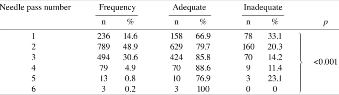
Data were summarized as the mean ± standard deviation, and median (range) for continuous variables and as percentage for categorical variables. Normal Table 1. Relationship of needle passes and sampling adequacy
Needle pass number Frequency Adequate Inadequate
n % n % n % p 1 236 14.6 158 66.9 78 33.1 2 789 48.9 629 79.7 160 20.3 3 494 30.6 424 85.8 70 14.2 4 79 4.9 70 88.6 9 11.4 5 13 0.8 10 76.9 3 23.1 6 3 0.2 3 100 0 0 ˝ ˛ ˛ ˙ ˙ ˙ ˙ ˙ ˙ <0.001
Table 2. Relationship of aspiration sites and sampling adequacy
Aspiration sites Frequency Adequate Inadequate
n % n % n % p 7 545 32 451 82.8 94 17.2 4R 415 24.4 356 85.8 59 14.2 11R 227 13.3 176 77.5 51 22.5 11L 195 11.5 135 69.2 60 30.8 4L 141 8.3 105 74.5 36 25.5
Lesion (lung, mediastinum, trachea) 96 5.6 74 77.1 22 22.9
10R 38 2.2 28 73.7 10 26.3 2R 18 1.1 14 77.8 4 22.2 10L 13 0.8 9 69.2 4 30.8 2L 4 0.2 3 75.0 1 25.0 3 3 0.2 2 66.7 1 33.3 12R 3 0.2 3 100 0 0 12L 1 0.1 1 100 0 0 Precarinal 1 0.1 0 0 1 100 Total 1700 100 1357 79.8 343 20.2 ˝ ˛ ˛ ˙ ˙ ˙ ˙ ˙ ˙ ˙ ˙ ˙ ˙ ˙ ˙ ˙ ˙ ˙ ˙ ˙˙ <0.005
distribution was tested with Kolmogorov-Smirnov test. The Pearson chi-square test was used to test categorical variables. The baseline predictors of specimen adequacy that showing an association (p<0.05) with outcome in univariate analysis were included in the multivariate forward stepwise model after adjustment for age and gender. P value of <0.05 was considered significant for all tests. Statistical Package for the Social Sciences (SPSS) version 11.0 software (SPSS Inc., Chicago, IL, USA) was used. RESULTS
In many patients, samplings were obtained from more than one site (one-five sites/patient). Overall 1,604 (94.4%) samples were obtained from LNs and 96 (5.6%) samples were obtained from lung, mediastinum or trachea lesions. Each aspiration site had averagely four (one-13) slides.
Of the 1,700 EBUS-TBNA specimens, 1,357 (79.8%) had adequate material. In a subset of 1,614 (95%) aspirates, needle pass number was available. The mean number of needle passes from each site was
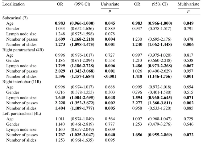
two (one-six). The average slides of each pass were two (one-three). A significant association between needle pass numbers and specimen adequacy (p≤0.001) were shown in Table 1. The specific measurement of the LN/lesion size (short axis diameter) was available for 1,583 (93%) of the aspirates. Short axis diameters of aspirated LNs and/or lesions were 0.3-7.9 cm (mean 1.5 cm). Specimen adequacy was 74.2% in LNs of <1 cm and 81.2% in LNs ≥1cm. Subcarinal (7), right paratracheal (4R), right interlobar (11R), left interlobar (11L), and left paratracheal (4L) LNs were the most frequently sampled LN stations. Sampling adequacy was analyzed according to aspiration site and results were listed in Table 2. There was a significant association between aspiration site and sampling adequacy (p≤0.005). Factors that affect sampling adequacy were reviewed by regression analysis in the frequently aspirated sites in various anatomical localizations (Table 3). The univariate analysis of subcarinal site, patient age (p=0.045), the number of passes (p=0.004), and number of slides (p=0.001), were associated with specimen adequacy. In multivariate analysis, patient age [odds ratio (OR), 0.983; 95% Table 3. Factors that affect sampling adequacy in various lymph node stations
Localization OR (95% Cl) Univariate OR (95% Cl) Multivariate
p p
Subcarinal (7)
Age 0.983 (0.966-1.000) 0.045 0.983 (0.966-1.000) 0.049
Gender 1.033 (0.652-1.636) 0.889 0.937 (0.578-1.517) 0.791
Lymph node size 1.248 (0.975-1.598) 0.078
Number of passes 1.609 (1.168-2.218) 0.004 1.230 (0.695-2.176) 0.478
Number of slides 1.273 (1.098-1.475) 0.001 1.240 (1.062-1.448) 0.006
Right paratracheal (4R)
Age 0.996 (0.976-1.017) 0.727 0.997 (0.975-1.020) 0.817
Gender 1.186 (0.671-2.094) 0.558 1.210 (0.660-2.218) 0.538
Lymph node size 1.799 (1.186-2.728) 0.006 1.486 (0.973-2.268) 0.067
Number of passes 2.029 (1.342-3.068) 0.001 1.026 (0.400-2.629) 0.957
Number of slides 1.396 (1.157-1.684) <0.001 1.418 (1.146-1.756) 0.001
Right interlobar (11R)
Age 0.996 (0.974-1.017) 0.688 0.995 (0.972-1.018) 0.654
Gender 0.716 (0.378-1.353) 0.303 0.796 (0.401-1.580) 0.515
Lymph node size 1.645 (1.004-2.695) 0.048 1.594 (0.960-2.645) 0.071
Number of passes 2.228 (1.352-3.672) 0.002 2.277 (1.360-3.811) 0.002
Number of slides 1.404 (1.109-1.777) 0.005 0.958 (0.533-1.720) 0.885
Left paratracheal (4L)
Age 1.011 (0.974-1.049) 0.564 1.007 (0.968-1.047) 0.729
Gender 1.140 (0.461-2.819) 0.777 1.253 (0.479-3.276) 0.646
Lymph node size 1.160 (0.657-2.049) 0.609
Number of passes 1.767 (1.025-3.047) 0.040 1.656 (0.955-2.869) 0.072
Number of slides 1.253 (0.961-1.635) 0.095
confidence interval (CI), 0.966-1.000; p=0.049] and the number of slides (OR, 1.240; 95% CI, 1.062-1.448; p=0.006) were observed to be independent determining factors of specimen adequacy in this site. According to these results, adequacy increased in the subcarinal localization as the patient age decreased, and more significantly, as the number of slides increased. In univariate analysis of the right paratracheal region, LN size (p=0.006), the number of passes (p=0.001) and number of slides (p≤0.001) were shown to be in a significant relationship with sampling adequacy. In univariate analysis of the right interlobar region, LN size (p=0.048), the number of passes (p=0.002) and number of slides (p=0.005) were shown to be in significant relationship with specimen adequacy.
In multivariate analysis of the right paratracheal region, LN size (OR, 1.486; 95% CI, 0.973-2.268; p=0.067), and the number of slides (OR, 1.418; 95% CI, 1.146-1.756; p=0.001); for right interlobar region, LN size (OR, 1.594; 95% CI, 0.960-2.645; p=0.071) and the number of passes (OR, 2.277; 95% CI, 1.360-3.811; p=0.002) were observed to be independent determinants of sampling adequacy. In the results of both univariate and multivariate analysis of the left paratracheal anatomical site, the number of passes (OR, 1.656; 95% CI, 0.955-2.869; p=0.072) was observed to be an independent determinant of specimen adequacy. In this localization, as the number of passes increased, adequacy also increased. No significant determinant was found in sampling Figure 1. Squamous cell carcinoma. Bizarre and elongated cells
with dens, orangeophilic cytoplasm (Papanicolaou ¥400).
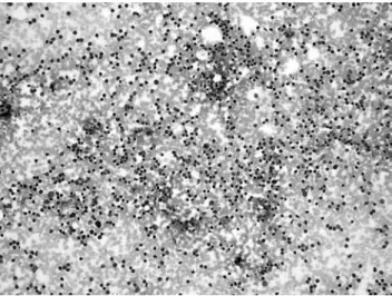
Figure 4. Lymphoid cells in benign “non metastatic” lymph node (May-Grünwald Giemsa ¥100).
Figure 3. Small cell carcinoma. Small, round to fusiform cells with evenly dispersed, powdery chromatin and scant cytoplasm (Papanicolaou ¥1000).
Figure 2. Adenocarcinoma. Glandular cells with large polarized nuclei, prominent nucleoli and lacy cytoplasm (Papanicolaou ¥1000).
adequacy in the univariate analysis of the left interlobar site.
Of the total 1,700 EBUS-TBNA specimens, 360 (21.2%) had malignant (Figures 1-3), and 953 (56%) had benign lesions. In LN specimens, 946 benign diagnoses included 650 “metastasis negative” LNs and 296 granulomatous inflammations (Figures 4-6). Benign diagnosis of the lesion from mediastinum, lung or trachea included five granulomatous inflammations and two intrathoracic thyroid tissues. Totally 57 EBUS-TBNA specimens had atypical cytologic findings, and 301 specimens were nondiagnostic.
DISCUSSION
Specimen adequacy is an important component of fine needle aspiration (FNA) performance, which is frequently reported in FNA studies. Correspondingly, specimen adequacy and factors that affect adequacy are the subjects of multiple reports of EBUS-TBNA. Studies have shown variation in the adequacy rates of EBUS-TBNA.[4,6,16,17,19,20] Because of the fact that EBUS-TBNA is a multistep process, many factors have potential to affect the adequacy of the specimen. The ROSE performed for EBUS-TBNA is one of the most questioned factors in this area.[20-22] During the aspiration procedure, the presence of a pathologist or cytopathologist increases the likelihood of obtaining adequate diagnostic material due to optimum sampling and correct pre-evaluation while decreasing complication rates.[21] However, the ROSE procedure increases the workload of pathologists/cytopathologists and in certain centers, such as ours, ROSE could not be performed due to its uncompensated daily workload. Whereas, it has been reported that as the
lesion and procedure can be observed in real-time in EBUS-TBNA, the necessity of ROSE is diminishing.[20] Nonetheless, manipulations such as changes in the intervention area and the depth or needle angle in accordance with the ROSE results may increase the specimen adequacy. In addition to ROSE, the effects of factors on specimen adequacy, such as operator’s experience, the needle used, sedation type and the pathologist’s/cytopathologist’s decision, have been investigated in EBUS-TBNA.[11,12,14,16,23] Groth et al.[14] revealed that the learning curve for EBUS-TBNA requires nearly 10 procedures In a study investigating 2414 LN aspirates, DePew et al.[16] commented that bronchoscopists and experience durations had no significant effects on the sample adequacy. Furthermore, in a study comparing sedation types, Yarmus et al.[12] showed that the diagnostic yield was higher in deep than in moderate sedation. Another study comparing aspirates with 21-gauge and 22-gauge needles found no difference between specimen adequacy and diagnostic yield.[11,12] In a study comparing the standard EBUS-TBNA and capillary sampling without aspiration, Casal et al.[24] revealed no difference in the adequacy rates. Moreover, a study evaluating the cytological diagnostic reproducibility among pathologists showed that appropriate experience is directly related to the diagnostic reproducibility.[23]
On the other hand, standard and well-defined adequacy criteria do not yet exist for LNs sampled with EBUS-TBNA, and the criteria used show inconsistencies among pathologists.[23] In some studies, specimen adequacy has been based on the presence of lymphocytes and tumor cells, but the criteria have not been sufficiently clarified.[4-6,13,19,20] In some studies, Figure 5. Pigmented macrophages in benign “non metastatic”
the criteria defined by Alsharif were used, while others found these to be difficult to apply.[7,17,21,25] Our study used Alsharif’s criteria; however, they were challenging to use in the case of some slides due to the aspiration and spreading techniques. The specimen adequacy rate observed in our study was 79.8% without ROSE. In the relevant literature, adequacy rates were reported to be between 77% and 95%.[4,6,16,19,20]
During our study period, the aspiration team, anesthesia type, needle diameter, preparation technique, and cytopathologist were constant. The EBUS-TBNA experience of the aspiration team started in October 2008, and the cytology experience of the microscopist began in December 2008. The other parameters for evaluating the EBUS-TBNA adequacy were the pass number, LN size and LN localization.
In many studies, between one and six passes were performed for each LN station.[4,6,10,16,17,19,20] Lee et al.[13] reported a study of investigating the LN aspirations of suspected or known NSCLC patients with adequacy rate of 92%. In their study, each LN station had four aspirations and cumulative specimen adequacy rates were 90.1% for a single pass, 98.1% for two passes, and 100% for three passes. Independent of the aspiration site, overall adequacy rates in our study were 66.9% for one, 79.7% for two, and 85.8% for three passes. The high specimen adequacy in Lee et al.’s study[13] may be defined with the aspiration technique, which provides qualified and quantitative material, including tissue cores, in each pass. Furthermore, cumulative specimen adequacy was evaluated for sufficient aspirates.
In the evaluation of cytologic material, preparation technique is one of the most important parameters that have an effect on pathologist/cytopathologist conclusion. Handling of the specimen can differ according to the aspiration quality and quantity. In the majority of the EBUS-TBNA studies (including ours), conventional smearing and both alcohol fixation and the air-dry technique have been used, and cell blocks have been prepared from the residual material.[7,13,17,19,21,25] In the others, liquid based cytology and/or combination of these techniques were preferred.[5,10,20,25] Different slide numbers have been reported in those studies. Some of them described smearing slides for each aspiration and the others per aspiration site or per case.[6,7,13,17,19,20] In Alsharif et al.’s study[17] with a mean of six slides prepared for each LN localization, independent of the LN station, the specimen adequacy rate was 84.3%. Similarly, in our study, four slides were prepared for each aspiration localization, and the adequacy rate was found to be 79.8%, independent of the aspiration site.
The EBUS-TBNA technique aids in the imaging and sampling of small LNs at difficult localizations. DePew et al.[16] revealed that LNs of <1cm have a lower specimen adequacy rate, while Jhun et al.[15] demonstrated that there was no significant difference in diagnostic performance in LNs of ≥1 cm.[15,16] In our study, the specimen adequacy rate was higher in LNs of ≥1 cm.
When the anatomical localization and the relationship with neighboring structures are considered, the material quality might be expected to differ for each LN station. Although DePew et al.[16] stated that there was no association between the LN station and the specimen adequacy in EBUS-TBNA, Jhun et al.[15] argued that for diagnostic performance, bronchoscopists must pay attention to the nodal station effect. It is difficult to sample the paratracheal region via conventional TBNA. Szlubowski et al.[8] revealed that the imaging and biopsy procedure for paratracheal (particularly left) LNs is technically more complicated than for subcarinal and hilar LNs. This was primarily the case because the left paratracheal LN is near the subaortic region and more deeply localized in the trachea. However, this researcher emphasized that the diagnostic yield was higher in paratracheal LNs. Jhun et al.[15] stated that mediastinal and hilar/interlobar node groups did not differ in diagnostic performance, but when LNs were categorized into three groups - the middle (3P and 7), right (1R, 2R, 4R, 10R and 11R) and left side (4L, 10L and 11L) - the negative predictive value was lower for the left nodal group than for the others, and the sensitivity showed a decreasing trend.
In our study, the adequate material rates obtained from most sampled regions, namely the subcarinal, right paratracheal, right interlobar, left interlobar, and left paratracheal LNs were 82.8%, 85.8%, 77.5%, 74.5%, and 69.2%, respectively. The adequacy rates in the right paratracheal and subcarinal regions were higher than those in the other regions. This difference might be explained by the right paratracheal LN’s more proximal localization, the subcarinal LN’s central anatomical position, and the left paratracheal LN’s relationship with the aortic arc and interlobar LNs, a more distal localization.
Ost et al.[18] defined the patient -and hospital-based markers of the EBUS-TBNA diagnostic yield in the multicentric AQuIRE (American Collage of Chest Physicians Quality Improvement Registry, Evaluation, and Education) study. He stated that LNs, smoking, and the number of sampled LNs were the variables affecting the diagnostic yield at the patient level. If a hospital had higher TBNA volume, this had
robust effects on the diagnostic yield. This reflects the combined results of team experience, anesthesia practice, and cytopathological decisions.
In our study, we focused on the variables that affected the specimen adequacy in LN stations. If the anatomical positions of the bronchial tree and LN stations and their relationships with surrounding structures were taken into account, it would not be surprising for the specimen adequacy to differ for each localization. When the variables affecting the adequacy in the frequently aspirated LN stations were examined using multivariate logistic regression analysis, distinct parameters were found as independent markers for each localization. Accordingly, in the multivariate analysis, the following variables were related to adequacy: for the subcarinal region, patient age and number of slides; for the right paratracheal region, LN size and number of slides; for the right interlobar region, LN size and number of passes; and for the left paratracheal region, number of passes.
We think that the reliability of our results is increased due to the large sample size, the inclusion of a single center (with its standardized aspiration team, microscopist, anesthesia, device, needle, and preparation technique), and the focus on specimen adequacy. However, due to the study being retrospective, with the LN size and number of passes not been reached in all cases and ROSE not been performed may have limited the results of our study. Therefore, prospective studies to be applied on larger populations of patients on which more standardized criteria will be applied may produce more leading results in clinical applications.
In conclusion, the criteria for adequacy have not yet been established for the endobronchial ultrasound-guided transbronchial fine needle aspiration materials. Similarly, methodological details, such as the aspiration number required for each target and the preparation technique, are still unclear. In this study, although rapid on-site evaluation was not performed, the adequacy rate was 79.8% independent of the aspiration site. According to lymph node stations, sampling adequacy was higher in the more proximal and central lymph nodes than more distal lymph nodes. Various parameters affecting the adequacy rate (such as needle pass number, lymph node size, slide number, and patient age) were reviewed for different lymph node stations. Consideration of aspiration localization and factors that affect sampling adequacy can increase the acquirement of sufficient material for cytologic evaluation, particularly in the absence of rapid on-site evaluation. It can save time and economic profits, and
decrease work load, without the process having to be repeated.
Declaration of conflicting interests
The authors declared no conflicts of interest with respect to the authorship and/or publication of this article.
Funding
The authors received no financial support for the research and/or authorship of this article.
REFERENCES
1. Krasnik M, Vilmann P, Larsen SS, Jacobsen GK. Preliminary experience with a new method of endoscopic transbronchial real time ultrasound guided biopsy for diagnosis of mediastinal and hilar lesions. Thorax 2003;58:1083-6. 2. Çetinkaya E, Eyhan Ş. Endobronşiyal ultrasonografi
rehberliğinde transbronşiyal iğne aspirasyonu. Turkiye Klinikleri J Thor Surg-Special Topics 2009;2:6-12.
3. Bulman W, Saqi A, Powell CA. Acquisition and processing of endobronchial ultrasound-guided transbronchial needle aspiration specimens in the era of targeted lung cancer chemotherapy. Am J Respir Crit Care Med 2012;185:606-11. 4. Yasufuku K, Chiyo M, Sekine Y, Chhajed PN, Shibuya K,
Iizasa T, et al. Real-time endobronchial ultrasound-guided transbronchial needle aspiration of mediastinal and hilar lymph nodes. Chest 2004;126:122-8.
5. Rintoul RC, Skwarski KM, Murchison JT, Wallace WA, Walker WS, Penman ID. Endobronchial and endoscopic ultrasound-guided real-time fine-needle aspiration for mediastinal staging. Eur Respir J 2005;25:416-21.
6. Herth FJ, Eberhardt R, Vilmann P, Krasnik M, Ernst A. Real-time endobronchial ultrasound guided transbronchial needle aspiration for sampling mediastinal lymph nodes. Thorax 2006;61:795-8.
7. Feller-Kopman D, Yung RC, Burroughs F, Li QK. Cytology of endobronchial ultrasound-guided transbronchial needle aspiration: a retrospective study with histology correlation. Cancer 2009;117:482-90.
8. Szlubowski A, Kuzdzał J, Kołodziej M, Soja J, Pankowski J, Obrochta A, et al. Endobronchial ultrasound-guided needle aspiration in the non-small cell lung cancer staging. Eur J Cardiothorac Surg 2009;35:332-5.
9. Cömert SS, Çağlayan B, Fidan A, Salepçi B, Doğan C, Demirhan R, et al. A comparision of endobronchial ultrasound-guided transbronchial needle aspiration and integrated positron emission tomography-computed tomography in the diagnosis of malignant mediastinal/hilar lymph nodes. Turk Gogus Kalp Dama 2012;20:843-9. 10. Karunamurthy A, Cai G, Dacic S, Khalbuss WE, Pantanowitz
L, Monaco SE. Evaluation of endobronchial ultrasound-guided fine-needle aspirations (EBUS-FNA): correlation with adequacy and histologic follow-up. Cancer Cytopathol 2014;122:23-32. 11. Yarmus LB, Akulian JA, Gilbert C, Mathai SC, Sathiyamoorthy
S, Sahetya S, et al. Comparison of moderate versus deep sedation for endobronchial ultrasound transbronchial needle aspiration. Ann Am Thorac Soc 2013;10:121-6.
12. Yarmus LB, Akulian JA, Gilbert C, Mathai SC, Sathiyamoorthy S, Sahetya S, et al. Comparison of moderate versus deep sedation for endobronchial ultrasound transbronchial needle aspiration. Ann Am Thorac Soc 2013;10:121-6.
13. Lee HS, Lee GK, Lee HS, Kim MS, Lee JM, Kim HY, et al. Real-time endobronchial ultrasound-guided transbronchial needle aspiration in mediastinal staging of non-small cell lung cancer: how many aspirations per target lymph node station? Chest 2008;134:368-74.
14. Groth SS, Whitson BA, D'Cunha J, Maddaus MA, Alsharif M, Andrade RS. Endobronchial ultrasound-guided fine-needle aspiration of mediastinal lymph nodes: a single institution's early learning curve. Ann Thorac Surg 2008;86:1104-9. 15. Jhun BW, Park HY, Jeon K, Koh WJ, Suh GY, Chung
MP, et al. Nodal stations and diagnostic performances of endobronchial ultrasound-guided transbronchial needle aspiration in patients with non-small cell lung cancer. J Korean Med Sci 2012;27:46-51.
16. DePew ZS, Edell ES, Midthun DE, Mullon JJ, Bungum AO, Decker PA, et al. Endobronchial ultrasound-guided transbronchial needle aspiration: determinants of sampling adequacy. J Bronchology Interv Pulmonol 2012;19:271-6. 17. Alsharif M, Andrade RS, Groth SS, Stelow EB, Pambuccian
SE. Endobronchial ultrasound-guided transbronchial fine-needle aspiration: the University of Minnesota experience, with emphasis on usefulness, adequacy assessment, and diagnostic difficulties. Am J Clin Pathol 2008;130:434-43. 18. Ost DE, Ernst A, Lei X, Feller-Kopman D, Eapen GA,
Kovitz KL, et al. Diagnostic yield of endobronchial ultrasound-guided transbronchial needle aspiration: results of the AQuIRE
Bronchoscopy Registry. Chest 2011;140:1557-66.
19. Jacob-Ampuero MP, Haas AR, Ciocca V, Bibbo M. Cytologic accuracy of samples obtained by endobronchial ultrasound-guided transbronchial needle aspiration at Thomas Jefferson University Hospital. Acta Cytol 2008;52:687-90.
20. Griffin AC, Schwartz LE, Baloch ZW. Utility of on-site evaluation of endobronchial ultrasound-guided transbronchial needle aspiration specimens. Cytojournal 2011;8:20. 21. Nakajima T, Yasufuku K, Saegusa F, Fujiwara T, Sakairi Y,
Hiroshima K, et al. Rapid on-site cytologic evaluation during endobronchial ultrasound-guided transbronchial needle aspiration for nodal staging in patients with lung cancer. Ann Thorac Surg 2013;95:1695-9.
22. Schmidt RL, Witt BL, Lopez-Calderon LE, Layfield LJ. The influence of rapid onsite evaluation on the adequacy rate of fine-needle aspiration cytology: a systematic review and meta-analysis. Am J Clin Pathol 2013;139:300-8.
23. Skov BG, Baandrup U, Jakobsen GK, Kiss K, Krasnik M, Rossen K, et al. Cytopathologic diagnoses of fine-needle aspirations from endoscopic ultrasound of the mediastinum: reproducibility of the diagnoses and representativeness of aspirates from lymph nodes. Cancer 2007;111:234-41. 24. Casal RF, Staerkel GA, Ost D, Almeida FA, Uzbeck MH,
Eapen GA, et al. Randomized clinical trial of endobronchial ultrasound needle biopsy with and without aspiration. Chest 2012;142:568-573.
25. Stoll LM, Yung RC, Clark DP, Li QK. Cytology of endobronchial ultrasound-guided transbronchial needle aspiration versus conventional transbronchial needle aspiration. Cancer Cytopathol 2010;118:278-86.