EXPERIMENTAL RESEARCH - NEUROSURGICAL ANATOMY
Falcine venous plexus within the falx cerebri:
anatomical and scanning electron microscopic
findings and clinical significance
Necati Tatarlı&Davut Ceylan&Hüseyin Canaz&
Mehmet Tokmak&Hüsniye Hacıoğlu Bay&Aşkın Şeker&
Evren Keleş&Türker Kılıç&Safiye Çavdar
Received: 15 December 2012 / Accepted: 22 August 2013 / Published online: 14 September 2013 # Springer-Verlag Wien 2013
Abstract
Background Only limited attention has been paid to the anat-omy and clinical importance of the falcine venous plexus. The aim of this study was to evaluate the falcine venous plexus anatomically using scanning electron microscopy (SEM), and to provide guidance for neurosurgical approaches.
Methods Latex or ink was injected into the superior and inferior sagittal sinus. The falcine venous plexus lying within the connective tissue of the falx cerebri was observed by dividing the falx into thirds (anterior, middle and pos-terior). Further, the SEM appearance of the falcine venous plexus was evaluated.
Results The anterior third of the falx cerebri consisted of small diameter falcine venous vessels. These vessels were localized close to either the superior or inferior sagittal sinus, and none extended as far as mid-falx cerebri levels in any of the 16 cases. They communicated with either superior or inferior sagittal sinuses, but not with both of these sinuses. In the middle third of the falx cerebri, the majority of the vessels of the falcine venous plexus had larger diameter compared to those of the anterior third. These vessels extended the length of the falx cerebri levels. They communicated with both superior and inferior sagittal sinuses. In the posterior third of the falx cerebri, the vessels of the falcine venous plexuses had
This study was presented at a conference at Turkish Neurosurgical Society, 26th Scientific Congress on 20–24 April 2012 in Antalya, Turkey.
N. Tatarlı
Department of Neurosurgery, Dr. Lutfi Kirdar Kartal Education and Research Hospital, Istanbul, Turkey
N. Tatarlı
:
H. H. Bay:
A.Şeker:
T. KılıçInstitute of Neurological Sciences, Marmara University, Istanbul, Turkey
D. Ceylan
Department of Neurosurgery, Sakarya University School of Medicine, Sakarya, Turkey
H. Canaz
Department of Neurosurgery, Haseki Education and Research Hospital, Istanbul, Turkey
M. Tokmak
Department of Neurosurgery, Medipol University School of Medicine, Istanbul, Turkey
H. H. Bay
Department of Anatomy, Marmara University School of Medicine, Istanbul, Turkey
A.Şeker
:
T. KılıçDepartment of Neurosurgery, Marmara University School of Medicine, Istanbul, Turkey
E. Keleş
Department of Neurosurgery, Koç University School of Medicine, Istanbul, Turkey
S. Çavdar (*)
Department of Anatomy, Koç University School of Medicine, Sarıyer, 34450 Istanbul, Turkey
cerebri was denser than in the anterior and middle portions. The SEM revealed small vessels whose diameter ranged between 42 and 138μm. The vessels of the falcine venous plexus in the anterior third had a mean diameter of 0.42± 0.1 mm, in the middle third a mean diameter of 0.87 ± 0.17 mm, and in the posterior third, 1.38±0.21 mm.
Conclusion The falcine venous plexus is a network of venous channels that exists within the connective tissue of the falx; the sizes and patterns of communication of these structures showed regional differences. Neurosurgeons should be aware of the regional differences when making an incision or punc-turing the falx during a surgical approach.
Keywords Falcine venous plexus . Falx cerebri venous anatomy . Falx meningioma . Inferior sagittal sinus . Superior sagittal sinus . Neuroanatomy
Introduction
Numerous anatomical and clinical studies have examined the superior and inferior sagittal sinuses, whereas less attention has been paid to the anatomy and clinical importance of the falx cerebri (cerebral falx) in various surgical approaches [1,2,6,8,
12,15,17,22,24,26–28]. Anatomy and surgical textbooks describe the attachments of the falx cerebri in detail, but do not describe the venous plexus that exists within the connective tissue of the falx. Netter [18] illustrated some venous structures within the falx cerebri and showed these venous channels draining into the inferior sagittal sinus; however, no such struc-tures were mentioned or described in the accompanying text. Rhoton [22] also stated that small plexiform channels exist within the falx cerebri, but provided no detailed description.
The intrauterine (20-mm embryo) stage of human devel-opment features an anastomotic sagittal venous plexus within the connective tissue of the falx cerebri, and this plexus develops from the mesenchyme in the mesencephalic flexure [9]. The large venous channels that form the dorsal aspect of the anastomotic sagittal venous plexus contribute to the supe-rior sagittal sinus, whereas venous channels on the ventral aspect ultimately form the inferior sagittal and straight sinuses [13,16,25]. Initially, some smaller channels of the anasto-motic sagittal venous plexus form within the falx cerebri; most of these disappear during development, but some persist and form the“falcine venous plexus”. Within the falcine venous plexus, a large venous channel that extends from the posterior third of the superior sagittal sinus to the beginning of the
superior sagittal sinus. However, their classification has cer-tain drawback; they do not describe the regional differences of the falcine venous plexus within the falx cerebri that can be more practical to neurosurgeons. The present study explores this topic and relates the findings to those of Tubbs et al. [27]. Further, we describe the internal structure of the venous chan-nels using scanning electron-microscopy (SEM).
Methods and materials
A total of 25 falx cerebri were used in the study. Of the 25 falx cerebri, latex (blue or red)/ink (dark blue) injections were made into 20 falx cerebri and five were used for SEM studies. Of the 20 latex/ink injected falx cerebri, four were excluded from the study due to blood clots within the superior and inferior sagittal sinus, which prevented the passage of ink/ latex. Of the 16 falx cerebri included in the gross anatomical part of the study, five were obtained from formalin-fixed heads of adult cadavers (one female and four males, age range 53–67 years) and 11 were from autopsies (three females and eight males, age range 39–62 years). The cadavers were donated to the Anatomy Department of Marmara University and the meninges were obtained from routine autopsies performed at the Ministry of Justice Council of Forensic Medicine in Istanbul, Turkey. All procedures were approved by the ethics committee of Marmara University.
Preparation of cadaveric specimens
Latex was manually injected into the venous systems of five formalin-fixed cadavers via the left and right internal jugular veins. After 24 h, the calvariae were removed using a high-speed drill. The falx cerebri was exposed and divided into thirds according to Olivecrona’s [20] parasagittal meningioma classi-fication, with the anterior third extending from the crista galli to the coronal suture, the middle third extending from the coronal suture to the lambdoid suture, and the posterior third extending from the lambdoid suture to the torcula. The distribution of latex within connective tissue of the falx cerebri was evaluated. Preparation of autopsy specimens
The calvariae were removed by routine autopsy dissection. The dura was detached from its bony attachments and was lifted with care. The superior and inferior sagittal sinuses were perfused with saline solution to clear all blood and blood clots. Dark blue
ink was directly injected into the superior and inferior sagittal sinuses and the distribution of the ink was evaluated.
Preparation of histological specimens
Five falx cerebri were used for the SEM part of the study. Six specimens from each falx cerebri were examined: two from the anterior third of the falx cerebri, two from the middle third, and two from the posterior third. Of the two tissue samples from each third of the structure, one was obtained 1 cm below the superior sagittal sinus and the other from 1 cm above the inferior sagittal sinus. To ensure clear visualization of the channels, the interior of the falx cerebri was opened by fine dissection prior to tissue fixation for SEM. Each histological specimen was approximately 0.5 cm2and was fixed in 10 % formalin for 48 h, dehydrated in an ascending ethanol series, dried with liquid CO2under pressure with a critical point dryer
(Bio-Rad E 3000, Hertfordshire, UK), and sputter-coated with gold (Bio-Rad SC 502). A total of 30 specimens were exam-ined using Zeiss (EVO LS-15). For SEM, SmartSEM soft-ware was used for measurements of channel diameters.
Results
Gross anatomical findings
In all 16 falx cerebri included in the study, the diameter of the superior sagittal sinus (range, 5.3–6.6 mm at the level of
bregma) was larger than that of the inferior sagittal sinus (range, 2.2–4.7 mm at the level of the body of the corpus callosum), and the diameter of both sinuses increased from anterior to posterior. Five of the 16 falx cerebri featured small fenestrations or cribriform structures of varying shapes and at differing locations (Fig.1a, b, c).
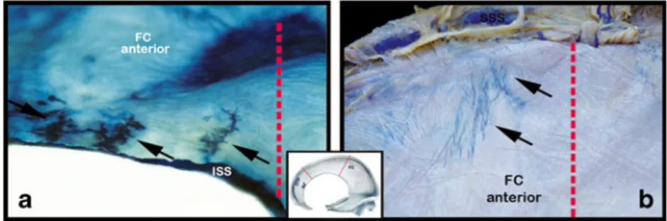
The latex/ink injections revealed a falcine venous plexus in all 16 falx cerebri. The venous plexus in the five latex-injected specimens was less dense than those in the 11 ink-injected specimens. The anterior third of the falx cerebri consisted of small diameter falcine venous vessels. These vessels were localized close to either superior or inferior sagittal sinus, and none extended as far as mid-falx cerebri levels in any of the 16 cases (Fig. 2a, b). They communicated with either superior or inferior sagittal sinuses, but not with both of these sinuses (Fig.2a, b). In the middle third of the falx cerebri, the majority of the vessels of the falcine venous plexus had larger diameter compared to anterior third. These vessels extended the length of the falx cerebri levels (Fig.3a, b). They commu-nicated with both superior and inferior sagittal sinuses (Fig.3a, b). In the posterior third of the falx cerebri, the vessels of the falcine venous plexuses had the largest diameter and were located at the junction of the inferior sagittal sinus and the straight sinus (Fig.4a, b). They were localized at the lower two-thirds of the falx cerebri. In all cases the dense venous networks communicated with the inferior sagittal sinus but not with the superior sagittal sinus. The falcine venous plexus observed in the posterior third of the falx cerebri was denser than in the anterior and middle portions.
Fig. 2 a, b The venous channels in the anterior third of the falx cerebri were small extensions from both the superior and inferior sagittal sinus that did not communicate: a A ink-injected falx cerebri showing venous channels extending from the inferior sagittal sinus; b A latex-injected
cadaver specimen showing venous channels extending from the superior sagittal sinus. A schematic illustration was added to show the localization of the venous channels in the anterior third of the falx cerebri
Fig. 1 a,b, c The falx cerebri had fenestrations of varying shapes at different locations: a An oval fenestration close to the inferior sagittal sinus located at the anterior third of the falx cerebri; b Multiple fenestra-tions close to the superior sagittal sinus and located at the middle third of
the falx cerebri; c A cribriform fenestration close to the inferior sagittal sinus and located at the posterior third of the falx cerebri. A schematic illustration was added to each figure to show the localization of the fenestrations
SEM findings
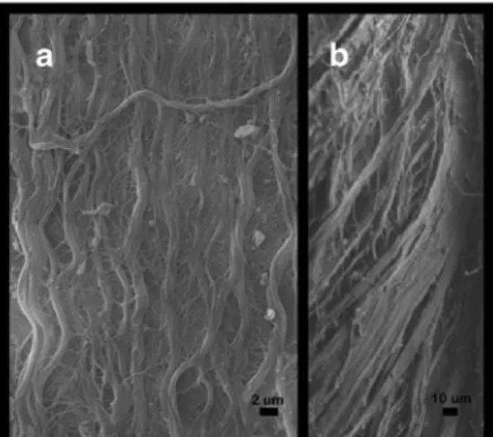
The falx cerebri specimens were composed of very dense connective tissue with numerous longitudinal bundles of col-lagen fibers, most of which ran vertically in the sagittal plane. Other bundles ran horizontally and obliquely, and were interconnected with the longitudinal collagen bundles (Fig.5a, b). The longitudinal collagen fibers formed gutters or channels in various regions of the falx cerebri where a falcine venous plexus was present, and these were vertically oriented and their surfaces in contact with blood vessels were smooth (Fig.6a ,b).
The specimens from the anterior third of the falx cerebri, both from the superior and the inferior section of tissue, fea-tured a falcine venous plexus and the mean diameter of these venous channels was 0.42±0.1 mm. Specimens from the mid-dle third of the falx cerebri had venous channels in both the superior and inferior tissue sections, with mean diameter 0.87± 0.17 mm. Specimens from the posterior third of the falx cerebri had numerous large channels in the inferior tissue section. The mean diameter of these channels was 1.38±0.21 mm, and they opened into the inferior sagittal sinus (Fig.7). In contrast, there
were no venous channels in the superior tissue sections from the posterior third (Fig.7).
Discussion
General considerations of the results
Our most important finding was that the falcine venous plexus exhibits different anatomical patterns in the anterior, middle, and posterior portions of the falx cerebri. These regional differences can be important for surgeons when making an incision or puncturing the falx cerebri during a procedure.
Tubbs et al. [27], examined 27 adult cadaveric specimens and categorized three types of this plexus based on communication between the plexus and the superior sagittal sinus: Type 1 had no communications with the superior sagittal sinus, type 2 had limited communications, and type 3 had significant communica-tions. In the present study, we have proposed a different classi-fication that may be more useful for the neurosurgeon. In our study, we reported findings for the falcine venous plexus in the anterior, middle, and posterior thirds of the falx cerebri. During
Fig. 4 a,b The venous plexus in the posterior third of the falx cerebri featured dense large venous channels extending from the inferior sagittal sinus and located at the junction of the inferior sagittal sinus and the straight sinus: a A ink-injected specimen showing dense venous channels
extending from the inferior sagittal sinus; b A latex-injected cadaver specimen showing dense venous channels extending from the inferior sagittal sinus. A schematic illustration was added to show the localization of the venous channels in the posterior third of the falx cerebri Fig. 3 a,b The venous plexus in the middle third of the falx cerebri
consisted of extensions from the superior and inferior sagittal sinuses, and most of these communicated: a A ink-injected falx cerebri showing venous extensions from the superior and inferior sagittal sinuses; b A
latex-injected cadaver specimen showing venous extensions from the superior and inferior sagittal sinuses. A schematic illustration was added to show the localization of the venous channels in the middle third of the falx cerebri
cranial surgery, the major external guide for neurosurgeons is the sutures. Therefore, we divided the falx cerebri into sectors using sutures of the skull as landmarks. Further, during surgery, neu-rosurgeons work through small windows in the region of interest; therefore, knowing the venous anatomy of specific regions of the falx cerebri can be more helpful than the percentage of veins draining into the superior sagittal sinus.
Tubbs et al. [27] described a“safe area” of the falx cerebri that contained no significant falcine venous plexus, and this section corresponds to the anterior sector of the falx cerebri in the present study. Where the falcine vessels are localized close to either superior or inferior sagittal sinus and do not extend to mid-falx cerebri levels, we agree that this area is“relatively safe,” although it is not free of venous channels. The falcine venous plexus in the middle third of the falx cerebri tended to be less dense than in the posterior third, where venous chan-nels form communications with both superior and inferior sagittal sinuses. The falcine venous plexus located at the middle sector of the falx cerebri can and it can become an important collateral venous outflow pathway when the supe-rior or infesupe-rior sagittal sinus is occluded. Our study and the research by Tubbs and coworkers [27] revealed a dense falcine venous plexus in the posterior third of the falx cerebri,
which points to the need for special care during transtentorial and posterior transcallosal surgical approaches, and when re-moving large meningiomas from this region of the falx cerebri. Tubbs et al. [27] stated that“the falcine venous plexus on the posterior aspect were usually more pronounced in the inferior thirds” of the falx cerebri. However, two of their photographs show weak communications with the superior sagittal sinus. In our study, we did not see relevant communi-cations with the superior sagittal sinus in this sector of the falx cerebri. This may be related to the different criteria used for defining the posterior falcine plexus in the two studies. Tubbs et al. [27] do not describe how they have defined the posterior falcine plexus. In the present study, we defined the posterior sector of the falx cerebri as extending from the lambdoid suture to the torcula. We may have included the communica-tions observed by Tubbs et al. in the middle third of the falx cerebri in the present study.
Our study is the first report of the SEM appearance of the venous gutters/channels that are located within the connective tissue of the falx cerebri. Protasoni et al. [21] examined the collagen architecture of the human dura mater using SEM. Our findings of interconnections between the thick longitudi-nal collagen bundles and the thin horizontal and oblique bundles in the falx cerebri were in accordance with these authors’ observations.
Fig. 5 a, b Scanning electron microscopy of the falx cerebri: a Dense longitudinal collagen bundles with scarce bundles also running horizon-tally; b Oblique-oriented collagen bundles
Fig. 6 a, b Scanning electron microscopy appearance of longitudinal collagen bundles forming gutters/channels within the falx cerebri
Fig. 7 Scanning electron microscopy appearance of venous channels in the falx cerebri opening into the superior sagittal sinus. FC: Falx cerebri, F: Fenestration, ISS: Inferior sagittal sinus, SSS: Superior sagittal sinus, TC: Tentorium cerebelli
giomas and noted that 45 % were anterior, 45 % were middle, and 10 % were posterior. These data show that most of these tumors are located in the anterior or middle portions of the falx cerebri, and that fewer occur in the posterior portion. Our anatomical results showed a dense falcine venous plexus in the posterior portion of falx cerebri. We suggest that special care should be taken in removing meningiomas that are pos-teriorly located.
A persistent falcine sinus is a large single venous channel that extends from the posterior third of the superior sagittal sinus to the beginning of the straight sinus, and has been extensively documented [4,10,14,16,23,25]. It has been frequently reported in the pediatric population, but has also been observed in adults [4,10,14,16,25]. None of our 16 falx cerebri specimens exhibited this anomaly.
Newer imaging techniques and improved technology can demonstrate the fine details of venous structures within the falx cerebri, and these can potentially help surgeons access this area more safely. However, many institutions do not have this technological capacity and in such situations it is valuable to have fine anatomical knowledge of the falcine venous plexus in the falx cerebri. This is particularly true regarding procedures for tumors that involve the dural sinuses, and for cerebral venous sinus thrombosis or falx meningiomas. Methodological considerations
We have used ink or a latex solution to mark the falcine venous plexus within the falx cerebri. The latex is a thicker solution compared to ink. Due to this, adequate amounts of latex may not have reached small diameter channels within the falx. Further, in the present study, the falx cerebri were obtained from cadavers and autopsies, although the superior and inferior sagittal sinus had been perfused before the dye injections. However, in four cases, thorough cleaning of the sinuses could not be achieved due to blood clots. Therefore, in some regions of the falx cerebri, there was limited labelling of the falcine venous plexus, and for this reason four cases were excluded from the study. Further, although all latex/ink injec-tions to the 16 falx cerebri were applied manually by a single observer, variations in the applied pressure may have oc-curred. The falx cerebri obtained from the cadavers were formaldehyde fixed and were more stiff compared to the autopsy specimens, and this may have produced some varia-tions in the amount of pressure applied. This may have resulted with leaving some unlabelled small diameter venous channels within the falx cerebri. Due to these limitations of the
specimen can be irreversibly damaged, as seen in the endo-thelial cells of the falcine venous structures [3,11].
Conclusion
The falcine venous plexus is a network of channels that are formed within the connective tissue of the falx cerebri during embryological development. Our findings reveal that in the posterior third of the falx cerebri, the falcine venous plexus is consistently dense with relatively large venous channels, whereas in the anterior and middle thirds, the plexus is less dense and more variable. Further, it is important for neurosur-geons to be aware of these regional differences so that safety can be maximized during procedures that involve or are close to this structure.
Acknowledgments The authors thank Dr. Uğur Ünal from Koç Uni-versity for his technical support with scanning electron microscopy. Conflicts of interest None.
References
1. Afifi AK BR (1998) Functional Neuroanatomy. McGraw-Hill, pp 62 2. Babu R, Barton A, Kasoff SS (1995) Resection of olfactory groove
meningiomas: technical note revisited. Surg Neurol 44:567–572 3. Brown RW (2009) Histologic preparations: common problems and
their solutions. College of American Pathologists, pp 56–57 4. Cai CQ, Zhang QJ, Yang WD, Wang CX, Shen CH (2009) Neuroimages
of persistent falcine sinus in children. World J Pediatr 5:63–64 5. EL Cushing H (1939) Meningiomas. Their classification, regional
behaviour, life history, and surgical end results. Br J Surg 26:957–957 6. Elsherbiny SM, Grunewald RA, Powell T (1997) Isolated inferior sagittal sinus thrombosis: a case report. Neuroradiology 39:411–413 7. Giombini S, Solero CL, Lasio G, Morello G (1984) Immediate and late outcome of operations for parasagittal and falx meningiomas. Report of 342 cases. Surg Neurol 21:427–435
8. Grunewald R, Powell T, Elsherbiny S (1998) Inferior sagittal sinus thrombosis. Neuroradiology 40:472
9. Hamilton WJMH (1972) Human embryology, 4th edn. The Williams and Wilkins Company, Baltimore, p 453
10. Kashimura H, Arai H, Ogasawara K, Ogawa A (2007) Persistent falcine sinus associated with obstruction of the superior sagittal sinus caused by meningioma–case report. Neurol Med Chir 47:83–84 11. Katelaris A, Kencian J, Duflou J, Hilton J (1994) Brains at necropsy:
to fix or not to fix? J Clin Pathol 47:718–720
12. Keogh AJ, Sharma RR, Vanner GK (1993) The anterior interhemispheric trephine approach to anterior midline aneurysms: results of treatment in 72 consecutive patients. Br J Neurosurg 7:5–12
13. Kesava PP (1996) Recanalization of the falcine sinus after venous sinus thrombosis. Am J Neuroradiol 17:1646–1648
14. Kim MS, Lee GJ (2010) Two Cases with Persistent Falcine Sinus as Congenital Variation. Journal of Korean Neurosurgical Society 48:82
15. Lacerte D, Gagne F, Copty M (1996) Intracranial chondroma. Report of two cases and review of the literature. Can J Neurol Sci 23:132– 137
16. Manoj KS, Krishnamoorthy T, Thomas B, Kapilamoorthy TR (2006) An incidental persistent falcine sinus with dominant straight sinus and hypoplastic distal superior sagittal sinus. Pediatr Radiol 36:65–67
17. Martin JH (1996) Neuroanatomy, 2nd edn. McGraw-Hill, pp 19–111 18. Netter FH (1983) Nervous system. Ciba-Geigy Cooperation, pp 56 19. Nikas DCBL, Black PM (2000) Parasagittal and falx meningiomas.
In: Kaye AHBP (ed) Operative Neurosurgery. Hartcourt Publishers Ltd, London, pp 505–521
20. Olivecrona H (1947) The parasagittal meningiomas. J Neurosurg 4: 327–341
21. Protasoni M, Sangiorgi S, Cividini A, Culuvaris GT, Tomei G, Dell’Orbo C, Raspanti M, Balbi S, Reguzzoni M (2011) The collagenic architecture of human dura mater. J Neurosurg 114:1723–1730 22. Rhoton AL Jr (2002) The cerebral veins. Neurosurgery 51:S159–205 23. Ryu CW (2010) Persistent falcine sinus: is it really rare? Am J
Neuroradiol 31:367–369
24. Sheerin F (2009) The imaging of the cerebral venous sinuses. Seminars in Ultrasound, CT, and MR 30:525–558
25. Strub WM, Leach JL, Tomsick TA (2005) Persistent falcine sinus in an adult: demonstration by MR venography. Am J Neuroradiol 26: 750–751
26. Toledo MM, Wilson TJ, Dashti S, McDougall CG, Spetzler RF (2010) Dural arteriovenous fistula associated with superior sagittal sinus occlusion secondary to invasion by a parafalcine meningioma: case report. Neurosurgery 67:205–207, discussion 207
27. Tubbs RS, Loukas M, Louis RG Jr, Shoja MM, Acakpo-Satchivi L, Blount JP, Salter EG, Oakes WJ, Wellons JC 3rd (2007) Anatomy of the falcine venous plexus. J Neurosurg 107:155–157
28. Williams PL, Warwick R, Dyson M, Banister LH (1989) Gray’s anatomy, 37th edn. Churchill Livingstone, London, pp 1086
Comment
The study by Tatarlı and Colleagues describing the macroscopic and microscopic anatomy of the venous system forming a "falcine venous plexus" is particularly relevant to neurosurgical practice. Actually, this falcine venous plexus could play an important role as a collateral venous outflow pathway when the superior sagittal sinus is occluded by a parasagittal meningioma.
When dealing with meningiomas occluding the middle or posterior third of the superior sagittal sinus, resection of the sinus en bloc with the tumor is commonly considered a safe procedure. Nevertheless, several cases of patients who deteriorated and died following en bloc resection of a completely occluded sinus with the tumor have been described. This circumstance could be reasonably explained on the grounds of lack of development of adequate collateralization of the venous outflow anterior to the site of occlusion. The implication is that in those cases without adequate collateralization of the venous outflow anterior to the site of occlusion, a complete en bloc resection of the tumor with the sinus could interrupt anastomotic pathways that run through the tumor, the capsule of the tumor, or even the walls of the sinus and along the falx [1]. The present study clearly demonstrates the existence of a venous plexus coursing along the falx that otherwise could be only supposed.
The resection of a parasagittal meningioma together with the occluded sinus should be therefore performed only if there is evidence that an adequate venous outflow from the sinus anterior to the area of occlusion has been developed. This can occur by drainage through the most distal patent veins prior to the occlusion into alternative routes like the trans-verse sinus through the vein of Labbé or the spheno-parietal sinus through sylvian veins [2].
References
1. Heros RC (2006) Meningiomas involving the sinus. J Neurosurg 105:511-513; discussion 513
2. Tomasello F, Conti A, Cardali S, Angileri FF (2013) Venous preservation-guided resection: a changing paradigm in parasagittal me-ningioma surgery. J Neurosurg 119(1):74-81
Alfredo Conti Francesco Tomasello Messina, Italy