The pathological findings of Rhodococcus equi infection and its
diagnosis with immunoperoxidase technique in foals
*Şule Yurdagül ÖZSOY1, Rıfkı HAZIROĞLU1
1Department of Pathology, Faculty of Veterinary Medicine, Mustafa Kemal University, Hatay; 2Department of Pathology, Faculty of
Veterinary Medicine, Ankara University, Ankara.
Summary: Twelve foals with difficulty in respiration that have died during spring-summer months between 2003-2006 years were necropsied. On necropsy, pneumonia was detected in all of the foals. In 5 animals, caseo-necrotic nodules were seen in the lungs. In histopathological examination, purulent bronchopneumonia was noticed. In immunohistochemical staining, Rhodococus
equi antigens were determined in the cytoplasm of macrophages and occasionally in neutrophils in lungs and mediastinal lymph
nodes by immunoperoxidase technique. No lesions were detected in the intestines confirming that this form of infection is rare compared to the lung form. In the present study, R. equi was microbiologically isolated only in two cases. In conclusion, R. equi infection was diagnosed in all of the twelve cases, and estimated to be the reason of pneumonia in the foals. It was also shown that immunoperoxidase technique can be successfully used for R. equi infection in the field.
Key words: Foal, immunoperoxidase technique, pathology, Rhodococcus equi.
Taylarda Rhodococcus equi enfeksiyonunun patolojik bulguları ve immunoperoksidaz tekniği ile tanısı
Özet: Klinik olarak solunum güçlüğü bulguları gösteren ve 2003-2006 yıllarının ilkbahar-yaz aylarında ölen 12 tayın nekropsisi yapıldı. On iki tayda da R. equi’ye bağlı pnömoni saptandı. Makroskobik olarak; 5 olguda akciğerde tipik kazeonekrotik odaklarla karşılaşıldı. Histopatolojik incelemede; irinli bronkopnömoni tablosu dikkati çekti. R. equi’ nin, yapılan immunoperoksidaz boyamalarla, akciğer ve mediastinal lenf düğümü kesitlerinde, çoğunlukla makrofajların, bazen de nötrofil lökositlerin sitoplazmasında yerleştiği görüldü. Bağırsaklarda lezyon görülmemesi, bu formun akciğer formuna göre ender şekillendiğini gösterdi. Sadece iki olguda mikrobiyolojik olarak R. equi izole edilebildi. Sonuç olarak; çalışmada kullanılan 12 tayda da R. equi’nin saptanmış olması, taylarda görülen pnömonilerin çoğunluğundan bu bakterinin sorumlu olduğunu ve ayrıca tanısında immunoperoksidaz tekniğinin başarıyla kullanılabileceğini gösterdi.
Anahtar sözcükler: İmmunoperoksidaz tekniği, patoloji, Rhodococcus equi, tay.
* This research has been summarized from Ph.D. thesis. It was supported by projects TÜBİTAK (VHAG-2013) and BİYEP
(2005K120140-7).
Introduction
Rhodococcus equi (R. equi) is a facultative
intracellular pathogen of macrophages (5, 12, 14). It is an opportunistic pathogen that causes purulent bronchopneumonia, pulmonary abscesses, ulcerative enteritis, and is associated with lymphadenitis in 1 to 6 months old foals (5, 15, 16). The pathogen has also been less frequently associated with osteomyelitis in foals (18). The infection occurs sporadically on some farms and morbility rate increases as high as 80% at newborns when the immune system is still immature and maternal antibodies have disappeared (19). However, the disease is very rare in adult horses (5, 17).
R. equi is a robust soil organism living widespread
in the environment. The organism potentially multiplies wherever there is horse manure. Temperature plays a major role in the growth of R. equi, which shows an
optimal growth at 30oC. A direct relationship exists
between the number of R. equi in the environment of young foals and the number of pneumonia. The pathogen is commonly found in loafing paddocks on horse breeding farms during summer time and reaches to the lung by inhalation (4, 23, 24).
The purpose of this study was to investigate pathological findings of R. equi infection, which causes high mortality in foals, and to evaluate the use of immunoperoxidase technique in the diagnosis of the infection.
Materials and Methods
The study materials were twelve foals suffering form respiratory difficulty and died during spring-summer months in 2003-2006. following routine necropsy, samples of all the organs were fixed in 10%
buffered formalin solution, embedded in parafin and cut into 5-6 µ thick sections that were stained with haematoxyline-eosin (HE), Brown-Brenn and Ziehl-Neelsen staining. Then, the sections were dewaxed and rehydrated by routine methods for immunohistochemical staining as follows. The preparations were incubated in 3% H2O2 solution for 5 minutes, then incubated with
normal goat serum for 20 min at 40°C. The sections were incubated with anti- Rhodococcus equi hyperimmune serum diluted 1:300 (AGIDT: 1/200+++) for 1 hour at room temperature (1:300 dilution). The sections were then incubated with streptavidin-peroxidase reagent for 20 min (Dako/Denmark). Colour labeling was developed by a final incubation step using 3-amino-9-ethyl-carbazole (AEC, Dako/Denmark) for 7 minutes. Finally, the sections were counterstained with Mayer’s haematoxyline and covered with glycerol-gelatin.
Results
High fever (41oC) (except in case 11), severe
coughing and difficulty in breathing (except in case 9) were seen as the general clinical signs in the foals. In case 9 ulseration in cornea and swollen joints were noticed.
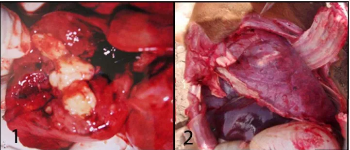
Significant pathological changes were observed only in the lungs. Nodules, which were yellowish-white in colour, hard or fluctuant in consistency, about walnut sized, and surrounded by severe and extensive consolidation and atelectatic areas, were detected in lung lobes. On cut surface, the nodules were irregular and well defined and showed white firm or caseous appearance (Figures 1, 2). In four cases (Cases 1, 8, 10, 12), the nodules were located in the cranial lung lobes. In case 7, nodules involved in all lung lobes while in case 2 the nodule was located in the cranio-ventral lobe. A serous
bloody and foamy fluid was seen at bronch and bronchial lumens. In case 9 and 11, hemorrhage and consolidation were the only findings in lungs. There were no lung lesions in cases of 3, 4, 5, and 6.
The mediastinal lymph nodes were swollen and the cut surface showed yellowish-green in colour and creamy appearance (Cases 7, 8, 11) (Figures 9, 10). Moreover, macro-pathologic changes were noted in liver (Cases 8, 11, 12), kidney (Cases 8, 11) and intestines (Case 8).
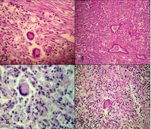
The macroscopic findings are listed in Table 1. Histologically, bronchopneumonia was observed in the lungs. Various degrees of hyperemia (Cases 3, 5, 7, 9, 11 and 12) and thrombosis were noted (Case 12). Macrophages and neutrophiles, which were observed at alveoli lumens, were seen at bronch and bronchiole and around the blood vessels. The degree of these inflammatory cells which were weak at Cases 3 and 6; were perivascular placement at Case 3. In cases 1, 4, 8 and 12, multinucleated giant cells accompanied inflammatory cell infiltration (Figures 3-6). In some cases (Cases 1, 2, 5 and 12), necrosis of inflammatory cells and alveoli walls were seen. Hyalinization of inflammatory exudate (Case 12) was determined in alveoli lumens. Inter-alveolar septums were dilated with fibrous tissue elements at cases 1, 2 and 4. In some alveoli lumens, pink homogeneous oedema fluid and erythrocytes were observed. Bronchus and bronchioles epitelheliums were hyperplastic, and occasionally desquamated in lumens (Cases 5, 8). Peribronchial lymph follicles were hyperplasic in case 4. Oedema (Case 8), macrophage and lymphocyte infiltration, and fibrous tissue proliferation were also seen in pleura (Case 7).
Between the severe inflammatory cell infiltrations and alveoli lumens, immunopositive macrophages and neutrophils were observed (Figures 7, 8).
Figure 1. Lung, case number 7; nodule; yellowish-white in colour and fluctuant in consistency. Şekil 1. Akciğer, olgu no 7; sarımtırak-beyaz renkli, fluktuan kıvamlı nodül.
Figure 2. Lung, case number 10; caseous nodule; hard in consistency, yellowish-white in colour.
Table 1.The macroscopic findings of infection. Tablo 1. Enfeksiyonda gözlenen makroskobik bulgular.
Case number The localization of nodules/ Other lesions
The consistency and appearance of nodules
The size of nodules
Mediastinal lymph nodes İntestine Liver Rens
1 Left cranial lobe
Hard in consistency, yellowish-white in colour, caseous nodule
4x3 cm - - - - 2 Cranioventral lobe 2x3 cm - - - - 3 - - - - - - 4 - - - - - - 5 - - - - - - 6 - - - - - - 7 Diffuse Hard in consistency, yellowish-white in colour, caseous nodule
Varried between 2x3 cm- 4x5.5
Swollen and the cut surface showed yellowish-green in colour
and creamy appearance
- - -
8 Right cranial lobe
Hard and some areas fluctuant in consistency, white in colour, capsuled
with fibrous tissue, caseous nodule
1.5x2 cm and 1x1.5
cm
Swollen and the cut surface showed yellowish-green in colour
and creamy appearance
Subserozal damarlar dolgun ve lumeninde sarı sulu renkte içerik ile mukozada hiperemi Swollen, cut surface was hemorrhagic İncrease in size 9 hemorrhage Petechial areas - - - - - -
10 Right cranial lobe Hard in consistency, yellowish-white in colour, caseous nodule
2.5x3 cm - - - - 11 Consodilation at cranial lobes and diffuse petechial hemorrhagies - -
Swollen and cut surfaaces
had hemorrhage areas -
Swollen, cut surface was hemorrhagic Diffuse, small, white in colour focuses located at cortex 12 Right cranial lobe Very hard in consistency, white in colour, capsuled with fibrous tissue caseous
nodule
3x4 cm - -
Swollen, cut surface was hemorrhagic -
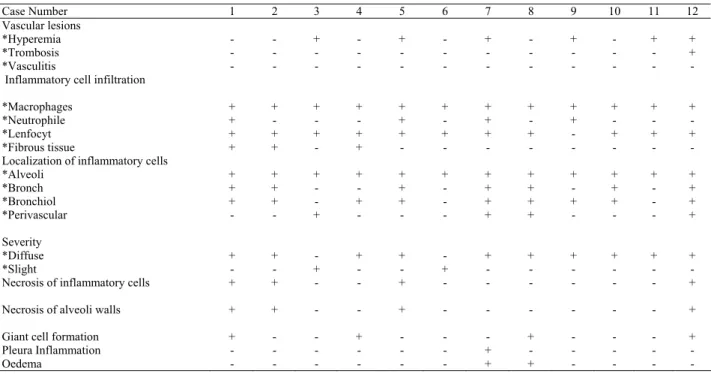
Table 2. The microscopic findings of infection. Tablo 2. Enfeksiyonda gözlenen mikroskobik bulgular.
Case Number 1 2 3 4 5 6 7 8 9 10 11 12 Vascular lesions *Hyperemia *Trombosis *Vasculitis - - - - - - + - - - - - + - - - - - + - - - - - + - - - - - + - - + + - Inflammatory cell infiltration
*Macrophages *Neutrophile *Lenfocyt *Fibrous tissue + + + + + - + + + - + - + - + + + + + - + - + - + + + - + - + - + + - - + - + - + - + - + - + - Localization of inflammatory cells
*Alveoli *Bronch *Bronchiol *Perivascular Severity *Diffuse *Slight + + + - + - + + + - + - + - - + - + + - + - + - + + + - + - + - - - - + + + + + + - + + + + + - + - + - + - + + + - + - + - - - + - + + + + + - Necrosis of inflammatory cells
Necrosis of alveoli walls
+ + + + - - - - + + - - - - - - - - - - - - + +
Giant cell formation + - - + - - - + - - - +
Pleura Inflammation
Figure 7. Lung, case number 5; immunopositive staining at macrophages against R. equi antiserum, ABC-P,1000x. Şekil 7. Akciğer, olgu no 5; makrofajlarda R. equi antiserumuna karşı IP pozitif boyanmalar, ABC-P, 1000x. Figure 8. Lung, case number 11; immunopositive staining at macrophages against R. equi antiserum, ABC-P,1000x. Şekil 8. Akciğer, olgu no 11; makrofajlarda R. equi antiserumuna karşı IP pozitif boyanmalar, ABC-P, 1000x.
Figure 3. Lung, case number 4; increase of multinucleated giant cell, macrophage, lymphocyte and fibrous tissue, H&E, 400x. Şekil 3. Akciğer, olgu no 4; çok çekirdekli dev hücreleri, makrofaj, lenfosit ve fibröz doku artışı, HE, 400x.
Figure 4. Lung, case number 5; macrophages and other imflammatory cell infiltration in alveoli and bronchial lumens, H&E, 40x. Şekil 4. Akciğer, olgu no; 5 alveol ve bronşiol lumenlerinde, makrofaj ve diğer yangısal hücreler, HE, 40x.
Figure 5. Lung, case number 8; multinucleated giant cells, macrophages and lymphocyte infiltration in alveoli lumens and paranchyma, H&E, 400x.
Şekil 5. Akciğer, olgu no 8; alveol lumenlerinde ve paranşimde çok çekirdekli dev hücresi, makrofaj ve lenfosit infiltrasyonu, HE, 400x. Figure 6. Lung, multinucleated giant cells and mononuclear cell infiltration in alveolar exudate, H&E, 400x
Vascular hyperemia and sinusoidal dilatation were observed in mediastinal lymph nodes. Lymphatic follicles were hyperplastic, and infiltrations of macrophage, neutrophile and lymphocyte cells were seen in sinus lumens (Cases 7, 8, 11), and necrosis was noted in Case 7 (Figure 11).
R. equi immunopositive macrophages were seen in
medullar sinuses of mediastinal lymph nodes (Case 7) (Figure 12).
Subacut enteritis was observed in the intestine of case 8. In liver, passive hyperemia (Cases 8, 9, 11, 12), degenerative changes (Cases 8, 9, 10, 11, 12) and serous
Figure 9. Mediastinal lymph node, case number 7; swollen and the cut surface showed yellowish-green and creamy appearance material.
Şekil 9. Mediastinal lenf düğümü, olgu no; 7, şişkin, kesit yüzü sarımtırak yeşil renkli ve krema benzeri görünüşte materyal.
Figure 10. Mediastinal lymph node, case number 8; swollen and the cut surface showed yellowish-green in colour and creamy appearance material.
Şekil 10. Mediastinal lenf düğümü, olgu no; 8, şişkin, kesit yüzü sarımtırak yeşil renkli ve krema benzeri görünüşte materyal. Figure 11. Mediastinal lymph node, case number 7; widen of sinuses, macrophage, neutrophile and lymphocyte cell infiltration was seen in sinus lumens. Lymphoidal necrosis was seen, HE, 400x.
Şekil 11. Mediastinal lenf düğümü, olgu no; 7, sinusta genişleme, lümeninde makrofaj, nötrofil ve lenfosit infiltrasyonu. Ayrıca kortekste lenfoid nekrozlar gözlenmekte, HE, 400x.
Figure 12. Mediastinal lymph node, case number 7; IP positive staining at macrophages, ABC-P, 400x. Şekil 12. Mediastinal lenf düğümü, olgu no; 7, makrofajlarda IP pozitif boyanmalar, ABC-P, 400x.
Table 3. The immunoperoxidase results at lungs and mediastinal lymph nodes.
Tablo 3. Akciğerler ve mediastinal lenf düğümlerinde gözlenen immunoperoksidaz sonuçlar.
Case number 1 2 3 4 5 6 7 8 9 10 11 12
Lungs ++ ++ + + ++ + + ++ + + ++ +
Mediastinal lymph
nodes - - - + - - - - -
hepatitis (Cases 9, 12), in rens tubulonephrosis (Cases 8, 10), interstitial nephritis and at glomerular capillaries and at interstitial areas bacterial gatherings were observed (Case 11). The bacterial gatherings were stained blue-purple with Brown-Brenn Gram and Ziehl-Neelsen. The microscopic and immunoperoxidase findings were listed in Tables 2 and 3.
For microbiological examination, tissue samples were taken from cases 8, 10, 11. Only in two cases (Cases 8, 11) R. equi was isolated microbiologically.
Discussion and Conclusion
In this study, gross and microscopic findings of R.
equi infection were evaluated in foals. To determine the
presence and localization of R. equi antigens, immunoperoxidase technique was used.
Factors such as age, season and sheltering condition are predisposing factors for R. equi infection. When the immune system is immature and maternal antibodies have disappeared, the infection causes death about 80% in new born foals (19). Temperature plays a major role in the growth of R. equi, as the optimum growth is succeeded at 30oC. A direct relationship
between the number of R. equi in the environment of young foals and the number of pneumonia exists. The pathogen is commonly found in loafing paddocks on horse breeding farms in summer and reaches to the lung by inhalation (4, 23, 24). In the present study, age of foals varied between 3 days old to 7 months old, and death of foals were seen between the months of April and August.
Clinical findings of high fever (41-41.5 oC), cough
and difficult breathing were similar with other reports (1, 15, 22). Infection at distant sites from the respiratory or gastrointestinal tract is likely to be associated with bacteremic dissemination and localization of bacteria at other sites. (2, 5, 15). Ulseration of cornea and swelling at joints were seen before foal 9 was died was also explained with these reports.
The main routes of infection are the respiratory and alimentary tracts. Inhalation of the organism is probably is the main route of exposure in all foals (22). Zink et all. (1986) reported that 96 % of pneumonias observed in foals which were died due to R. equi infection (27) and this infection accounted for 45 % of all foals with pneumonia (25). The lesions are usually more extensive in the right lung than the left (7). Similar to the report of Hillidge (1986) (7), most of the lesions in cases 8, 10 and 12 were located in the right lung in the present study. Although congestion and consolidation was the only findings in cases 9 and 11, in some cases (Cases 3, 4, 5, 6)no macroscopic findings were observed. These findings thought that at peracute form of infection death
was occured quickly. The isolation of R. equi from case 11 is supported this opinion.
The macroscopic appearance of caseous nodules in lungs and the swollen mediastinal lymph nodes, as well as morphological features in the cut surfaces of nodules and lymph nodes were similar with previous reports (6, 11, 14).
Infiltration of numerous bacteria laden macrophages and neutrophils with multinucleated giant cells and necrosis of these inflammatory cells were seen in lungs. The microscopic appearance of lungs seemed as in previous reports (6, 11, 20, 21). In mediastinal lymph nodes, inflammatory cell infiltration and necrosis of lymphoid follicles were also observed similarly to the previous reports (8,14).
R. equi induced enterocolitis has been described in a
few case reports. The findings of this form ulcerative enteritis and associated with lymphadenitis (3, 5, 10, 15, 16) were not seen in the present study. Only subacute enteritis was observed in case 8. No findings any lesions in intestine confirmed that this form of the infection is rarely encountered compared to the lung form.
When R. equi agents spread from lungs, pathological lesions may form in parenchymal organs such as liver and kidney (9). In this study, gross and histopathologic changes were seen in liver (Cases 8, 11, 12) and kidney (Cases 8, 11)
Morphological identification of R.equi in tissue samples is difficult, since spesific histochemical stains are not available. Gram and ZN stains can be regarded only as diagnostic aids. But immunohistochemical tests which are highly sensitive and specific, detect intracellular and extracellular antigen (13). The immunohistechemical method is relatively inexpensive and, unlike PCR, does not require special laboratories or specilalist personel. In contrast to indirect immunofluorescense (IIF), immunohistochemical preparations can be examined with a simple light microscope and can be stored for extended periods, and immunohistochemistry enables the study of cell and tissue morphology at the same time (20). R. equi has been detected by immunohistochemical methods, using polyclonal and monoclonal antibodies in formalin-fixed and parafin-embedded sections. R. equi antigens were demonstrated with immunoperoxidase technique usually in cytoplasm of macrophages and rarely in cytoplasm neutrophils and giant cells at lung and mediastinal lymph node sections (8, 13, 14, 20, 21, 26). In the present study the R. equi antigens were located in the center of suppurative inflammation, in the bronchiole lumina, and in the cytoplasm of macrophages and rarely neutrophils. In the mediastinal lymph nodes near the necrotic lenfoid follicles, R. equi positive immunostained few macrophages were seen (Case 7).
In this study, presence of macrophages, some of them were bacteria laden, within the lesions could be explained by role of these cells as the main defender against agents that are inhaled as previously reported (5, 8, 13, 24 ).
In conclusion, R. equi infection was diagnosed in twelve foals. It was concluded that R. equi might be one of the most important factor in foal pneumonias. We have shown that immunoperoxidase technique could be successfully used as a diagnostic mean of the infection on the field cases.
Acknowledgements
I’m grateful to Professor Doctor Yakut ÖZGÜR (University of İstanbul, Faculty of Veterinary Medicine, Department of Microbiyology) for assurance of virulent
R. equi which was containing an 85 kb type I plasmid,
was used for preparing immunization antigen. References
1. Aucoin S, Eades CS (2000): Rhodococcus equi
pneumonia in foals.
http://eurp./Isu.edu/healthtips/Rhodococcus-equi-Pneumonia.htm. Accessed April 10, Accessed 18.12.2002
2. Chaffin MK, Honnas CM, Crabill, MR, Schneiter HL, Brumbaugh GW, Beiner RP (1995): Cauda equina
syndrome, diskonpondylitis and a paravertebral abscess caused by R.equi in a foal. JAVMA, 206, 215-220.
3. Cimprich RE, Rooney JR (1977): Corynebacterium equi
enteritis in foals. Vet Pathol, 14, 95-102.
4. Cohen ND, O’conor MS, Chaffin MK, Martens RJ (2005): Farm characteristics and management practices
associated with development of Rhodococcus equi pneumonia in foals. JAVMA, 226, 404-413.
5. Giguere S (2000): Rhodococcus equi infections.
http://www.ivis.org ,Accessed 13.01. 2003
6. Hazıroğlu R (2001): Solunum sistemi. 97-98. In: R. Hazıroğlu, Ü.H. Milli (Ed), Veteriner Patoloji.2. Baskı, Medipres Yayıncılık, Malatya.
7. Hillidge CJ (1986): Review of Corynebacterium
(Rhodococcus) equi lung abscesses in foals: pathogenesis, diagnosis and treatment. Vet Rec, 119, 261-264.
8. Ishino S, Kumagai K, Kuniyoshi S, Nakazawa M, Matsuda I, Oka M (1992): Immunohistochemical
observations on pneumonic lesions caused by Rhodococcus equi in foals. J Vet Med Sci, 54, 509-515.
9. Johnson JA, Prescott JF, Markham RJF (1983): The
pathology of experimental Corynebacterium equi infection in foals following intrabronchial challenge. Vet Pathol, 20,
440-449. 10. Johnson JA, Prescott JF, Markham RJF (1983): The
pathology of experimental Corynebacterium equi infection in foals following intragastric challenge. Vet Pathol, 20,
450-459.
11. Karadaş E, Gülcü HB, Beytut E, Kahraman EM (1997): İki arap tayında R.equi (Corynebacterium equi)
enfeksiyonu. FÜ Sağlık Bil Derg, 11, 321-325.
12. Kedlaya I (2007): Rhodococcus equi. http://emedicine.
com/med/topic3378.htm. Accessed 01.12.2005
13. Madarame H, Takai S, Morisawa N, Fujii M, Hidaka D, Tsubaki S, Hasegawa Y (1996): Immunohistochemical
detection of virulence-associated antigens of Rhodococcus equi in pulmonary lesions of foals. Vet Pathol, 33,
341-343.
14. Mariotti F, Cuteri V, Takai S, Renzoni G, Pascucci L, Vitellozi G (2000): Immunohistochemical detection of
virulence- associated R.equi antijens in pulmonary and intestinal lesions in horses. J Comp Path, 123,186-189.
15. Özgür NY, Ilgaz A (2000): Tayların R.equi pnömonisi. İnfeksiyon Derg, 15, 405-408.
16. Özgür NY, İkiz S, Carioglu B, Ilgaz A, Takai S (2000):
Two cases of dead foals associated with R.equi pneumonia in Turkey. J Equine Sci, 11, 1-5
17. Özgür NY, İkiz S, Bagcigil F, Carioglu B, Ilgaz A, Takai S (2002). Rhodococcus equi pneumonia in a mare
in Turkey. Vet Rec, 151, 613
18. Price CS, Rush BR, Gaughan EM, Cox JH (2003):
Osteomyelitis of the pelvis caused by Rhodococcus equi in a two-year-old horse. JAVMA, 222, 969-972.
19. Riedesel EA (1996): What’s Your Radiogaphic
Diagnosis? College of Veterinary Medicine. Iowa State
Univ Vet, 58, 31-32.
20. Szederi L, Makrai L, Denes B (2001): Rapid
ımmunohistochemical detection of Rhodoccoccus equi in impression smears from affected foals on postmortem examination. J Vet Med B, 48, 751-758.
21. Szederi L, Molnar T,Glavits R, Takai S, Makrai L, Denes B, Del Piero F (2006): Two cases of equine
abortion caused by Rhodoccoccus equi. Vet Pathol, 43,
208-211.
22. Takai S, Sasaki Y, Tsubaki S (1995): Rhodoccoccus equi
infections in foals-current concepts and implication for future research. J Equine Sci, 6, 105-119.
23. Thomas HS (2000): Foal pneumonia caused by
Rhodoccoccus equi. http://www.Jully.htm. Accessed
13.01.2003
24. Wada R, Kamada M, Anzai T, Nakanishi A, Kanemaru T, Takai S, Tsubaki S (1997): Pathogenecity and
virulence of Rhodococcus equi in foals following intratracheal challenge. Vet Microbiol, 56, 301-312.
25. Wright B (2006): Rhodococcus equi pneumonia of foals.
http://www.omafra.gov.on.ca/english/livestock/horses/facts /90-056.htm]. Accessed 02.05.2006
26. Zink MC, Yager JA, Prescott JF, Wilkie BN (1985): In
vitro phagocytosis and killing of Corynebacterium equi by alveoler macrophages of foals. Am J Vet Res, 46,
2171-2174.
27. Zink MC, Yager JA, Smart NL (1986):
Corynebacterium equi infections in horses, 1958-1984: a review of 131 cases. Can Vet J, 27,213-217.
Geliş tarihi: 13.05.2008 / Kabul tarihi: 07.07.2008
Address for correspondence:
Dr.Şule Yurdagül Özsoy Department of Pathology Faculty of Veterinary Medicine
Mustafa Kemal University, Hatay, Turkey E-mail: [email protected]