Aydın, E et al 2016 Vascular Leiomyoma Presenting as Anterior Knee Pain (Case Report). Journal of the Belgian Society of Radiology, 100(1): 50, pp. 1–3, DOI: http://dx.doi.org/10.5334/jbr-btr.976
CASE REPORT
Vascular Leiomyoma Presenting as Anterior Knee Pain
(Case Report)
Elçin Aydın
*, Gokcen Coban
*, Esra Zeynep Coşkunoğlu
*and Mehmet Tükenmez
*Vascular leiomyomas or angioleiomyomas are rare benign solitary smooth muscle tumors that origin usually in the extremities. Most of these tumors are composed of venous vessels. Here in, we report a rare case of subcutaneous vascular leiomyoma of the right knee of a 38 year old woman who was presented with recurrent anterior right knee pain and soft tissue swelling. Clinical findings, magnetic resonance imaging and histopathologic findings of the tumor is discussed. Leiomyomas are not mostly considered in the dif-ferential diagnosis by radiologist due to its rarity. Typical imaging and clinical findings of a tumor is an important clue for an accurate and early diagnosis.
Keywords: vascular leiomyoma; angioleiomyoma; subcutaneus tumours; MRI Background
Angioleiomyomas or vascular leiomyomas are rare subcu-taneus benign tumours arising from smooth muscle cells of arterial or venous walls [1]. They are most frequently seen in women and in the lower extremities [2,3]. The typ-ical presentation is a painful subcutaneus mass. Magnetic resonance imaging (MRI) and clinical findings help with the diagnosis. Our aim is to present a rare case of vascular leiomyoma in the right knee of a 38-year-old woman with MRI, clinical, and pathological findings.
Case Presentation
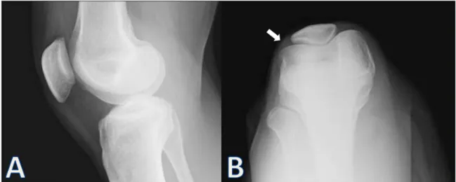
A 38-year-old woman presented with painful, mobile swelling in the right knee. There was no trauma history, and the onset of pain was sporadic and had reduced her mobility. On physical examination, there was a mobile painful swelling on the lateral aspect of her right knee. The laboratory findings were normal. Plain orthogonal radiographs including lateral and skyline views of the knee demostrated no bone abnormalities (Figure 1A–B).
But there was a soft tissue swelling on the lateral aspect of the patella (Figure 1B). MRI examination with and
without contrast was performed. An MRI of the right knee revealed a subcutenous, ovoid, well-circumscribed, homogenous, capsulated soft tissue mass adjacent to the lateral patellar retinaculum (Figure 2A–F). The tumor was
hyperintense on proton density (PD) images and hypoin-tense on T1-weighted images according to muscles. On post-contrast T1-weighted (Figure 1B) and PD-weighted
(C) images there was a sharp thin hypointense rim
sur-rounding the lesion. The tumor showed marked homoge-neous gadolinium enhancement after contrast adminis-tration (Figure 1B, E–F). There was no joint effusion or
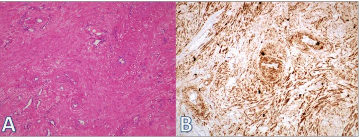
soft tissue edema. The muscles and bones were normal. The patient underwent surgery and complete excision of the tumor was performed. Gross examination revealed a 10×10 mm firm, well-circumscribed, bean-shaped mass with a white-beige cut surface. Histological sections demonstrated a solid tumor composed of intersecting fascicles of mature smooth muscle cells surrounding vas-cular structures (Figure 3A). The smooth muscle cells
showed no cellular atypia, and mitotic figures were rare. Immunohistochemical analysis showed diffuse positiv-ity for smooth muscle actin (SMA) (Figure 3B). After the
operation, her symptoms disappeared.
Discussion
Vascular leiomyomas or angioleiomyomas are benign sub-cutaneus tumours that originate from the smooth mus-cles of blood vessels. They commonly influence the lower extremities, but they seldom affect the knee [1,2]. Females are more likely to be affected than males; vascular leiomy-omas generally occur in the third or fourth decads [2,3]. Angioleiomyomas are generally seen in the deep layers of the dermis or in the subcutaneus tissue. Histologically, angioleiomyomas consist of smooth muscle bundles, vas-cular channels, and a thin fibrous capsule.
Morimoto defined three subtypes: solid or capillary, cav-ernous, and venous [3,4]. Solid is the most frequent, three times as common as in females and typically seen in the lower extremities. The cavernous subtype is more common in males than females and involves the head and upper extremities [4,5]. The most characteristic complaints are pain and tenderness. A painful subcutaneus mass in the lower extremities is the typical finding in the solid
* Başkent University, TR [email protected] Corresponding author: Elçin Aydın
Aydın et al: Vascular Leiomyoma Presenting as Anterior Knee Pain (Case Report) Art. 50, pp. 2 of 3
histological subtype. Differential diagnoses of a leiomyoma are glomus tumor, hemangiomas, angiolipoma, ganglion, schwannoma, giant cell tumor, neurilemoma, traumatic neuromas, and eccrine spiradenoma [4]. Small malignant lesions cannot be excluded by clinic or MRI alone.
Ultrasound remains the first-line imaging tool to confirm the presence of a mass lesion and its solid
nature. MRI is the best choice for imaging because it can better delineate the lesion and define its relation-ship to the adjacent structures. Hwang et al. defined that hyperintens areas on T2-weighted MR images show the smooth muscle and numerous vessels in vas-cular leiomyomas [6]. The fibrous capsule defined as a hypointense rim on T2-weighted MRI, with contrast
Figure 2: This is a preoperative MRI of the right knee. Axial (A) and coronal (D) T1-weighted pre-contrast images show
well-circumscribed, hypointense soft tissue lesion adjacent to the lateral patellar retinaculum. The axial PD-weighted imaging with fat suppression (C) image shows a well-circumscribed, marked homogenous hyperintense subcutaneus lesion. Post-contrast axial (B), fat-supressed coronal (E), and sagital (F) T1-weighted images show prominent enhance-ment of the tumor and a sharp outline.
Figure 1: Lateral (A) and skyline (B) plain orthogonal radiographs of the right knee demostrate no bone abnormalities.
Aydın et al: Vascular Leiomyoma Presenting as Anterior Knee Pain (Case Report) Art. 50, pp. 3 of 3
it has marked enhancement that shows its vascular origin [4–7].
In conclusion, when a painful subcutaneus swelling in lower extremities is seen, vascular leiomyoma must be kept in mind for the differential diagnosis. MRI is the best choice for imaging that can delineate the lesion and define its relationship to the adjacent structures, allowing better pre-excisional planning.
Leiomyomas are not often considered in the differential diagnosis by radiologists due to their rarity. They are usu-ally well-circumscribed, sharply outlined, homogenous, and show prominent enhancement. Typical imaging and clinical findings of a tumor are important clues for an accurate and early diagnosis.
Competing Interests
The authors declare that they have no competing interests.
References
1. Stout, AP. Solitary cutaneus and subcutaneus
leio-myoma. Am J Cancer. 1937; 29: 43569. DOI: http:// dx.doi.org/10.1158/ajc.1937.435
2. Hachisuga, T, Hashimoto, H and Enjoji, M.
Angi-oleiomyoma: a clincopathologic reappraisal of 562 cases. Cancer. 1984; 54: 125–30. DOI: http://dx.doi. org/10.1002/1097-0142(19840701)54:1<126::AID-CNCR2820540125>3.0.CO;2-F
3. Angiomyoma, MN. A clinicopathologic study. Med J Kagoshima Univ. 1973; 24: 663–83.
4. Cantisani, V, D’Ambrosio, OM, Marsecano, GI,
et al. Vascular leiomyoma presenting asmedial joint line pain of the knee. Journal of Ultrasound. 2009; 12(4): 163–5. DOI: http://dx.doi.org/10.1016/j. jus.2009.09.009
5. Duhig, JT and Ayer, JP. Vascular leiomyoma: a study
of 61 cases. Arch Pathol Lab Med. 1959; 68: 424–30. 6. Hwang, JW, Ahn, JM, Kang, HS, et al. Vascular
leiomyoma of an extremity: MR-imaging-pathology correlation. AJNR. 1998; 171: 981–5. DOI: http:// dx.doi.org/10.2214/ajr.171.4.9762979
7. Gulati, MS, Kapor, A and Maheswari, J.
Angio-myoma of the knee: value of magneticresonance imaging. Aust Radiol. 1999; 43: 353–4. DOI: http:// dx.doi.org/10.1046/j.1440-1673.1999.433671.x
Figure 3: The tumor is composed of well-differentiated smooth muscle cells and blood vessels ((A), H&E; 200).
Immu-nohistochemical staining for SMA shows positive reaction in smooth muscle bundles ((B)×200).
How to cite this article: Aydın, E, Coban, G, Coşkunoğlu, E Z and Tükenmez, M 2016 Vascular Leiomyoma Presenting as Anterior
Knee Pain (Case Report). Journal of the Belgian Society of Radiology, 100(1): 50, pp. 1–3, DOI: http://dx.doi.org/10.5334/ jbr-btr.976
Published: 15 March 2016
Copyright: © 2016 The Author(s). This is an open-access article distributed under the terms of the Creative Commons
Attribution 4.0 International License (CC-BY 4.0), which permits unrestricted use, distribution, and reproduction in any medium, provided the original author and source are credited. See http://creativecommons.org/licenses/by/4.0/.