http://journals.tubitak.gov.tr/medical/ © TÜBİTAK
doi:10.3906/sag-1711-3
Effect of mometasone furoate nasal spray on the DNA of nasal mucosal cells
Hakan AKKAŞ1*, Erdinç AYDIN1, Seda TÜRKOĞLU BABAKURBAN1, Erkan YURTCU2, Özlem YILMAZ ÖZBEK3
1Department of Otolaryngology, Faculty of Medicine, Başkent University, Ankara, Turkey 2Department of Medical Biology, Faculty of Medicine, Başkent University, Ankara, Turkey 3Department of Pediatric Allergy, Faculty of Medicine, Başkent University, Ankara, Turkey
1. Introduction
Allergic rhinitis (AR) is a major chronic respiratory disease (1). Its prevalence, impact on quality of life, and association with comorbidities, such as asthma, are important (1). AR is caused by inflammation in the nasal mucosa, and appears as a type-1 hypersensitivity reaction. Approximately 50%–80% of all cases of rhinitis are due to AR (2). AR appears to be more prevalent in urban than in rural areas (2,3). Worldwide prevalence of AR is 25%–35% (4), whereas it is 15% in Turkey, and tends to be higher in children and young adults (5).
AR is characterized by sneezing, rhinorrhea, nasal itching, and nasal congestion (6). These symptoms occur because of mediator (histamine, prostaglandins, leukotrienes, cytokines, chemotactic factors) release from inflammatory cells that migrate to the nasal mucosa (6). The effects of topical corticosteroids are inhibition of aggregation of inflammatory cells in the airway, suppression of local production of cytokines, prevention of mediator release, and repair of the structure of the nasal mucosa (7).
Corticosteroids administered within the nasal cavity ((intranasal corticosteroids (ICs)) have been used as an effective and safe treatment for various diseases of nasal sinuses (e.g., AR, vasomotor rhinitis) since the 1970s (8). AR incidence is increasing in tandem with the use of ICs. Many patients use ICs for months or years (9). Side effects are often reported for ICs, and include burning sensations, dryness, bleeding, and septal perforation within the nose (8,10–12). Atrophic changes in the nasal mucosa have been noted, especially in the first 4 weeks of IC treatment (13). However, the mechanism of action of this side effect is not known.
Deoxyribonucleic acid (DNA) is very sensitive to damage and is the target of various reactive molecules. DNA damage, also known as genotoxic damage, can be spontaneous during DNA metabolism or can be attributed to the effects of environmental factors. It has been implicated in numerous diseases, such as cancer and inflammatory diseases, and is evaluated when monitoring chronic degenerative diseases, effectiveness of chemotherapy and radiotherapy, and genotoxic effects of medications.
Background/aim: Allergic rhinitis (AR) is a respiratory disease caused by inflammation of the nasal mucosa. Intranasal corticosteroids
(ICs) are an effective treatment for AR; however, their use has been associated with atrophy in nasal mucosae. Because DNA damage has been linked to several chronic diseases, we hypothesize that use of ICs could cause DNA damage in nasal mucosa cells, leading to mucosal atrophy and septal perforation.
Materials and methods: Sixty patients with moderate or severe AR were divided randomly into two groups. Mometasone furoate (MF)
and antihistamine tablets (desloratadine) were given to the study (IC) group. Physiologic saline and desloratadine were given to the control ((serum physiologic (SP)) group. Nasal irrigation fluid was taken from patients before study commencement and after 4 weeks of treatment. The comet assay was applied to detect DNA damage in nasal mucosa cells.
Results: Nineteen patients were excluded, leaving a study population of 41 patients (IC group: 17 patients; SP group: 24 patients).
Genotoxic damage was evaluated by comet assay.
Conclusion: Treatment with MF spray for 4 weeks does not cause DNA breaks within cells in the nasal mucosa. These results could form
the basis of clinical trials involving treatment with different ICs over longer treatment periods.
Key words: Allergic rhinitis, allergic rhinitis and its impact on asthma, mometasone furoate, comet assay, intranasal corticosteroids Received: 01.11.2017 Accepted/Published Online: 01.02.2018 Final Version: 30.04.2018
The comet assay, also known as alkaline single cell gel electrophoresis (SCGE), is a noninvasive, rapid, precise fluorescent microscopic method. It can be applied to detect DNA damage and different cell types as well as determining the extent and type of damage.
Our hypothesis is that side effects in the nose, such as mucosal atrophy and septal perforation, are dependent on DNA damage, which can lead to apoptosis. The purpose of our study was to investigate the use of mometasone furoate (6) (MF; an active ingredient of an IC, commonly used in Turkey), its efficacy, and the pathophysiology of its side effects.
2. Materials and methods
The present study was conducted in collaboration with the Departments of Otolaryngology, Pediatric Allergy, and Medical Biology of the Faculty of Medicine, Başkent University, Ankara, Turkey. This study was approved by the Institutional Review Board and Ethics Committee of Başkent University (Project No. KA14/203). It was a prospective, controlled, double-blind clinical study. All participants were informed about the methods and potential risks of the study. Written informed consent was provided by all subjects.
2.1. Patients
The study was carried out between September 2014 and April 2015. The inclusion criteria were as follows: diagnosis of moderate or severe AR, according to allergic rhinitis and its impact on asthma (ARIA) criteria (14); positive (>3 mm) skin-prick test or epicentral leather-prick test, undertaken by the Pediatric Allergy Department and indicating medical treatment; males or females aged 18– 65 years; history of AR symptoms (sneezing, rhinorrhea, nasal itching, nasal congestion); no use of systemic or topical corticosteroids in the previous month; and no viral or bacterial infections of the upper airways in the previous month.
We excluded patients who consumed alcohol or smoked tobacco; were undergoing immunotherapy or systemic immunosuppressive therapy; were using antihistamines, leukotriene antagonists, anti-lgE monoclonal antibodies, or decongestant drugs; were suffering from diabetes mellitus, asthma, cancer, cystic fibrosis, or other chronic inflammatory diseases; had advanced deviation of the nasal septum, nasal polyps, nasopharyngeal disease, or who had undergone nasal surgery previously; had infection of the upper respiratory tract during the first month of treatment; were suffering from epistaxis; or were pregnant.
Sixty patients met the inclusion criteria. The patients had not used any treatment for allergic rhinitis before. They were randomly divided into two groups using the sealed envelope method. IC and antihistamine tablets (desloratadine) were given to the study (IC)
group. Physiologic (0.9%) saline and antihistamine tablets (desloratadine) were given to the control (serum physiologic (SP)) group. Identical antihistamine tablets were given to both groups, so that patients diagnosed with AR were not left untreated and the homogeneity of the two groups was ensured. Mechanical trauma was supplied by administration of IC spray to the IC group and physiologic serum spray to the SP group. Patients used the IC spray or physiologic serum spray every day for 4 weeks at an identical frequency and in equal doses. Each group used a single dose of the same antihistamine on 1 day during the 4 weeks. In the IC group, patients administered two puffs of IC spray (MF) into each nasal passage in the morning and evening. Each spray provided 100 µg of MF to the nasal passage. In the SP group, patients administered two puffs of 0.9% sodium chloride into each nasal passage in the morning and evening.
Cells from the nasal mucosae of patients were obtained before study commencement and at the end of 4 weeks of treatment. Patients were asked not to wipe their noses on the morning that samples were to be taken. Patients were seated, 3.5 mL of saline was injected (using a 5-mL injector) into the right nasal passage, and a sterile container was used to collect the nasal wash. Then the curettage procedure was performed 2–3 times to the right nasal passage to release cells, and 3.5 mL of saline was injected into the right nasal passage by providing flow-exposed cells in the container. The same process was applied to the left nasal passage, and all nasal wash fluid was collected in the same container. The resultant nasal wash fluid was taken to the Medical Biology Laboratory on the same day. Absence or presence of genotoxic damage to cells (15,16), obtained from nasal wash fluid from both groups, was determined using CA (17–21) by a researcher who was not involved in sample procurement.
2.2. Alkaline comet assay (alkaline single cell gel electrophoresis)
For the determination of genotoxic damages of patients, alkaline SCGE was performed as previously described (22). In brief, nasal wash fluids were centrifuged at 500 ×
g for 10 min. Supernatants were removed and cell pellets
were resuspended in phosphate-buffered saline (PBS). Cell suspension, mixed with low melting point agarose (LMPA; Sigma–Aldrich, USA), was added onto the slides, which had been precoated with normal melting point agarose (NMPA; Sigma–Aldrich, USA). Coverslips were placed and were removed after the solidification of the agarose. Finally, a third layer of NMPA was added over the slides. After solidification of the third layer of NMPA, slides were incubated in a lysis solution at 4 °C (dark) for 2 h. Slides were incubated in an alkaline electrophoresis buffer for 20 min in the dark, and electrophoresis was performed at 24 V (300 mA) for 30 min. Slides were neutralized in a
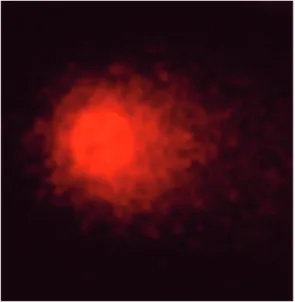
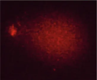
neutralization buffer and stained with 2 µg/mL ethidium bromide. Cells were scored under fluorescence microscope (Nikon, Eclipse 600, Japan). A minimum of two SCGE slides were prepared from each sample and 300 nuclei were evaluated by visual scoring. Nuclei were scored as 0, 1+, 2+, 3+, and 4+ by a blinded observer, according to the apparent relative proportion of DNA in the tail and head, as shown in Figures 1–5. Each counted nucleus was multiplied by its score, and total scores were expressed as AU.
2.3. Statistical analyses
Data analyses were undertaken using SPSS 11.5 (SPSS Inc., Chicago, IL, USA). Continuous and discontinuous numerical variables were investigated for normal and nonnormal distribution, using the Kolmogorov–Smirnov test. Significance of differences among groups for mean and median values was ascertained with the Mann–Whitney U-test. Categorical variables were analyzed with Pearson’s chi-square test or Fisher’s exact test. Correlation between continuous and discontinuous variables was ascertained using Spearman’s correlation test. A value of P < 0.05 was considered significant.
3. Results
Among the 60 patients at study commencement, 9 patients did not attend for treatment, epistaxis was observed in one patient, and respiratory tract infection was observed in 9 patients. Hence, 19 patients were excluded from the study, leaving a study population of 41 patients. Seventeen patients (41.46%) were in the IC group ((8 female (19.51%) and 9 male (21.95%)). Twenty-four patients (58.54) were in the SP group ((14 female (34.15%) and 10 male (24.39%)). The sex distribution of patients is shown in Table 1. There was no significant difference between groups in terms of sex (P > 0.05). The age range of 41 patients was 20–58 years (median, 27; mean, 30.70 ± 9.31). The age range of the IC group was 20–51 years (median, 27; mean, 30.18 ± 9.07), whereas that of the SP group was 20–58 years (28; 31.08 ± 9.65). The age distribution of the patients is shown in Table 2. There was no significant difference between the groups in terms of age (P > 0.05).
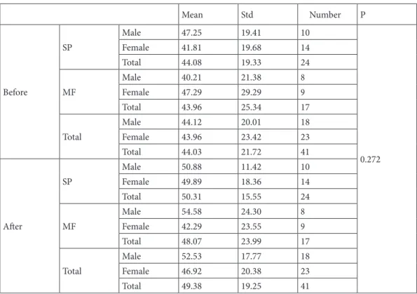
Mean comet score for 41 patients was 44.03 ± 21.72 before treatment and 49.38 ± 19.25 after treatment. Mean comet score was 43.96 ± 25.34 before treatment and 48.07 ± 23.99 after treatment in the IC group. Mean comet score was 44.08 ± 19.33 before treatment and 50.30 ± 15.55 after treatment in the SP group. No significant difference was observed before and after treatment for mean comet scores within or between groups (P = 0.272). The comet scores of the patients are shown in Table 3.
4. Discussion
ICs have been used for the treatment of AR since the 1970s. The clinical effects of topical corticosteroids are
Figure 1. Appearance of a cell with comet scoring 0.
Figure 2. Appearance of a cell with comet scoring 1+.
associated with inhibition of aggregation of inflammatory cells in the airway, suppression of local production of cytokines, prevention of mediator release, and repair of nasal mucosal structure. Intranasal glucocorticosteroids are well-tolerated drugs and carry a low prevalence of side effects compared with placebo (23–25). The synthetic corticosteroids MF, beclomethasone dipropionate, betamethasone, dexamethasone, and hydrocortisone have been shown to be potent against AR (26). MF is a topical corticosteroid used commonly in AR treatment (27). In our study, MF was used for 1 month by AR patients.
Intranasal glucocorticosteroids ameliorate AR symptoms, but use of certain ICs can lead to local histopathologic changes (28,29).
Side effects in the nasal cavity are often seen with intranasal glucocorticosteroids. These include epithelial irritation, burning sensations, dryness, crusting, epistaxis, and septal perforation (8,10–12,30,31). In our study, no patients suffered from headaches, nasal dryness, or nasal irritation, but epistaxis was seen in one patient, which merited exclusion from the study. It is thought that epistaxis after IC use is due to dryness and thinning of nasal mucosae (32,33). However, in a clinical study by Waddell et al. (34), ICs and placebo were compared and the prevalence of epistaxis was similar. Therefore, epistaxis after IC use is thought to be due to exposure of the nasal septum or anterior nasal concha to nasal applicators.
Figure 4. Appearance of a cell with comet scoring 3+. Figure 5. Appearance of a cell with comet scoring 4+. Table 1. Sex distribution of patients.
Male Female Total
Number % Number % Number %
SP 10 24.39 14 34.15 24 58.54
MF 9 21.95 8 19.51 17 41.46
Total 19 46.34 22 53.66 41 100
Table 2. Age distribution of patients.
Mean Number Std. [±] Median Minimum Maximum
SP 31.08 24 9.65 28.00 20.00 58.00
MF 30.18 17 9.07 27.00 20.00 51.00
Another problem considered to be associated with IC use is mucosal atrophy. Davies et al. (35) showed no atrophy in the nasal mucosa due to long-term use of MF nasal spray, and no reduction in epithelial thickness was observed in biopsies of nasal mucosae. In another study, atrophy in nasal mucosae was not detected after treatment with MF nasal spray for 12 months in AR patients (31). Minshall et al. (36) administered MF for 12 months to 69 patients with perennial rhinitis, and did not detect changes in epithelial thickness, distribution and density of goblet cells, morphologic characteristics of glands and vessels in the lamina propria, or integrity of basal membranes. They found that use of nasal spray improved the appearance of the epithelium and reduced the prevalence of inflammatory cell infiltrates (especially those containing eosinophils and mast cells).
Several scholars have investigated whether ICs cause histopathologic changes. Pipkorn et al. (37) showed that short-term application of sprays containing MF or budesonide to rat nasal mucosae did not cause significant histopathologic changes. Güngör et al. (38) did not observe significant differences in edema, ciliary loss, or intraepithelial loss by use of budesonide in rat nasal mucosae. Benninger et al. (39) reported that ICs did not cause mucosal atrophy or nasal septal perforation. Cervin et al. (8) reported that IC sprays used for AR treatment
cause atrophy of nasal mucosae and increase the risk of septal perforation.
In our study, we wished to ascertain whether MF nasal spray causes mucosal atrophy or not by observing DNA damage in nasal mucosa cells. We did not find a significant difference between the SP and IC groups in terms of DNA damage, which suggests that MF does not cause mucosal atrophy.
IC (e.g., MF, fluticasone propionate, fluticasone furoate) use elicits minimal systemic exposure (<1%) due to their pharmacokinetic properties, and so systemic side effects are very limited (40). MF is an effective local antiinflammatory topical glucocorticosteroid and is not active systemically. Systemic effects have not been shown in children, adolescents, or adults. MF suspensions show little absorption in the gastrointestinal system, and absorbed MF is excreted from urinary and biliary systems by first-pass metabolism (31) Smith et al. (41) showed that MF has high affinity for glucocorticoid receptors and that its systemic absorption is minimal; thus it has minimal side effects. In our study, MF nasal spray was used for AR treatment because it would not lead to systemic side effects.
Studies have shown that MF does not lead to DNA damage in rat liver cells and that it is not genotoxic (31). We found that MF did not cause DNA damage to nasal
Table 3. Comet scores of patients.
Mean Std Number P Before SP Male 47.25 19.41 10 0.272 Female 41.81 19.68 14 Total 44.08 19.33 24 MF Male 40.21 21.38 8 Female 47.29 29.29 9 Total 43.96 25.34 17 Total Male 44.12 20.01 18 Female 43.96 23.42 23 Total 44.03 21.72 41 After SP Male 50.88 11.42 10 Female 49.89 18.36 14 Total 50.31 15.55 24 MF Male 54.58 24.30 8 Female 42.29 23.55 9 Total 48.07 23.99 17 Total Male 52.53 17.77 18 Female 46.92 20.38 23 Total 49.38 19.25 41
mucosa cells in either group after 4 weeks of treatment. Hence, MF nasal spray seems to be safe for AR treatment.
In our study, SCGE was used for the determination of DNA breaks because it is rapid, inexpensive, and only a small number of cells was available for analyses. SCGE is a simple, rapid, and sensitive method that can be applied to different types of cells and DNA damage. It does not require radioactive labeling, so it is often the preferred method of measurement of DNA damage (18). Another advantage is that SCGE allows the determination of DNA breaks with small amounts of cells from human biopsies. Moreover, the cost of equipment is lower than that of other test methods of genotoxicity (21). In comparison to other methods of genotoxicity testing, it can detect DNA damage at lower levels and with higher sensitivity. Use of SCGE in genotoxicity studies has increased gradually thanks to such advantages (42).
Nevertheless, SCGE has disadvantages. Certain differences (agarose concentration, number of applied
cells, electrophoresis time) at technical implementation can affect results, and there can be differences among results from different laboratories even if the same protocol is applied (43). These differences occur primarily because of the subjective nature of comet classification. Therefore, comet counting must be undertaken by the same experienced researcher throughout the study (18), as was achieved in the present study.
In conclusion, we have shown, for the first time, that treatment with MF spray for 4 weeks does not cause DNA breaks within cells in the nasal mucosa. These data need to be confirmed by clinical trials involving treatment with different ICs over longer periods.
Acknowledgment
This study received financial support from the Research Council of the Faculty of Medicine, Başkent University (Project No. KA 14/203).
References
1. Bousquet J, Van Cauwenberge P, Khaltaev N. Allergic rhinitis and its impact on asthma. J Allergy Clin Immunol 2001; 108: 147-334.
2. Keleş N, Ilicali C, Değer K. The effects of different levels of air pollution on atopy and symptoms of allergic rhinitis. Am J Rhinol 1999; 13: 185-190.
3. Settipane RA, Lieberman P. Update on nonallergic rhinitis. Ann Allergy Asthma Immunol 2001; 86: 494-507.
4. Cingi C, Songu M. Nasal steroid perspective: knowledge and attitudes. Eur Arch Otorhinolaryngol 2010; 267: 725-730. 5. Kalyoncu AF, Demir AU, Ozcakar B, Bozkurt B, Artvinli M.
Asthma and allergy in Turkish university students: two cross sectional surveys 5 years apart. Allergol Immunopathol (Madr) 2001; 29: 264-271.
6. Ciprandi G, Pronzato C, Ricca V, Baqnasco M, Canonica GW. Evidence of ICAM-1 expression on nasal epithelial cells in acute rhinoconjunctivitis due to pollen exposure. J Allergy Clin Immunol 1994; 4: 738-746.
7. Minshall E, Ghaffar O, Cameron L, O’Brien F, Quinn H, Rowe-Jones J, Davies RJ, Prior A, Lund VJ, Mackay IS et al. Assessment by nasal biopsy after long-term use of mometasone furoate aqueous nasal spray (Nasonex) in the treatment of perennial rhinitis. Otolaryngol Head Neck Surg 1998; 118: 648-654.
8. Cervin A, Andersson M. Intranasal steroids and septum perforation – an overlooked complication? A description of the course of events and a discussion of the causes. Rhinology 1998; 36: 128-132.
9. Bende M, Mark J. Long-term effects of topical corticosteroids in the nose. J Laryngol Otol 1992; 106: 810-812.
10. Trangsrud AJ, Whitaker AL, Small RE. Intranasal corticosteroids for allergic rhinitis. Pharmacotherapy 2002; 22: 1458-1467.
11. Cervin A, Hansson C, Greiff L, Andersson M. Nasal septal perforations during treatment with topical nasal glucocorticosteroids are generally not associated with contact allergy to steroids. ORL J Otorhinolaryngol Relat Spec 2003; 65: 103-105.
12. Eweiss A, Dogheim Y, Hassab M, Tayel H, Hammad Z. VCAM-1 and eosinophilia in diffuse sino-nasal polyps. Eur Arch Otorhinolaryngol 2009; 266: 377-383.
13. Verret DJ, Marple BF. Effect of topical nasal steroid sprays on nasal mucosa and ciliary function. Curr Opin Otolaryngol Head Neck Surg 2005; 13: 14-18.
14. Bousquet J, Khaltaev N, Cruz AA, Denburg J, Fokkens WJ, Toqias A, Zuberbier T, Baena-Caqnani CE, Canonica GW, van Weel C. Allergic Rhinitis and its Impact on Asthma (ARIA) 2008 update. Allergy 2008; 63(Suppl 86): 8-160.
15. do Amaral RJ, Pedrosa Cda S, Kochem MC, Silva KR, Aniceto M, Claudio-da-Silva C, Borojevic R, Baptista LS. Isolation of human nasoseptal chondrogenic cells: a promise for cartilage engineering. Stem Cell Res 2012; 8: 292-299.
16. Koehler C, Ginzkey C, Friehs G, Hackenberg S, Froelich K, Scherzed A, Burghartz M, Kessler M, Kleinsasser N. Aspects of nitrogen dioxide toxicity in environmental urban concentrations in human nasal epithelium. Toxicol Appl Pharmacol 2010; 245: 219-225.
17. Yurtcu E, Iseri OD, Sahin FI. Effects of ascorbic acid and β-carotene on HepG2 human hepatocellular carcinoma cell line. Mol Biol Rep 2011; 38: 4265-4272.
18. Dinçer Y, Kankaya S. Comet assay for determining of DNA damage: review. Turkiye Klinikleri J Med Sci 2010; 30: 1365-1373.
19. Green MH, Lowe JE, Delaney CA, Green IC. Comet assay to detect nitric oxide-dependent DNA damage in mammalian cells. Methods Enzymol 1996; 269: 243-266.
20. Collins AR. The comet assay for DNA damage and repair: principles, applications, and limitations. Mol Biotechnol 2004; 26: 249-261.
21. McKelvey-Martin VJ, Green MH, Schmezer P, Pool-Zobel BL, De Meo MP, Collins A. The single cell gel electrophoresis assay (comet assay): a European review. Mutat Res 1993; 288: 47-63. 22. İşeri ÖD, Yurtcu E, Sahin FI, Haberal M. Corchorus olitorius
(jute) extract induced cytotoxicity and genotoxicity on human multiple myeloma cells (ARH-77). Pharm Biol 2013; 51: 766-770.
23. Rosenblut A, Bardin PG, Muller B, Faris MA, Wu WW, Caldwell MF, Fokkens WJ. Long-term safety of fluticasone furoate nasal spray in adults and adolescents with perennial allergic rhinitis. Allergy 2007; 62: 1071-1077.
24. Kim KT, Rabinovitch N, Uryniak T, Simpson B, O’Dowd L, Casty F. Effect of budesonide aqueous nasal spray on hypothalamic-pituitary-adrenal axis function in children with allergic rhinitis. Ann Allergy Asthma Immunol 2004; 93: 61-67.
25. Fokkens WJ, Jogi R, Reinartz S, Sidorenko I, Sitkauskiene B, van Oene C, Faris MA, Ellsworth A, Caldwell MF. Once daily fluticasone furoate nasal spray is effective in seasonal allergic rhinitis caused by grass pollen. Allergy 2007; 62: 1078-1084. 26. Barton BE, Jakway JP, Smith SR, Siegel MI. Cytokine inhibition
by a novel steroid: mometasone furoate. Immunopharmacol Immunotoxicol 1991; 13: 251-261.
27. Onrust SV, Lamb HM. Mometasone furoate. A review of its intranasal use in allergic rhinitis. Drugs 1998; 56: 725-745. 28. Meltzer EO. An overview of current pharmacotherapy in
perennial rhinitis. J Allergy Clin Immunol 1995; 95: 1097-1110.
29. Van Henke E, Temmerman L. Contact allergy to the corticosteroid budesonide. Contact Dermatitis 1980; 6: 509. 30. Garzaro M, Pecorari G, Pezzoli M, Arrondini M, Novero D,
Nadalin J, Giordano C. Mucous membrane plasmacytosis of the nose in a patient affected by B-cell chronic lymphocytic leukemia. Eur Arch Otorhinolaryngol 2009; 266: 1651-1654. 31. Product information for Nasonex® aqueous nasal spray 0.05%
alcohol free (serial online). Available online at: www.medicines. org.au/files/mkpnanua.pdf; accessed 12 October 2016.
32. Scadding G, Erkan AN, Chau H, Maskell S. Audit of nasal steroid use and effectiveness in a rhinitis clinic. Expert Rev Pharmacoecon Outcome Res 2010; 10: 87-90.
33. Blaiss MS. Safety considerations of intranasal corticosteroids for the treatment of allergic rhinitis. Allergy Asthma Proc 2007; 28: 145-152.
34. Waddell AN, Patel SK, Toma AG, Maw AR. Intranasal steroid sprays in the treatment of rhinitis: is one better than another? J Laryngol Otol 2003; 117: 843-845.
35. Davies RJ, Nelson HS. Once-daily mometasone furoate nasal spray: efficacy and safety of a new intranasal glucocorticoid for allergic rhinitis. Clin Ther 1997; 19: 27-38.
36. Minshall E, Ghaffar O, Cameron L, O’Brien F, Quinn H, Rowe-Jones J, Davies RJ, Prior A, Lund VJ, Mackay IS. Assessment by nasal biopsy of long-term use of mometasone furoate aqueous nasal spray (nasonex) in the treatment of perennial rhinitis. Otolaryngol Head Neck Surg 1998; 118: 648-654.
37. Pipkorn U, Pukander J, Suonpää J, Mäkinen J, Lindqvist N. Long-term safety of budesonide nasal aerosol: a 5.5-year follow-up study. Clin Allergy 1998; 18: 253-255.
38. Güngör A, Poyrazoğlu E, Yıldırım Ş, Basutcu S, Candan H. The effect of budesonide that is the topical corticosteroid on the rat nasal mucosa (experimental study). Turk Arch Otorhinol 2002; 40: 23-27.
39. Kridel RWH. Considerations in the etiology, treatment and repair of septal perforations. Facial Plast Surg Clin North Am 2004; 12: 435-450.
40. Sastre J, Mosges R. Local and systemic safety of intranasal corticosteroids. J Investig Allergol Clin Immunol 2012; 22: 1-12.
41. Smith CL, Kreutner W. In vitro glucocorticoid receptor binding and transcriptional activation by topically active glucocorticoids. Arzneimittelforschung 1998; 948: 956-960. 42. Tice RR, Agurell E, Anderson D, Burlinson B, Hartmann A,
Kobayashi H, Miyame Y, Rojas E, Ryu JC, Sasak, YF. Single cell gel/comet assay: guidelines for in vitro and in vivo genetic toxicology testing. Mutat Res 2000; 35: 206-221.
43. Forchhammer L, Bräuner EV, Folkmann JK, Danielsen PH, Nielsen C, Jensen A, Loft S, Friis G, Møller P. Variation in assessment of oxidatively damaged DNA in mononuclear blood cells by the comet assay with visual scoring. Mutagenesis 2008; 23: 223-231.