78
DAHİLİ BİLİMLER / MEDICAL SCIENCES
Olgu Bildirisi / Case Report
Acute viral acalculous cholecystitis due to viral
hepatitis A
Hepatit A’ya bağlı akut taşsız kolesistit
Nazan Dalgıç
1, Erdal İnce
1, Ergin Çiftçi
1, Selim Öncel
1, Meltem Güneş
1, Suat Fitöz
2, Ülker Doğru
11Ankara University, School of Medicine, Department of
Pediatric Infectious Diseases
2Ankara University, School of Medicine, Department of
Radiology Ankara, Turkey.
Hepatitis A virus (HAV) is a self-limiting, usually asymptomatic infection that occurs predomi-nantly among children. Although some gallbladder abnormalities such as increased thickness of the gallbladder wall and sludge formation were defined during HAV infection, acute cholecystitis has very rarely been reported. We here report an eleven-year old female patient with acute viral acalculous cholecystitis due to hepatitis A virus infection.
Key words: Acute viral acalculous cholecystitis, hepatitis A infection
Hepatit A sıklıkla çocukluk çağında oluşan, genellikle asemptomatik geçirilen, kendi kendini sı-nırlayan bir enfeksiyon tablosudur. Hepatit A enfeksiyonu süresince safra kesesi duvarında kalın-laşma ve safra çamuru gibi safra kesesi anormallikleri bildirilmesine rağmen akut kolesistit nadir olarak rapor edilmiştir. Hepatit A enfeksiyonuna bağlı akut taşsız kolesistiti olan onbir yaşında bir kız hasta sunulmaktadır.
Anahtar kelimeler: Akut viral taşsız kolesistit, hepatit A enfeksiyonu
A
cute hepatitis A virus (HAV) infection is frequently encountered in deve-loping countries especially in children (1). Extra-hepatic manifestations of hepatitis A include arthalgias, cutaneous vasculitis, cryoglobulinemia, and hemophagocytic syndrome. These manifestations are rare; when they do occur, they resolve with the resolution of hepatitis (2).During HAV infection, the gallbladder may undergo changes that include decreased fasting volume, increased wall thickening and appearance of biliary sludge (3). Gallbladder involvement has been described in 50 to 98% of adults with acute viral hepatitis, mild gallbladder wall thickening (GBWT) being the most common sonographic finding (4). However this feature, especially acute viral cholecystitis due to viral hepatitis A, has rarely been described in children (5, 6).
Here we report an eleven-year old girl with acute viral acalculous cholecystitis due to hepatitis A virus infection.
Case report
An eleven-year old girl was admitted to the emergency department with the complaints of fever, fatigue, nausea, vomiting, abdominal pain and loss of ap-petite. Her complaints had begun one week ago. In the last four days, she was having dark urine and pale stool. Her medical history was unremarkable except for an episode of urinary system infection in the past year. There was no history of medication or drug abuse.
Corresponding author Nazan Dalgıç
Ankara Üniversitesi Tıp Fakültesi Çocuk Sağlığı ve Hastalıkları Anabilim Dalı, Çocuk Enfeksiyon Bilim Dalı, Ankara Tel : (312) 362 3030/ 6589
Faks : (312) 362 0581 E-mail : [email protected] Received: 04.30.2004 • Accepted: 10.11.2004
79
Nazan Dalgıç, Erdal İnce, Ergin Çiftçi, Selim Öncel et al. Journal of Ankara University Faculty of Medicine 2005; 58(2)
Physical examination showed body temperature of 37,5°C, heart rate of 78/minute, breath rate of 20/min-ute and blood pressure of 100/60 mmHg. She appeared fatigued and mild skin and scleral icterus was present. Her liver was painful and palpable 4 cm under right costal margin. Laboratory studies revealed the following: hemo-globin, 12.7 mg/dL; white blood cell (WBC), 5200/mm3; platelets, 165 000/mm3; alanine aminotransferase (ALT), 2915 U/L (7-40 U/L); aspartate aminotransferase (AST), 3830 U/L (7-40 U/L); total serum bilirubin, 4.38 mg/dL (0.3 mg/dL); with a direct fraction of 2.62 mg/dL, alka-line phosphates 574 U/L (38-155 U/L); gamma-glutam-yltranspeptidase (GGT), 194 U/L (15-60 U/L), albumin 3.3 g/dl, prothrombin time 18.7 seconds, erythrocyte sedi-mentation rate 30 mm/h, C-reactive protein 0.74 mg/dL (0-5 mg/dL), HBsAg (-), anti-HBcIgM (-), anti-HCV (-), anti-HAV IgM (+) and anti-HAV IgG (+).
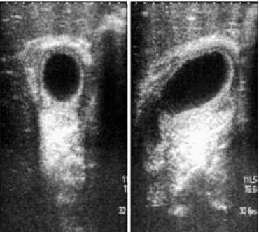
Within two days of admission to the hospital, her jaun-dice, abdominal pain, vomiting frequency, temperature, skin icterus and abdominal tenderness increased. The right side of the abdomen was tender with painful fullness in the right hypochondrium (a positive Murphy’s sign). Repeat-ed biochemical study showRepeat-ed total bilirubin 12.9 mg/dL, with a direct fraction of 7,8 mg/dL, ALT: 5025 U/L and AST: 4233 U/L. Abdominal ultrasound revealed hepato-megaly, hydropic gallbladder without calculus, thickened gallbladder wall (12 mm) and pericholecystic fluid (Figure 1A and 1B). During follow-up, she remained febrile for three days. On follow up with in four days, her tender-ness regressed, five days after the onset of her symptoms, serum total bilirubin and its direct fraction levels decreased to 10.7 mg/dL and 6 mg/dL, respectively; there was also a marked decrease in serum AST and ALT levels.
She was discharged on the 10th day of admission in good clinical condition and with considerable improve-ment in biochemical tests. During a follow-up of 6 months, she was in good condition without any complaint.
Discussion
Hepatitis A virus is a self-limiting, usually asymptomat-ic infection that occurs predominantly among children. In some patients, gallbladder abnormalities such as increased thickness of the gallbladder wall and sludge formation were described (1). Hermier et al. (6) described three children with acute cholecystitis due to HAV infection. These chil-dren presenting with acute hepatitis had an initial clinical onset suggestive of acute cholecystitis (pain and guarding in the right hypochondrium, fever and delayed jaundice) associated with important ultrasonographic findings in-cluding: gallbladder wall thickness greater than 10 mm [3
cases], the presence of 2 or 3 layers of different echogenici-ties [3 cases], presence of an ultrasonographic Murphy’s sign [one case], echogenic contents of the gallbladder [one case]. In another study of thirty-nine children hospitalized for hepatitis A virus infection were evaluated by ultrasound and pseudo surgical gallbladder wall of 10 mm or more with striation was found in 10 patients. In all of them the abnormalities had normalized within 4 weeks (7).
The presented case had acute HAV infection docu-mented by biochemical, serologic, and clinical features. Viral acalculous cholecystitis developed during the course of the disease. Ultrasonographic examination revealed the diagnosis of acalculous cholecystitis. Surgical intervention did not require in our patient. A repeated imaging with ultrasonography after 4 days revealed regression, such as thickening and edema of gallbladder wall and complete resolution of the pericholecystic fluid.
The pathophysiology of the GBWT during acute viral hepatitis is not clear: hypoalbuminemia, local extension of the hepatic inflammatory process, and elevated portal pres-sure all could be reflected as the edema of the gallbladder wall (7). A direct invasion of the gallbladder by the hepa-titis virus has been documented by Mourani et al (8); in a 68-year-old man with HAV in whom sonography showed marked GBWT, and HAV IgM was eventually found, and the viral antigen was demonstrated in most epithelial cells of the gallbladder wall of the patient. They suggested that acute cholecystitis may be a part of the spectrum of HAV infection. The sonographic finding of striation in the
gall-Figure 1 a,b. Axial (a) and sagittal (b) images of abdominal ultrasonography
demonstrate abnormally thickened and multilayered gall-bladder wall (arrows).
80 Acute viral acalculous cholecystitis due to viral hepatitis A Ankara Üniversitesi Tıp Fakültesi Mecmuası 2005; 58(2)
bladder reflects the distribution of fluid throughout all lay-ers of the wall.
Early studies have shown that during viral hepati-tis A, the gallbladder may undergo changes that include decreased fasting volume, increased wall thickening and appearance of biliary sludge. These morpho-functional events are transient and gradually disappear when viremia becomes low. Gallbladder wall thickness returns to normal in these patients within few days. These patients do not require surgical intervention (3).
Only few cases of gallbladder involvement during HAV infection were reported in medical literature. It should be kept in mind that although rare, like adults acute viral cho-lecystitis can develop during the course of acute HAV infec-tion in children. We suggest that the right upper quadrant pain, high temperature, severe vomiting, severe jaundice during acute hepatitis A is, at least in part, may be caused by gallbladder involvement. Paediatricians and paediatric surgeons must be familiar with the possibility of gallblad-der and pancreatic involvement during HAV infection to avoid unnecessary invasive procedures.
References
1. Ozaras R, Mert A, Yılmaz MH, et al. Acute viral cholecystitis due to hepatitis A virus infection. J Clin Gastroenterol 2003; 37: 79-81.
2. Bell BP, Shapiro CN. Hepatitis A Virus. In: Principles and Practice of Pediatric Infectious Diseases, Long SS, Plckering LK, Prober CG (eds), 2nd ed, Churchill-Livingstone , New York, 2003; 1118-1194.
3. Portincasa P, Moschetta A, Di Ciaula A, et al. Changes of gallbladder and gastric dynamics in patients with acute hepatitis A. Eur J Clin Invest 2001; 31: 617-622.
4. Sharma MP, Dasarathy S. Gallbladder abnormalities in acute viral hepatitis: a prospective ultrasound evaluation. J Clin Gastroenterol 1991; 13: 697-700.
5. Black MM, Mann NP. Gangrenous cholecystitis due to hepatitis A infection. J Trop Med Hyg 1992; 95: 73-74.
6. Hermier M, Descos B, Collet JP, et al. Acute cholecystitis disclosing virus hepatitis. Arch Fr Pediatr 1985; 42: 525-529. 7. Klar A, Branski D, Nadjari M, et al. Gallbladder and pancreatic
involvement in hepatitis A. J Clin Gastroenterol 1998; 27: 143-145.
8. Mourani S, Dobbs SM, Genta RM, et al. Hepatitis A virus associated cholecystitis. Ann Intern Med 1994; 120: 398-400.