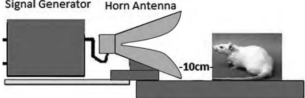
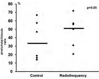
The effect of non-ionizing radiation on the ovarian reserves of female rats
Tam metin
Şekil
Benzer Belgeler
Figure 2 LH levels before and after the ovarian manipulation of all regularly ovulating
Species Length of Length of Ovulation Length of Estrous Cycle Estrus Pregnancy.. Bitch 6 months 9 days 4 - 24 days
Ancak, temel örgütlenme biçimi patriarkal olan tüm diğer toplumlarda olduğu gibi Türkiye’de de namus kavramı esas olarak kadının cinselliğinin denetlenmesi ile bir
(b) Intraperi- toneally administration of paracetamol, at a dose of 300 mg/kg, before PTZ injection, decreased the mean frequency of PTZ-induced epileptiform ECoG activity
Thus, the aim of this study was to investigate the effect of embryo collection on the pregnancy, clinical pregnancy and live birth rates in poor responder women, as defined according
Objective: To report the effects of the torsion attacks on the ovarian reserve, and to present the surgical approach in cases of ovary torsion that develop after preventive
This study aims to investigate isotretinoin’s effects on anti-Mullerian hormone (AMH), follicle stimulating hormone (FSH) and total antral follicle count (TAFC) as
As shown in the analysis based on variables of administrative task, academic title and age, young female academics who have low level administrative tasks and who
