CATARACT
Evaluating relaxed ciliary muscle tone in presbyopic eyes
Erhan Özyol1&Pelin Özyol2Received: 2 June 2016 / Revised: 29 January 2017 / Accepted: 9 February 2017 / Published online: 25 February 2017 # Springer-Verlag Berlin Heidelberg 2017
Abstract
Purpose Studies of age-related changes in ciliary muscle (CM) morphology and contractility have variously reported that CM weakens or strengthens with age. In response, the aim of this study was to evaluate relaxed CM tone in vivo in pre-presbyopic and presbyopic patients using a predictor val-ue (PCM).
Methods Two groups of eyes—40 eyes of 40 healthy volun-teers with a mean age of 28.1 ± 5.8 years and 40 eyes of 40 healthy volunteers with a mean age of 56.6 ± 7.3 years— formed the sample for this prospective, observational cross-sectional study. Used to evaluate relaxed CM tone, PCMwas calculated as the difference between the change in mean ante-rior chamber depth (ACD) and lens thickness (LT) before and after cycloplegia, as measured with swept-source optical biometry.
Results The PCMfor relaxed CM tone was 0.04 ± 0.04 mm in pre-presbyopic participants, 0.06 ± 0.03 mm in presbyopic ones, and significantly greater in presbyopic patients (p = .018).
Conclusion The statistical significance of PCMbetween pre-presbyopic and pre-presbyopic eyes might not signify clinical sig-nificance, since the difference was close to the repeatability limits for swept-source optical biometry. When relaxed, CM tone does not diminish with presbyopia according to changes in anterior chamber parameters due to cycloplegia.
Keywords Ciliary muscle . Cycloplegia . Presbyopia
Introduction
To accommodate increased optical power, young crystalline lenses can change shape, thereby allowing them to adapt their power to visualize objects at different distances. According to the traditionally accepted Helmholtz’s theory of accommoda-tion, the contraction of the ciliary muscle (CM) reduces zonular tension around the lens equator, thereby relaxing the lens. As zonular tension dissipates, the young crystalline lens thickens, and its radius of curvature steepens. When the CM is relaxed, it pulls the lens radially, thereby flattening it [1,2].
Decreased accommodation becomes a problem for most people in later life, especially when they can no longer see clearly enough to perform tasks requiring nearsightedness. Known as presbyopia, the condition remains an ophthalmic mystery, although several reviews have summarized theories attempting to explain its mechanisms [3–6]. As those reviews show, two classic theories explaining the mechanics of pres-byopia have surfaced as the product of longstanding debate [3,
6,7], and differ with regard to CM involvement and predicted relationships between CM contraction and changes in lens shape. On the one hand, according to Gullstrand’s theory, or lenticular theory, developed from concepts first described by Helmholtz in the mid-19th century, presbyopia is caused by the lens’s decreased ability to change shape. The theory holds that throughout life, a constant amount of CM contraction is necessary for each diopter of accommodative change. Accordingly, as the amplitude of accommodation reduces due to lenticular changes, an increased proportion of potential CM contraction becomes latent in the fully accommodated eye, insofar as further contraction will not produce any change in accommodation.
* Erhan Özyol
1
Department of Ophthalmology, Mugla Sitki Kocman University, Training and Research Hospital, Mugla 48000, Turkey 2
Department of Ophthalmology, Mugla Sitki Kocman University, Faculty of Medicine, Mugla, Turkey
On the other hand, Duane–Fincham’s theory, or extralenticular theory, assumes that as the eye ages, the force required to produce a given change in accommodation in-creases and that the CM will be maximally contracted when the near point is reached. Whereas Duane [8,9] argued that the CM weakens with age, in agreement with Fisher’s [10] later findings, Fincham [11] did not. Studies like Fisher’s [12–14] and Glasser and Campbell’s [2,15] suggested that as the eye ages, greater force needs to be exerted on the lens and capsule in order to achieve the same change in accommodation, which led to the Duane–Fincham model as a result.
To understand the dynamic of accommodative behavior, dynamic biomechanical properties of the components of the accommodation mechanism have been identified [16]. Using that biomechanical model, Beers and van der Heijde [17] pro-posed that changes in the dynamic response are characteristics of a function of age, thereby indicating that changes in the lens’s elastic properties are the chief cause of presbyopia.
As recent studies have demonstrated, the most accepted hypothesis for presbyopia is the progressive loss of lens elas-ticity [15,18], although changes in the CM [19,20], choroid [21], and vitreous body [22] probably contribute as well. Researchers have elucidated presbyopia in terms of lenticular changes that reduce lens elasticity [2,13,15,23], whereas others have concluded that, in weakening with age, the CM becomes unable to perform the required release of zonular tension for accommodation [13,21,24]. By contrast, Fisher has proposed that the CM strengthens with age in order to produce accommodative changes [10].
In any case, due to the CM’s anatomical location, its exact accommodative movement remains difficult to determine. In fact, functional theories of the CM’s role in accommodation often draw upon research involving rhesus monkeys [21,24], in-vitro studies [19], or imaging methods [10, 25–27]. Although imaging techniques do not provide a dynamic in-vivo image, they do provide an opportunity to analyze indi-vidual muscle fiber groups for age-related differences and ac-commodative changes.
Generally, the CM and zonular fibers suspend the lens in its normal position. In contrast to accommodation, cycloplegia, or CM paralysis, results in the backward movement of the crystalline lens and decreased lens thickness (LT). The chief determining factor in decreased LT is lens elasticity, not loss of CM tone. Due to changes in crystalline lens dynamics, ante-rior chamber depth (ACD) increases [28,29], meaning that the difference between the change in mean ACD and LT with cycloplegia reflects the contributory effect of CM tone upon ACD. In physiology, relaxed muscle tone refers to continuous, passive, incomplete contraction or the muscle’s resistance to passive strength during rest [30].
In response to all of the above, the aim of this study was to evaluate relaxed CM tone in vivo in pre-presbyopic and pres-byopic patients using a predictor value (PCM).
Methods
Two groups of eyes—40 eyes of 40 healthy volunteers (23 men, 17 women) with a mean age of 28.1 ± 5.8 years (range, 21–38 years) and 40 eyes of 40 healthy volunteers (20 males, 20 females) with a mean age of 56.6 ± 7.3 years (range, 47–62 years)—formed the sample for this prospective, cross-sectional study. The exclusion criteria were spherical equivalent refrac-tive error greater than ±1.0 D, previous ocular surgery, corneal opacities, severe dry eye, history of contact lens wear, retinal disease (e.g., cystoid macular edema or elevated scars), glau-coma, narrow iridocorneal angle, cataract, inability to open eyelids widely, ocular inflammatory disease, and poor ocular fixation. The study was approved by the ethics committee of Muğla University and conducted according to the Declaration of Helsinki. The aim of the study was explained to each patient, and his or her written informed consent was obtained.
To eliminate the effect of diurnal variation, all mea-surements were taken in room light during the period 9:00–11:00 am. All noncycloplegic and cycloplegic mea-surements were performed within 1 h on the same day by the same experienced operator and according to the man-ufacturer’s recommendations. Three measurements were a c q u i r e d u s i n g s w e p t - s o u r c e o p t i c a l b i o m e t r y ( I O L M a s t e r 70 0 ; C a r l Z e i s s Me d i t ec A G , J en a , Germany) before and after cycloplegia. Quality control criteria were fulfilled in accordance with the manufac-turer’s recommendations. With the IOLMaster 700, the entire scan image was viewed, and the eye geometry and axis of measurements were visually assessed. Foveal scans confirmed the correct fixation. The entire cross-sectional images generated by the IOLMaster 700 of each participant before and after cycloplegia were
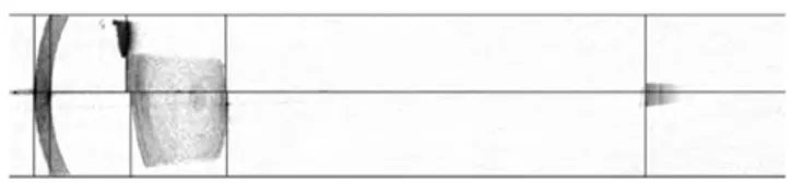
Fig. 1 The entire cross-sectional images generated by the IOLMaster 700 of a participant aged 30 years before (upper half) and after cycloplegia (lower half) are superimposed. The lower part demonstrates that decrease in lens thickness is more evident than the backward movement of the crystalline lens due to cycloplegia
Fig. 2 The entire cross-sectional images generated by the IOLMaster 700 of a participant aged 54 years before (upper half) and after cycloplegia (lower half) are superimposed. The lower part demonstrates that the backward movement of the crystalline lens is more evident than the decrease in lens thickness due to cycloplegia
I
'
l
~!
~
J ,...
superimposed in consideration of the locations of corneas and foveas. The proper images were subsequently record-ed for analysis (Figs.1 and 2).
To create an optically smooth tear film over the cornea, participants were asked to blink immediately before mea-surements were taken. Cycloplegia was induced by ad-ministering three drops of an eye solution containing 1%
cyclopentolate every 5 min. After cycloplegia, pupillary light reflex was assessed, and about 45 min later, mea-surements were taken. LT and ACD meamea-surements were recorded before and after pupil dilation.
The PCMused to evaluate relaxed CM tone was formulated as the difference between the change in mean ACD and LT before and after cycloplegia, as follows:
Statistical analysis
The Statistical Package for the Social Sciences version 18.0 was used for statistical analysis. Normality was analyzed with Kolmogorov–Smirnov’s test, and Levene’s test was used for equality of variances. After acquiring normal distribution and equality of variances, changes before and after dilation were evaluated with a dependent samples t-test, and parameters be-tween groups of pre-presbyopic and presbyopic participants were compared with Hotelling’s two-sample T-squared test. For standard deviation of repeatability, three measurements of the ACD and LT parameters with and without cycloplegia taken by the same operator for both groups were analyzed. The
standard deviation of repeatability was estimated by the square root of estimated variance due to measurement error, based on the random effects analysis of variance model. The coefficient of variability was calculated by the quotient of the standard devia-tion of repeatability and the mean of all measurements used. A p value of <.05 was considered to be statistically significant.
Results
Tables1,2, and3show ACD and LT measurements with and without cycloplegia in pre-presbyopic and presbyopic participants.
Table 1 The mean ACD and LT measurements obtained using swept source optical biometry with and without cycloplegia in pre-presbyopic subjects Parameter Noncycloplegia mean ± SD Cycloplegia mean ± SD Mean difference ± SD P value* ACD 3.42 ± 0.37 3.55 ± 0.34 0.13 ± 0.08 <0.001 LT 3.72 ± 0.25 3.63 ± 0.23 0.09 ± 0.04 <0.001
* Dependent samples t-test, p value for comparison of parameters in noncycloplegia and cycloplegia states ACD, anterior chamber depth; LT, lens thickness
Table 2 The mean ACD and LT measurements obtained using swept source optical biometry with and without cycloplegia in presbyopic subjects
Parameter Noncycloplegia mean ± SD Cycloplegia mean ± SD Mean difference ± SD P value*
ACD 3.09 ± 0.28 3.17 ± 0.29 0.08 ± 0.03 <0.001
LT 4.55 ± 0.44 4.53 ± 0.43 0.02 ± 0.01 <0.001
*Dependent samples t-test, p value for comparison of parameters in noncycloplegia and cycloplegia states ACD, anterior chamber depth; LT, lens thickness
PcM = ~ACD -
~LTPcM = Predictor value of relaxed CM tone
~ACD = Change in mean ACD after cycloplegia
Firstly, the change in mean ACD with cycloplegia was 0.13 ± 0.08 mm in pre-presbyopic participants and 0.08 ± 0.03 mm in presbyopic ones, with a statistically significant difference in each group (p < .001). The change in mean ACD with cycloplegia between pre-presbyopic and presbyopic patients was 0.05 ± 0.02 mm, which was also statistically significant (p = .004).
Secondly, the change in mean LT with cycloplegia was 0.09 ± 0.04 mm in pre-presbyopic participants and 0.02 ± 0.01 mm in presbyopic ones, with a statistically significant difference in each group (p < .001, for each). The change in mean LT with cycloplegia between pre-presbyopic and pres-byopic patients was 0.07 ± 0.01 mm, which was also statisti-cally significant (p < .001).
Lastly, the PCMwas 0.04 ± 0.04 mm in pre-presbyopic par-ticipants, 0.06 ± 0.03 mm in presbyopic ones, and significantly greater in presbyopic participants (p = .018)..
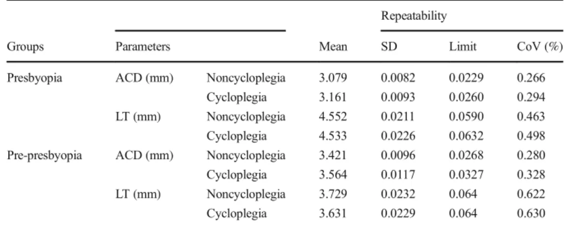
Table 4 summarizes the standard deviation of repeat-ability, the limits of the measurements, and the coeffi-cient of variability for ACD and LT in both groups. The standard deviations and the limits of repeatability were small and acceptable for both parameters with and with-out cycloplegia.
Discussion
Several authors have described changes in CM contrac-tility with age, yet often with conflicting results [19,31]. Duane [8, 9] proposed that accommodative loss occurs d u e t o t h e C M’s progressive weakening. In a
postmortem human study, Nishida and Mizutani [32] re-ported that the age-related increase of connective tissue and decrease of nuclei in the CM, together with de-creased circular fiber, suggest the possibility of CM at-rophy with age-related deterioration of accommodation. In a study involving rhesus monkeys that sought to iden-tify the pathophysiologic characteristics of presbyopia, which occurs in both humans and rhesus monkeys on a comparable relative time scale, researchers demonstrated that the contractile responses of CM to pilocarpine di-minish with age [24]. In agreement with earlier studies using impedance cyclography [25], Pardue and Sivak [19] found that the CM retains its ability to contract throughout an individual’s lifespan. Using anterior seg-ment optical coherence tomography, Sheppard and Davies [33] suggested that no significant decrease in the contractile ability of the muscle occurs, even in eyes with established presbyopia. In a magnetic resonance imaging study, Strenk [27] found that CM contractile activity persisted in all participants, including presbyopic ones. Fisher [10, 34] suggested that the maximum force of contraction of the entire CM increases with age, as well as that lenticular sclerosis and increased size and weight with age might simply require extra force from the muscle to produce accommodative changes. According to Fisher, the CM’s force of contraction is proportional to the square of the resultant accommoda-tive change and not linearly related. In sum, most pub-lished data indicate that the CM maintains its contractile ability long after the onset of presbyopia, although
Table 3 The comparison of the change in mean ACD, LT, and predictor values between pre-presbyopic and pre-presbyopic subjects
Parameter Pre-presbyopia Presbyopia Mean difference ± SD P value*
Change in ACD 0.13 ± 0.08 0.08 ± 0.03 0.05 ± 0.02 0.004
Change in LT 0.09 ± 0.04 0.02 ± 0.01 0.07 ± 0.01 <0.001
Predictor value 0.04 ± 0.04 0.06 ± 0.03 0.02 ± 0.01 0.018
* Hottelling’s two sample T-squared test, p value for comparison of parameters in pre-presbyopia and presbyopia. ACD, anterior chamber depth; LT, lens thickness
Table 4 Repeatability of
parameters Repeatability
Groups Parameters Mean SD Limit CoV (%)
Presbyopia ACD (mm) Noncycloplegia 3.079 0.0082 0.0229 0.266
Cycloplegia 3.161 0.0093 0.0260 0.294
LT (mm) Noncycloplegia 4.552 0.0211 0.0590 0.463
Cycloplegia 4.533 0.0226 0.0632 0.498
Pre-presbyopia ACD (mm) Noncycloplegia 3.421 0.0096 0.0268 0.280
Cycloplegia 3.564 0.0117 0.0327 0.328
LT (mm) Noncycloplegia 3.729 0.0232 0.064 0.622
Cycloplegia 3.631 0.0229 0.064 0.630
whether the nature of the response varies with age is not as clear [35, 36]. In light of a PCM obtained from IOLMaster 700 measurements reflecting relaxed CM tone, CM tone was significantly greater—by 20 μm— in presbyopic eyes. However, the difference between presbyopic and pre-presbyopic eyes was close to the limits of standard deviations of repeatability for the de-vice, which the manufacturer reports to be 10 μm and 19 μm for the ACD and LT respectively. Therefore, the statistically significant difference might not signify clin-ical significance. In any case, our results demonstrate that the relaxed CM tone of presbyopic patients is not inferior to that of pre-presbyopic ones.
High repeatability for all biometric parameters with IOLMaster 700 has been reported [37, 38]. For catarac-tous eyes, the standard deviations of repeatability of ACD and LT measurements were reported as 9.8 μm and 19.5μm by Kunert et al. [37] and 9 μm and 29 μm by Srivannabonn et al. [38]. The present study found stan-dard deviations of repeatability of ACD and LT measure-ments in presbyopic participants to be 8.2 μm and 21.1 μm without cycloplegia and 9.3 μm and 22.6 μm with cycloplegia; in pre-presbyopic participants, those values were 9.6 μm and 23.2 μm without cycloplegia and 11.7 μm and 22.9 μm with cycloplegia. Values of standard deviation of repeatability were fairly close to previously reported values [37,38].
Not only muscle tone, but also changes in geometrical factors with age, including the width, length, and anatomic location of CM, the size and curvature of lens, and the positions of CM and zonules relative to the lens, can affect the PCMof the current study. Quantitative and morphomet-ric studies of aging eyes have reported geometmorphomet-rical chang-es, and that the CM of older participants contains greater amounts of connective tissue, is shorter and wider, and has a forward-moving internal apical edge [19,32]. With age, an antero–inward displacement of muscle mass [33] and decrease in the diameter of the relaxed CM ring [27] cause the anterior movement of zonular insertions over the en-larged presbyopic lens. As a result, zonular insertions be-come more tangential to the lenticular surface, which can partly disable zonules from imparting tension upon the capsule [15, 18, 39, 40]. Although the width of the circumlental space in the relaxed eye diminishes with age, its effect on the zonule’s tension can be compensated for by the axial movement of attachment points of zonular fibers to the lens capsule [5,41,42]. In the present study, even if the difference of PCMwas close to the limits of repeatability, it was clearly greater in presbyopic patients than in pre-presbyopic ones. In the light of the aforemen-tioned studies, aging eyes demonstrate changes in the geo-metrical design of the accommodative system. However the role of zonules on the lens dynamics reduces or could
be compensated with some anatomical variations of zonule insertions. Therefore, the findings may not be explained by age-related geometrical changes.
Various studies have reported changes in anterior seg-ment parameters following pupil dilation with different devices—Pentacam [43], AL-Scan [44], Lenstar LS900 [45, 46], ultrasound biomicroscopy [47], A-scan ultraso-nography [47], and IOLMaster 500 [46]—and significant
increases in mean ACD have been detected with cycloplegia in all of these studies. Similarly to the above studies, we found a significant increase in mean ACD in cycloplegic individuals with a swept-source optical bio-metric device. However, participants with an ocular spher-ical refractive error above ±1 D or with other exclusion criteria (e.g., narrow iridocorneal angle, glaucoma, and cataract) were excluded from our sample, and the fact that the effects of cycloplegia in such eyes were not investi-gated represents a limitation of our study.
In summary, this study demonstrates that CM tone does not appear to diminish in aging presbyopic eyes. In future studies, researchers should use imaging techniques to correlate ana-tomical and functional changes of CM with age.
Acknowledgements The authors thank Mr Kursad Tosun (PhD, MSK University, Faculty of Medicine, Department of Biostatistics) and Mr. Ercan Baldemir (PhD, MSK University, Faculty of Medicine, Department of Biostatistics) for their assistance with statistical analysis. Compliance with ethical standards
Funding No funding was received for this research.
Conflict of interest All authors certify that they have no affiliations with or involvement in any organization or entity with any financial interest, or non-financial interest in the subject matter or materials discussed in this manuscript.
Ethical approval All procedures were in accordance with the ethical standards of the institutional research committee and with the 1964 Helsinki Declaration and its later amendments or comparable ethical standards.
Informed consent Informed consent was obtained from all individual participants included in the study.
References
1. Schachar RA (2012) The mechanism of accommodation and pres-byopia. Kugler Publishers, Amsterdam, The Netherlands 2. Glasser A, Campbell MC (1999) Biometric, optical and physical
changes in the isolated human crystalline lens with age in relation to presbyopia. Vis Res 39:1991–2015
3. Atchison DA (1995) Accomodation and presbyopia. Ophthalmic Physiol Opt 15:255–272
4. Strenk SA, Strenk LM, Koretz JF (2005) The mechanism of pres-byopia. Prog Retin Eye Res 24:379–393
5. Weale RA (1989) Presbyopia toward the end of the 20th Century. Surv Ophthalmol 34:15–30
6. Pierscionek B (1993) What we know and understand about presby-opia. Clin Exp Optom 76:83–90
7. Stark L (1998) Presbyopia in light of accommodation. Am J Optom Physiol Opt 65:407–416
8. Duane A (1922) Studies in monocular and binocular accommoda-tion with their clinical applicaaccommoda-tions. Am J Ophthalmol 5:865–877 9. Duane A (1925) Are the current theories of accommodation
cor-rect? Am J Ophthalmol 8:196–202
10. Fisher RF (1977) The force of contraction of the human ciliary muscle during accommodation. J Physiol 270:51–74
11. Fincham EF (1932) The mechanism of accommodation and the re-cession of the near point. Report of a Joint Discussion on Vision held at Imperial College. The Physical Society, London, pp 294–308 12. Fisher RF (1969) The significance of the shape of the lens and
capsular energy changes during accommodation. J Physiol 201: 21–47
13. Fisher RF (1971) The elastic constants of the human lens. J Physiol 212:147–180
14. Fisher RF (1973) Presbyopia and the changes with age in the hu-man crystalline lens. J Physiol 228:765–779
15. Glasser A, Campbell MCW (1998) Presbyopia and the optical changes in the human crystalline lens with age. Vis Res 38:209–229 16. Beers APA, Van der Heijde GL (1994) In vivo determination of the biomechanical properties of the component elements of the accom-modation mechanism. Vis Res 34:2897–2905
17. Beers APA, van der Heijde GL (1996) Age-related changes in ac-commodation. Optom Vis Sci 73:235–242
18. Charman WN (2008) The eye in focus: accommodation and pres-byopia. Clin Exp Optom 91:207–225
19. Pardue MT, Sivak JG (2000) Age-related changes in human ciliary muscle. Optom Vis Sci 77:204–210
20. Tamm E, Lütjen-Drecoll E, Jungkunz W, Rohen JW (1991) Posterior attachment of ciliary muscle in young, accommodating old, presby-opic monkeys. Invest Ophthalmol Vis Sci 32:1678–1692
21. Tamm E, Croft MA, Jungkunz W, Lütjen-Drecoll E, Kaufman PL (1992) Age-related loss of ciliary muscle mobility in the rhesus monkey: role of the choroid. Arch Ophthalmol 110:871–876 22. Harocopos GJ, Shui YB, McKinnon M, Holekamp NM, Gordon
MO, Beebe DC (2004) Importance of vitreous liquefaction in age-related cataract. Invest Ophthalmol Vis Sci 45:77–85
23. Pau H, Kranz J (1991) The increasing sclerosis of the human lens with age and its relevance to accommodation and presbyopia. Graefes Arch Clin Exp Ophthalmol 229:294–296
24. Lütjen-Drecoll E, Tamm E, Kaufman PL (1988) Age-related loss of morphologic responses to pilocarpine in rhesus monkey ciliary muscle. Arch Ophthalmol 106:1591–1598
25. Swegmark G (1969) Studies with impedance cyclography on hu-man ocular accommodation at different ages. Acta Ophthalmol 47: 1186–1206
26. Schachar RA, Anderson DA (1995) The mechanism of ciliary mus-cle function. Ann Ophthalmol 27:126–132
27. Strenk SA, Semmlow JL, Strenk LM, Munoz P, Gronlund-Jacob J, DeMarco JK (1999) Age-related changes in human ciliary muscle and lens: a MRI study. Invest Ophthalmol Vis Sci 40:1162–1169 28. Palamar M, Egrilmez S, Uretmen O, Yagci A, Kose S (2011)
Influences of cyclopentolate hydrochloride on anterior segment pa-rameters with Pentacam in children. Acta Ophthalmol 89:e461–e465 29. Wendt M, Croft MA, McDonald J, Kaufman PL, Glasser A (2008) Lens diameter and thickness as a function of age and
pharmacologically stimulated accommodation in rhesus mon-keys. Exp Eye Res 86:746–752
30. O’Sullivan SB (2007) Examination of motor function: motor con-trol and motor learning. In: O’Sullivan SB, Schmitz TJ (eds) Physical rehabilitation, 5th edn. F. A. Davis Company, Philadelphia, pp 233–234
31. Tamm S, Tamm E, Rohen JW (1992) Age–related changes of the human ciliary muscle. A quantitative morphometric study. Mech Ageing Dev 62:209–221
32. Nishida S, Mizutani S (1992) Quantitative and morphometric stud-ies of age-related changes in human ciliary muscle. Jpn J Ophthalmol 36:380–387
33. Sheppard AL, Davies LN (2011) The effect of aging on in vivo human ciliary muscle morphology and contractility. Invest Ophthalmol Vis Sci 52:1809–1816
34. Fisher RF (1986) The ciliary body in accommodation. Trans Ophthalmol Soc UK 105:208–219
35. Koeppl C, Findl O, Kriechbaum K, Drexler W (2005) Comparison of pilocarpine-induced and stimulus-driven accommodation in phakic eyes. Exp Eye Res 80:795–800
36. Kriechbaum K, Findl O, Koeppl C, Menapace R, Drexler W (2005) Stimulus-driven versus pilocarpine-induced biometric changes in pseudophakic eyes. Ophthalmology 112:453–459
37. Kunert KS, Peter M, Blum M et al (2016) Repeatability and agree-ment in optical biometry of a new swept-source optical coherence tomography-based biometer versus partial coherence interferome-try and optical low-coherence reflectomeinterferome-try. J Cataract Refract Surg 42:76–83
38. Srivannaboon S, Chirapapaisan C, Chonpimai P, Loket S (2015) Clinical comparison of a new swept-source optical coherence tomography-based optical biometer and a time-domain optical co-herence tomography-based optical biometer. J Cataract Refract Surg 41:2224–2232
39. Atchison DA, Collins MJ, Wildsoet CF, Christensen J, Waterworth MD (1995) Measurement of monochromatic ocular aberrations of human eyes as a function of accommodation by the Howland aberroscope technique. Vis Res 35:313–323
40. Dubbelman M, Van der Heijde GL, Weeber HA (2005) Change in shape of the aging human crystalline lens with accommodation. Vis Res 45:117–132
41. Farnsworth PN, Shyne SE (1979) Anterior zonular shifts with age. Exp Eye Res 28:291–297
42. Sakabe I, Oshika T, Lim SJ, Apple DJ (1998) Anterior shift of zonular insertion onto the anterior surface of human crystalline lens with age. Ophthalmology 105:295–299
43. Arıcı C, Turk A, Ceylan OM, Kola M, Hurmeric V (2014) Effects of 1% cyclopentolate hydrochloride on anterior segment parameters obtained with Pentacam in young adults. Arq Bras Oftalmol 77: 228–232
44. Can E, Duran M, Çetinkaya T, Arıtürk N (2016) The effect of pupil dilation on A L-sc an biometric parameters. Int Ophthalmol 36:179–183
45. Arriola-Villalobos P, Diaz-Valle D (2014) Effect of pharmacologic pupil dilation on OLCR optical biometry measurements for IOL predictions. Eur J Ophthalmol 24:53–57
46. Huang J, McAlinden C, Su B, Pesudovs K, Feng Y, Hua Y, Yang F, Pan C, Zhou H, Wang Q (2012) The effect of cycloplegia on lenstar and the IOLMaster biometry. Optom Vis Sci 89:1691–1696 47. Marchini G, Babighian S, Tosi R, Perfetti S, Bonomi L (2003)
Comparative study of the effects of 2% ibopamine, 10% phenyl-ephrine, and 1% tropicamide on the anterior segment. Invest Ophthalmol Vis Sci 44:281–289