CASE REPORT
Medium Cut-Off Membrane Can Be a New Treatment
Tool in Amanita phalloides Poisoning
Bülent Huddam, MD1; Alper Alp, MD1; ˙Ismail Kırlı, MD2; Mehmet Yılmaz, MD2; Aytu˘g Ça˘gırtekin, MD2; Hakan Allı3; Sultan Edebali, MD2
1
Department of Nephrology, Faculty of Medicine, Mugla University, Mugla, Turkey;2Department of Internal Medicine, Faculty of Medicine, Mugla
University, Mugla, Turkey;3Department of Biology, Faculty of Science and Arts, Mugla University, Mugla, Turkey
Mushroom poisoning is a common health problem that can be seen seasonally and geographically. Most mushroom poisoning requiring treatment worldwide is due to Amanita phalloides. Although liver failure and kidney injury are frequent, poisoning can also lead to more serious clinical situations, such as shock, pancreatitis, encephalopathic coma, cardiac failure, disseminated intravascular coagulation, and multiple organ dysfunction syndrome, and may cause death. In addition, when standard treatment approaches fail, extracorporeal treatment methods are often used. We report 2 cases in which hemodialysis with me-dium cut-off membrane was performed. We observed an improvement in liver and kidney function in
both of our cases. Thefirst case recovered, but the second case proved fatal owing to Acinetobacter
sepsis, despite an improvement in renal function. Medium cut-off membrane hemodialysis may be an alternative option in the treatment of Amanita phalloides poisoning.
Keywords: mushroom poisoning, hemodialysis, dialyser, extracorporeal treatment, acute kidney injury, intoxication
Introduction
Amanita phalloides (AP) is responsible for more than 90% of mushroom poisonings resulting in death.1 The presentation of AP poisoning can vary from subclinical status to fulminant hepatitis and death. Typically, the presentation follows a 4-stage clinical course: a symp-tomatic period (symptoms occur 6-8 h after oral intake); severe gastrointestinal symptoms, with nausea, vomiting and watery diarrhea, dehydration, renal injury, and elec-trolyte disturbances (up to thefirst 24 h); the development of hepatorenal syndrome and liver damage (24-48 h); and,finally, the terminal period, in which fulminant liver failure, hemorrhage, coma, and death can occur.
In the standard treatment approach, oral decontami-nation with activated charcoal, intravenous hydration,
N-acetyl cysteine (NAC), silibinin, and penicillin are recommended. Numerous extracorporeal methods have been described in the literature, with varying results. Initiating these treatments in the first 48 h is extremely important in terms of clinical prognosis.2Medium cut-off (MCO) membranes are a new type of hemodialysis membranes that have been used more frequently in past decade. They have the advantage of clearing higher-molecular-weight toxins and offer beneficial effects in the management of sepsis and inflammation. It has been shown that the mortality rate may decrease by up to 9% in patients who have received intervention in thefirst 48 to 72 h in mushroom poisonings.3 Despite standard treatments, most patients require liver transplantation.
Case 1
Mugla is situated in the southwest of Turkey and is a province dominated by the Mediterranean climate (37◦12’54.8”N, 28◦21’49.4”E). It is estimated that about 50 species of mushrooms are present in this region.
An 81-y-old local woman presented to the emergency department with nausea, vomiting, and diarrhea. She had
Corresponding author: Alper Alp, MD, Mugla Sitki Kocman Uni-versity, School of Medicine, Department of Nephrology, Kötekli
Mahallesi, Marmaris Yolu üzeri, No: 48, Mentes¸e /MU ˘GLA,
Mugla, Turkey; e-mail:[email protected].
Submitted for publication June 2020. Accepted for publication December 2020.
a history of eating mushrooms she collected from the Dikmen mountain (near the village of Fadıl, at an altitude of 660 m [2165 ft] above sea level) 4 d prior. On physical examination, her blood pressure was 110/70 mm Hg, heart rate was 105 beats⋅min-1, respiratory rate was 17 breaths⋅min-1, and body temperature was 36.9◦C. She was lethargic, and auscultation of the lungs revealed bilateral rales in the lower lung fields and bilateral 1+ pretibial edema.
On admission, laboratory findings revealed elevated hepatic enzymes and acute kidney injury
(Table 1, Figures 1 and 2). When we examined the
mushrooms that were collected, we found that they were an AP type. During follow-up, progression was observed in some laboratory values: aspartate aminotransferase (AST) 10564 IU⋅L-1, alanine trans-aminase (ALT) 4922 IU⋅L-1, total bilirubin 2.5 mg⋅dL-1, direct bilirubin 1.8 mg⋅dL-1, international normalized ratio (INR) 3.99, and activated partial thromboplastin time (APTT) 44.2 s. According to the progression and physical examination findings, the patient, who had an oliguric course, underwent he-modialysis (HD) with MCO membrane (Theranova, Baxter Healthcare, Deerfield, IL, USA) and other standard treatments (oral activated charcoal, intrave-nous hydration, NAC, penicillin, silymarin). The pa-tient received a total of 17 sessions of HD, and significant improvement in clinical status and labo-ratory values was seen. The patient’s urine output increased, and she was followed up for 22 d without additional HD. She was later removed from HD totally and discharged. On discharge, laboratory values were as follows: urea 100 mg⋅dL-1, creatinine
2.0 mg⋅dL-1, AST 11 IU⋅L-1, ALT 5 IU⋅L-1, total bilirubin 1.2 mg⋅dL-1, direct bilirubin 0.79 mg⋅dL-1, INR 1.12, and APTT 32 s.
Case 2
A 57-y-old woman presented to the emergency depart-ment with nausea and vomiting. The patient, who was the daughter-in-law of the first case and had been living in the same house, had consumed the same mushrooms. She presented to our hospital 2 d (5 d after eating the mushroom) after her mother-in-law’s hospital admission. She had no history of chronic disease.
On admission, her blood pressure was 120/80 mm Hg, heart rate was 85 beats⋅min-1, body temperature was 36.5◦C, and respiratory rate was 13 breaths⋅min-1. Lab-oratory values revealed high serum transaminase levels, but renal function tests were within normal limits
(Table 1,Figures 3and4). On the second day of
follow-up, her renal function deteriorated and she became anuric. In addition to other standard treatments (oral activated charcoal, intravenous hydration, NAC, penicillin, sily-marin), HD was initiated with MCO membrane (Ther-anova, Baxter Healthcare). The patient was intubated on the third day.
A total of 4 sessions of HD were performed. From the fourth day of treatment, the patient’s transaminases and renal function tests started to improve, and urine output started to increase. Although recovery in liver and kidney function was observed, the patient’s acute-phase reactants progressed and fever developed. Acinetobacter baumani reproduction occurred in the tracheal aspirate culture, and antibiotherapy was modified. During follow-up, septic conditions could not be controlled, and on the seventh day of treatment she died of cardiac arrest related to septic shock.
Table 1.Laboratory values on admission
Values Case 1 Case 2
Glucose (74–109 mg⋅dL-1 ) 172 115 Urea (16.6–48.5 mg⋅dL-1) 76.3 19.9 Na (136–145 mmol⋅L-1 ) 134 142 K (3.5–5.1 mmol⋅L-1 ) 3.8 4.43 Calcium (8.6–10 mg⋅dL-1) 7.9 7.9 Chloride (98–107 mmol⋅L-1 ) 92 105.6 Albumin (35–52 g⋅L-1) 31 32 GGT (6–42 U⋅L-1 ) 72 55 ALP (35–105 U⋅L-1) 129 86 LDH (135–214 U⋅L-1 ) 687 472 d-dimer (0–500 ng⋅mL-1 ) 882 -Hemoglobin (11.2–19.9 g⋅dL-1 ) 10.2 14.2 APTT (22–34.5) 75 25.5
ALP, alkaline phosphatase; APTT, activated partial thromboplastin time; GGT, gamma-glutamyl transferase; LDH, lactate dehydrogenase.
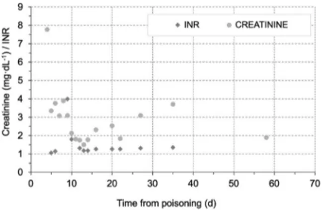
Figure 1.The course of creatinine and INR levels in case 1.
9 . . . .
cı:: 8 ---.---.--- .--- • INR • CREATININE >\
~ 7 : : ' ' ' ' : ··-····-···t·-···i··-···+···~---~---; ···ı =., 6 "O crı 5 _§, : : : : : ; i Q) 4 C ·c ~ 3 _. . . 1 ! ! • ! ! t l ---••: ---:.---. -: ---: ---:---·-·· : ---: ~ Ü 2 ' ' ' ' ' . .
----
••
h:::
___I : ____ • __ ! ----• _____ ! --- ! ________ • ! __________j
o o 10 20 30 40 50 60 70Discussion
Mushroom consumption is increasing in both rural and urban areas secondary to developments in gastronomy. In particular, mushrooms consumed in rural areas can lead to fatal poisonings owing to careless and misinformed approaches to collection.
Amanitin group mushrooms are the most poisonous mushroom species and are assumed to comprise 90% of the species that cause human death globally. AP consti-tutes the majority of these. In a retrospective study analyzing 93 mushroom poisonings between 1990 and 2008 in Portugal, 63% were dependent on the amatoxin type and 12% ended in mortality.4 AP contains 3 toxin groups: phallotoxins, virotoxins, and amatoxins. Ama-toxins, which are cyclic octapeptides, consist of 9 different components. In the main mechanism of action of the main toxin,α amatoxin is to bound covalently to RNA polymerase II, causing transcription inhibition and leading to protein synthesis disorders. The lethal human dose of amatoxins is 0.1 to 0.3 mg⋅kg-1. This is a very low concentration and can often be found in a single mushroom. Amatoxin is absorbed through the gastroin-testinal system; it is rapidly eliminated from the blood and disperses to the liver and kidneys within 48 h.
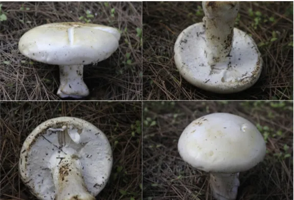
Because of this rapid absorption and distribution, it may be difficult to detect in plasma after 36 to 48 h. Approximately 60% of circulating amatoxins pass into bile and enterohepatic circulation, and the remaining 40% are cleared by the renal route. This metabolism explains the renal and hepatotoxicity commonly observed with these types of amitoxin mushroom in-gestions.5 Both of our patients consumed this type of mushroom by cooking it in boiling water, which was then drunk. The mushrooms were collected by the pa-tients’ relatives, brought to the emergency department, and later identified as AP by an expert mycologist (Figure 5).
Survival is primarily related to the degree of liver damage, but other potential complications are also very important. The major effect is due to liver necrosis (centrilobular and periportal, hemorrhage), which rapidly develops into both renal and liver dysfunction (hep-atorenal syndrome). An increase in ammonia levels can lead to coma and convulsions. This may be followed by respiratory failure and hemorrhage.
In rat studies, it has been shown that concentration in renal tissue is much higher than that in liver tissue. In autopsy studies, macroscopically, cortical extravasation in the kidneys, diffuse hemorrhagic stasis, acute tubular necrosis (ATN), and tubular dense hyaline cylinders were observed histopathologically. Degenerativefindings such as necrosis, vacuolization and edema, atrophy in the distal tubule, intratubular protein-rich cylinders, and thickening in the Bowman’s outer capsule were found in renal biopsy studies. Experimentally, polymyxin B, by inhibiting the effect of α a-amanatin, significantly pre-vented the occurrence of these findings in rats and decreased mortality.5 There were various levels of in-flammatory and oxidative changes in rat studies (lym-phocytic infiltrations, hydropic degeneration) and histopathologically brushy border loss as the applied dose increased, and necrotic areas have been shown to increase even more.6
Figure 2.The course of hepatic enzymes in case 1.
Figure 3.The course of creatinine and INR levels in case 2.
Figure 4.The course of hepatic enzymes in case 2.
::--.:., ~ '::; ~ ı-(/) <( 12000 10000 8000 6000 4000 2000 8 10 12 14 16 18 20 22
Time from poisoning (d)
3 ...•... 2.5 ... • INR • CREATININE : :
•
-~--i
i
+ 1.5 ---·--- -·--- .;... _! _______ , ________________ ....
1
...
1
... .... : ...
1 ... 1 ... i 0.5i
i •
i
i
i
i
O+----..---+---+---+---+---; o 4 10 12Tıme from poisoning (d)
6000 5000 4000 ::.. ~ 3000 ~ ~ 2000 ı-(/) <( 1000 o 5 ---,---,---.---,---. ' . ' l 1
i
/•',,,,
i --·AST i- ALT 1 / ; ~,,, r !---i---,,/··---i--- ',, ....
J...---:---: I : : ' : ---:---f---:---l ,/ ---ı,,/---6 7 8Time !rom poisoning (d)
In the standard treatment of mushroom poisoning, activated charcoal, penicillin-G, corticosteroid, NAC, and silibine treatments are advised to reduce absorption from the gastrointestinal tract in the acute period.2 Because excretion is mainly through urine, force diuresis has been recommended by some studies, especially to provide renal clearance in the first 48 h. However, considering the clinical conditions that reduced renal perfusion (hepatorenal syndrome, ATN), in these particular cases, we advised against this intervention.
Regarding extracorporeal treatment options for pa-tients with acute liver failure in AP ingestions, successful treatment with the molecular adsorbent recirculation system (MARS) and fractional plasma separation and adsorption system (FPSA) has been reported.7,8 It was shown that MARS treatment can decrease mortality and decrease urea-creatinine levels in this patient group.9 Positive results with FPSA also were reported.8A retro-spective analysis of 81 patients with both liver and renal injuries secondary to AP intoxication who underwent different dialysis methods (HD, hemoperfusion [HP], plasmapheresis [PF]) found that although 16 fatal cases had ATN, none died of renal causes.10Again, in a study comparing patients with fatal and nonfatal intoxication, no significant difference in creatinine levels was observed between the groups.11
In recent decades, a new-generation membrane type called an MCO membrane has been popular owing to its new potential utilities in HD.12 These membranes, by diffusion and convection, increase the permeability of medium-large toxins (up to 45 kDa molecular weight) owing to their higher pore sizes, allowing more and different uremic toxins to be removed. It has been re-ported that MCO membranes are more advantageous than high-flux HD in removal of some medium-molecular-weight toxins.13 In recent studies of the use of MCO dialyzer in HD patients, molecules such as β2-micro-globulin, light chains without kappa/lambda, complement factor D, and α1-microglobulin have been effectively removed, as well as expression of sepsis/in flammation-associated inflammatory markers such as TNF-α, mRNA-α, and IL-6 mRNA; levels of STNFR1 also have been shown to be reduced.
We intervened with emergent HD with an MCO membrane in both of our cases and successfully treated thefirst patient. In our second case, improvement in renal and liver function was achieved, but the patient died of sudden cardiac arrest related to septic shock. Because of the late clinicalfindings (at least 6–8 h) in AP poisonings, there is often a delay in hospital admission. Both of our patients delayed their presentation to our emergency department after consumption of the mushroom. How-ever, in ourfirst case, full resolution was achieved 48 h
after the first case, and the second case proved fatal owing to the late admission.
In a report of 2 patients with acute intoxication, 1 was treated for 72 h with continuous veno-venous hemo fil-tration (CVVH) and 3 plasma perfusions and the other with 100 h of CVVH and 5 doses of activated charcoal HP, and both cases fully healed.14 Again, 2 cases who had combined therapy (HP + HD) completely recov-ered.15In a study in which 21 patients were evaluated, PF was applied to all, and mortality was nearly 5%, lower than the average in the literature.16 In another report, beneficial effects of the combined use of extracorporeal therapies were emphasized. In this report, the effective-ness of FPSA and PF was demonstrated; continuous renal replacement therapy was used only to treat uremic status and elevated ammonia.2 Many extracorporeal toxin removal treatments (HD, HP, PF) appear to be effective, especially in thefirst 48 h of exposure. The effectiveness of systems such as MARS and FPSA has been demon-strated in AP poisoning, especially in liver failure. However, an important disadvantage is that these types of interventions can only be performed at major tertiary care centers, and as the highest clinical efficacy is within the first hour after ingestion, this level of care cannot be applied in every center.
Mushroom poisoning is a common and possibly fatal health problem. It is extremely important to diagnose early and begin treatment as soon as possible, especially because early HD has been reported to significantly reduce mortality resulting from liver and kidney failure.17 There are currently no specific antidotes or guidelines for treatment, so starting extracorporeal treatment methods as early as possible in addition to standard treatments can be considered as a method that reduces mortality and morbidity. Since the 1960s and 1970s, dialysis modalities have been tried in AP poisoning. Although the treatment of AP ingestion with extracorporeal methods seems rational, the most effective modality has not yet been clarified. Considering that special extracorporeal detoxi-fication methods can be performed in a tertiary or qua-ternary care center, it is very important to start treatment in thefirst 24 to 48 h, until transport is provided. Addi-tionally, we recommend that the HD MCO membrane be considered as a life-saving treatment modality for these types of mushroom poisonings. To our knowledge, these 2 cases represent thefirst documentation in the medical literature describing HD with MCO membrane in acute AP mushroom poisoning.
Acknowledgments: We are grateful to Doruk Demircioglu for his technical support.
Author Contributions: All authors contributed equally to the writing
of this manuscript and take responsibility for the final form of the
manuscript.
Financial/Material Support: None. Disclosures: None.
References
1. Ye Y, Liu Z. Management of Amanita phalloides
poisoning: a literature review and update. J Crit Care.
2018;46:17–22.
2. Kieslichova E, Frankova S, Protus M, Merta D,
Uchytilova E, Fronek J, et al. Acute liver failure due to Amanita phalloides poisoning: therapeutic approach and
outcome. Transplant Proc. 2018;50(1):192–7.
3. Saviuc P, Flesch F. Acute higher funghi mushroom
poisoning and its treatment. Presse Med.
2003;32(30):1427–35.
4. Brand˜ao JL, Pinheiro J, Pinho D, da Silva DC, Fernandes E,
Fragoso G, et al. Intoxicaç˜ao por cogumelos em Portugal [Mushroom poisoning in Portugal]. Acta Med Port.
2011;24(Suppl 2):269–78.
5. Garcia J, Costa VM, Carvalho ATP, Silvestre R, Duarte JA,
Dourado DFAR, et al. A breakthrough on Amanita phal-loides poisoning: an effective antidotal effect by polymyxin
B. Arch Toxicol. 2015;89(12):2305–23.
6. Ergin M, Dundar ZD, Kilinc I, Colak T, Oltulu P,
Girisgin AS. Alpha-amanitin poisoning, nephrotoxicity and oxidative stress: an experimental mouse model. Iran Red
Crescent Med J. 2015;17(8):e28068.
7. Stankiewicz R, Lewandowski Z, Kotulski M, Patkowski W,
Krawczyk M. Effectiveness of fractionated plasma separa-tion and absorpsepara-tion as a treatment for Amanita phalloides
poisoning. Ann Transplant. 2016;21:428–32.
8. Bergis D, Friedrich-Rust M, Zeuzem S, Betz C, Sarrazin C,
Bojunga J. Treatment of Amanita phalloides intoxication by
fractionated plasma separation and adsorption.
J Gastrointestin Liver Dis. 2012;21(2):171–6.
9. Sorodoc L, Lionte C, Sorodoc V, Petris O, Jaba I. Is MARS
system enough for A phalloides-induced liver failure
treat-ment? Hum Exp Toxicol. 2010;29(10):823-32.
10. Mydlik M, Derzsiova K. Liver and kidney damage in acute
poisonings. BANTAO J. 2006;4(1):30–3.
11. Escudié L, Francoz C, Vinel JP, Moucari R, Cournot M,
Paradis V, et al. Amanita phalloides poisoning: reas-sessment of prognostic factors and indications for
emer-gency liver transplantation. J Hepatol.
2007;46(3):466–73.
12. Boschetti-de-Fierro A, Voigt M, Storr M, Krause B. MCO
membranes: enhanced selectivity in high-flux class. Sci
13.Kirsch AH, Lyko R, Nilsson LG, Beck W, Amdahl M, Lechner P, et al. Performance of hemodialysis with novel medium cut-off dialyzers. Nephrol Dial Transplant.
2017;32(1):165–72.
14.Splendiani G, Zazzaro D, Di Pietrantonio P, Delfino L.
Continuous renal replacement therapy and charcoal plas-maperfusion in treatment of amanita mushroom poisoning.
Artif Organs. 2000;24(4):305–8.
15. Mullins ME, Horowitz BZ. The futility of hemoperfusion
and hemodialysis in Amanita phalloides poisoning. Vet
Hum Toxicol. 2000;42(2):90–1.
16. Jander S, Bischoff J. Treatment of Amanita phalloides
poisoning: I. retrospective evaluation of plasmapheresis in
21 patients. Ther Apher. 2000;4(4):303–7.
17. Mendonca S, Gupta S, Gupta A. Extracorporeal management