Correspondence: Sükrü Sanli MD, Istanbul University, Medical Faculty of Istanbul University, Istanbul, Turkey. 76
Acta Medica Anatolia
Introduction
Intestinal lipomatosis (IL) is a rare encountered condition which is characterized by the infiltration of fatty tissue in the intestinal submucosa. Although diffuse forms of intestinal lipomatosis may lead to intestinal sub-occlusion, digestive hemorrhage or diarrhea, the localized forms are generally tended to be asymptomatic. The isolated form of IL in the appendix has been reported by Antoci in 1956 as a histopatologically finding [1]. As our knowledge, there is no previously published paper about the radiologic findings of lipomatosis of appendix in English literature. This article is the first case of the literature discussing the computed tomography (CT) finding of the isolated form of intestinal lipomatosis which involves the appendix, mimicking the acute appendicitis.
Case
A 50-year-old male patient presented with the complaint of abdominal pain to the emergency department of hospital. Clinical history of the patient was unremarkable, except for the right lower quadrant abdominal tenderness was detected on physical examination. Performed laboratory blood tests were unremarkable. Abdominal ultrasound examination revealed of diffuse thickening of the appendix wall. With the suspect possibility presence of acute appendicitis computed tomography (CT) scans were performed in order to suspicion of acute appendicitis. CT imaging showed the diffuse thickening of the appendix wall with no inflammation in peri-appendicular fat tissue and no enlarged lymph node was detected (Fig. 1). The measurement value
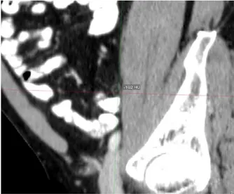
of the appendix wall was calculated as -102 HU which was representing the submucosal adipose tissue infiltration (Fig. 2).
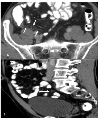
Fig 1
. Contrast Enhanced Computed Tomography
Axial plane (a,b) sagittal plane (c) coronal plane (d) images shows the fatty infiltration in the submucosa of the appendix resulting in the thickening of the wall. (Toshiba Aquilion 64 Detector CT. Protocol: 50 mAs, 120 kV, 5 mm Slice thickness, 350 ml iodine contrast).
Case Report
Acta Medica Anatolia
Volume 3Issue 22015
Isolated submucosal lipomatosis of appendix mimicking acute
appendicitis: computed tomography findings
Sükrü Sanli1, Hadi Sasani2, Eray Darici3, Baris Bakir1
1 Department of Radiology, Istanbul University, Medical Faculty of Istanbul University, Istanbul, Turkey 2 Department of Radiology, Istanbul Bilim University, Florence Nightingale Hospital, Istanbul, Turkey 3 Department of Radiology, Istanbul University, Cerrahpaşa Medical Faculty, Istanbul, Turkey
Abstract
Acute appendicitis is one of the more common surgical emergencies, and it is one of the most common causes of acute abdominal pain. Intestinal lipomatosis is a rare condition particularly the isolated form of lipomatosis of the appendix which may mimic or present as an acute appendicitis, that frequently requires the surgical exploration. In this paper, we report computed tomography findings of a case wıth isolated form of submucosal lipomatosis of appendix.
Keywords: Isolated, lipomatosis of appendix, computed tomography.
Case Report
Sanli S et al.
Acta Med Anatol 2015;3(2):76-77 77
No additional pathology was detected in the other visceral organs. Due to these findings, the patient was observed for one day duration with hydration support and oral intake cessation. On follow up no alternation was detected in laboratory and clinical findings of the patient. Due to no change of the present condition of the patient and no change in the clinical and laboratory findings, the patient was discharged from the hospital.
Fig 2. Contrast Enhanced Computed Tomography shows the measurement value of the appendix was calculated as -102 HU, representing fatty infiltration in the submucosa of the appendix. (Toshiba Aquilion 64 Detector CT. Protocol: 50 mAs, 120 kV, 7 mm Slice thickness, 350 ml iodine contrast)
Discussion
Intestinal lipomatosis (IL) is an unusual condition seen which is characterized by the anomalous fatty infiltration in the intestinal submucosa. Although diffuse forms of intestinal lipomatosis may lead to intestinal sub-occlusion, digestive hemorrhage or diarrhea, the localized forms are generally tend to be asymptomatic [2]. The isolated form of IL in the
appendix has been reported by Antoci in 1956 as a histopathological finding [1]. In the differential diagnosis of intestinal lipomatosis with other malignant pathologies of the colon or appendix, it frequently requires surgical exploration and the histopathological verification [2]. In our patient, due to the steady state clinical condition and laboratory findings and the CT appearance which revealed the isolated lipomatosis of the appendix, the patient did not undergo surgical procedure.
In the literature, the diffuse form of colonic lipomatosis has been reported as an extremely rare condition [3]. Appendiceal lipomatosis is an extremely rare condition and as far as our knowledge there is only one case report in English literature which did not discuss the radiologic findings.
In lipomatosis form of the appendix due to the anomalous adipose tissue infiltration in the submucosa resulting in the thickening of the appendix wall that may present and mimic the acute appendicitis as clinical finding. Laboratory tests and results of our patient were in the normal ranges that did not need any surgical procedure. Depending on the clinical and laboratory findings in the differential diagnosis sometimes it may require surgical exploration.
Conclusion
Although lipomatosis of appendix is an extremely rare condition, it should be considered in the differential diagnosis of acute appendicitis. In the isolated lipomatosis forms of the appendix, physician should be aware of considering such this condition to prevent the unnecessary surgical procedure whether the clinical and the laboratory findings are stable. In the presence of clinical and ultrasonographic suspicion of acute appendicitis in patients with appendiceal lipomatosis, CT examination is reasonably diagnostic.
References
1. Antoci B. Submucous lipomatosis of the appendix.Riv Anat Patol Oncol. 1956;11(9):127-133.
2. Leone N, Debernardi-Venon W, Marzano A, De Paolis P, Fronda GR, Rizzetto M. Lipohyperplasia or intestinal lipomatosis. Minerva Gastroenterol Dietol. 1998;44(4):207-10.
3. Catania G, Petralia G.A, Migliore M, Cardi F. Diffuse colonic lipomatosis with giant hypertrophy of the epiploic appendices and diverticulosis of the colon. Dis Colon Rectum. 1995;38(7):769-75.