Association Between Bietti Crystalline Fundus
Dystrophy and Retinitis Pigmentosa
Bietti Kristalin Fundus Distrofisi ile Retinitis Pigmentosa Birlikteliği
Özgür ARTUNAY1, Amber ŞENEL2, Alper ŞENGÜL2, Rıfat RASİER2, Erdal YÜZBAŞIOĞLU1, Halil BAHÇECİOĞLU3ABSTRACT
We described a case of Bietti’s Crystalline Fundus Dystro-phy (BCFD) who was also diagnosed with Retinitis Pigmen-tosa (RP). A full ophtalmologic examination was performed to a 29 years old female whose night vision has been wors-ening over the past few years. She was positive for RP65 mutation and yellow sparkling crystals and peripheric bony specules have been detected in her retina. According to her fundus fluorescein angiography, electroretinogram and visual field results, the patient was diagnosed with both BCFD and RP. We recommend to keep in mind that al-though RP may be seen associated with BCFD, BCFD may also present with RP signs and symptoms.
Key Words: Bietti’s crystalline dystrophy, retinitis pigmentosa.
ÖZ
Çalışmamızda Retinitis Pigmentosa ve Bietti’nin kristalin fundus distrofisi (BKFD) tanısı konulan bir olgu sunulmuş-tur. Son yıllarda kötüleşen gece görüşü şikayetiyle klini-ğimize başvuran 29 yaşında kadın hastanın RPE65 gen mutasyon pozitifliği mevcuttu. Hastanın yapılan tam of-talmolojik muayenesinde retinada parlak sarı kristaller ve periferik kemiksi çıkıntı şekilli pigmentasyonlar tespit edildi. Fundus floresan anjiyografi, elektroretinogram ve görme alanı sonuçlarına göre hastaya RP ve BKFD birlikteliği ta-nısı koyuldu. RP’nin BFKD ile beraber görülebileceği gibi, BFKD’nin de RP semptomları ve bulguları ile prezante ola-bileceği akılda tutulmalıdır.
Anahtar Kelimeler: Bietti’nin kristalin distrofisi, retinitis pig-mentosa.
Ret-Vit 2010;19:209-212
Received : March 15, 2010 Accepted : April 08, 2010
1- M.D.Assistant Professor., İstanbul Bilim University, Avrupa Florance Nighthingale Hospital Eye Clinic İstanbul/TURKEY
ARTUNAY Ö., [email protected] YÜZBAŞIOĞLU E., [email protected]
2- M.D.Assistant, İstanbul Bilim University, Avrupa Florance Nighthingale Hospital Eye Clinic İstanbul/TURKEY
SENEL A., [email protected] ŞENGÜL A., [email protected] RASİER R., [email protected]
3- M.D. Professor, İstanbul Bilim University, Avrupa Florance Nighthingale Hospital Eye Clinic İstanbul/TURKEY
BAHÇECİOĞLU H., [email protected]
Correspondence: M.D.Assistant Professor., Özgür ARTUNAY
İstanbul Bilim University, Avrupa Florance Nighthingale Hospital Eye Clinic İstanbul/TURKEY
Geliþ Tarihi : 15/03/2010 Kabul Tarihi : 08/04/2010
1- İstanbul Bilim Üniversitesi, Göz Hastalıkları A.D., İstanbul, Yrd. Doç. Dr. 2- İstanbul Bilim Üniversitesi, Göz Hastalıkları A.D., İstanbul, Asist. Dr. 3- İstanbul Bilim Üniversitesi, Göz Hastalıkları A.D., İstanbul, Prof. Dr.
210 Association Between Bietti Crystalline Fundus Dystrophy and Retinitis Pigmentosa
INTRODUCTION
Bietti’s crystalline dystrophy is first described by Bietti in 1937 as a tapatoretinal degeneration characterized by crystalline deposits in limbus and retina.1 It is a
progres-sive disease where yellow sparkling crystal deposits con-sisting of complex lipid inclusions in limbal keratocytes, retina with greater amount in its posterior pole, choroi-dal, conjunctival and limbal fibroblasts and atrophy in retina pigment epithelium (RPE), choriocapillaris and choroid.1-4 It causes symptoms like paracentral scotomas,
night blindness, visual field constriction and progressive vision loss in 2nd-4th decades and causes legal blindness
in 5th-6th decades.5-9 It is seen commonly in asian patients
and has an autosomal recessive inheritance pattern.3,6,8
The diagnosis of BCFD is based on clinical findings; bio-microscopic and ophthalmoscopic appearance are usu-ally sufficient to make diagnosis.6-9
Retinitis Pigmentosa (RP) is one of the most common forms of inherited retinal degenerations. It is a
progres-sive retinal dystrophy causing deterioration of the night vision in early phase and constriction of the visual field leading to tunnel vision in late phase.10-12 Hence it causes
photoreceptor loss, affected people experience legal blindness in childhood or in some cases in 4th-5th
de-cades. Mottling of the RPE with black bony specule pig-mentation seen in peripheral retina is patognomanic for RP. RP can be inherited as autosomal dominant, autoso-mal recessive, and X-linked manner, digenetic and mi-tochondrial forms have been also described.10-13 At least
35 different genes or loci mutation are known to cause nonsyndromic RP. Mutations in RPE65 is well known to cause autosomal recessive RP.11-13 We represent here a
patient with both BCFD and RP. CASE REPORT
A 29 years old woman referred to our clinic with a complaint of decreased night and day vision that has been worsening over the past few years.
Figure 1,2: Right and left colour fundus photos revealing intraretinal sparkling yellow crystals distributed in the posterior pole, choriocapillaris atrophy, pigmentary clumping in the form of bone spicules and salt-pepper like appearance in peripheral retinal.
Figure 3,4: FA obtained from right and left eye demostrated focal lobular areas of choriocapillary atrophy with salt-pepper like appearance correspond to concomitant diffuse RPE changes at the peripheral retina.
Ret-Vit 2010;19:209-212 Artunay et al. 211
She denoted that she was using statins because her cholesterol and triglyceryde levels were high. She also ex-plained that she was being followed by another clinic be-cause of atypical RP and she was positive for RP65 gene mutation. It has been noted that her family members have also RP features. She was performed a full ophthalmologic examination. Her intraocular pressure was 16 mmHg in the right eye and 13 mmHg in the left. Her visual acquity was 0.92 log MAR in the right eye and 0.88 log MAR in the left, reaching 0.78 log MAR in both eyes with correction.
On slit lamp examination, the cornea, lens and anterior chamber were normal. There were no limbal, corneal, conjunctival crystalline deposits. She did not have any history of associated other medical conditions or drug us-age. Fundus examination showed intraretinal sparkling yellow crystals distributed in the posterior pole and also midperiphery. There was midperipheral RPE and chorio-capillaris atrophy, pigmentary clumping in the form of bone spicules and salt-pepper like appearance in pe-ripheral retinal (Figure 1,2).
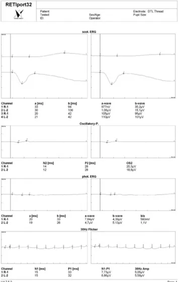
FA revealed the focal geographic appearance showing transmission hyperfluorescence in the crystal-line retina and characteristic lobuler choriocapillaris at-rophy in the adjacent noncrystalline retina (Figure 3,4). Choriocapillaris atrophy and hyper-reflective nodules at the level of RPE-Bruch’s membrane and choriocapillaris complex correspond to crystalline deposits were seen on optical coherence tomography (Figure 5). Her electro-retinogram (ERG) showed decreased scotopic a and b-wave amplitudes (Figure 6). Furthermore, the photopic a and b wave amplitudes were both severely attenuated. Visual field testing was performed and showed constric-tion of visual field bilaterally. She was diagnosed with both BCFD and RP on the basis of clinical findings, ge-netic and diagnostic tests, biomicroscopic and ophthal-moscopic appearance. She refused any further genetic examination or blood or cell culture.
DISCUSSION
Bietti first reported cases of tapetoretinal degenera-tion characterized by yellow glistering retinal crystals, choroidal sclerosis, and marginal crystalline dystrophy of the cornea.1-3 Similar cases with no limbal crystalline
deposits have been reported and called Bietti crystalline fundus dystrophy or crystalline retinopathy.
BCFD is an autosomal recessive inherited disorder associated with the CYP4-V2 gene polymorphism on 4th chromosome. This gene is responsible for synthesis of cy-tochrome p450 4 V2 (CYP 4 V2) enzymes functioning in fatty acid synthesis.2,6,8 CYP 4 enzymes are microsomal fatty acid Ω-hydroxylases working with mitochondrial and peroxisomal a-oxidation enzymes to degrade cel-lular lipids The pathogenesis of crystalline formation in BCFD is not fully understood but assumed malfunction in lipid metabolism of patients like defects in lipid binding proteins or in enzymes. The ocular and peripheral cell cultures of BCFD patients show high amounts of triglyc-eride and cholesterol deposition.2-4 The abnormal
inclu-sions are similar to those found in circulating lympho-cytes, keratolympho-cytes, and conjunctival and skin fibroblasts in electron microscopy. The patient in the present study also had no corneolimbal crystals. Althuogh our patient has high cholesterol and triglyceride levels, she refused to do genetic examination or biopsy of retina for electron microscopy trial or blood sampling.
Figure 5: Choriocapillaris atrophy and hyper-reflective nod-ules at the level of RPE-Bruch’s membrane and choriocapillaris complex correspond to crystalline deposits were seen on optical coherence tomography. The white arrows show hyper-reflective area consistent with crystallin deposits.
Figure 6: Her electroretinogram showed decreased scotopic a and b-wave amplitudes and the photopic a and b wave, 30 Hz flicker amplitudes were severely attenuated.
212 Association Between Bietti Crystalline Fundus Dystrophy and Retinitis Pigmentosa
RP diagnosis is based on visual field findings and genetic analysis. In 1989 a mutation in a gene encod-ing rhodopsin pigment was found, one of the main pro-teins of the outer segments of photoreceptors and having an important role in visual transduction cascade. More than 100 other mutations have been demonstrated in the same gene since that date. The main biochemical reason for RP is protein misfolding that is caused by rhodopsin gene mutations.5 Mutations in RPE65 is well known to
cause autosomal recessive RP.11-13 Visual field and ERG
results of RP and BCFD patients are similar.1,7,10 The
vi-sual field demonstrates predominantly a progressive loss of side vision in both of the disorders. Furthermore there may be another signs as blind spot enlargement, para-central scotomas or altitudinal defects that seem less typi-cal for the said disorders.11-13
ERG shows a marked reduction of both rod and cone signals, with general predomination of rod loss in RP pa-tients. BCFD patients’ ERG findings are mostly subnormal. Whereas some of BCFD patients have nonrecordable ERG’s, patients having recordable ERGs have reduced amplitudes in scotopic, photopic and 30 Hz flicker ERG.6,7
Our patient’s visual field and ERG findings are indistin-guishable between an mild stage case of RP or BCFD. An advanced BCFD case shows similarities with a RP case. Mataftsi et al., conducted a research which’s aim was to determine the prevalence of BCFD in RP patients.7 The
207 RP patients included in the study have been ophthal-mologicaly examined that incorporated fluorescent and indocyanine green angiography for three years.
The study resulted in 6 patients being diagnosed with BCFD, furthermore it has been established that BCFD has a prevalence rate of 3% in nonsyndromic RP.8
In Jiao et al., study it is said that BCFD is a form of auto-somal recessive RP and accounts for 3% of nonsyndromic RP.9 Our patient had RPE 65 mutation as an RP spesiphic
mutation. Furthermore, FA findings, fundus apperance and high lipid levels in blood lead us to believe that this patient had also BCFD.
Nevertheless, the patient refused to do further ge-netic examination or biopsy of retina for electron micros-copy trial or blood sampling. To the best of our knowl-edge, this is the first case report showing one patient having both RP and BCFD from Turkey.
We recommend that clinicians should keep in mind that while RP cases may show BCFD manifestations, RP and BCFD may be seen together in same patient. If there is any suspicious condition, genetic analysis should be performed. Further clinic and genetic investigations are needed to evaluate this clinical association.
REFERENCES/KAYNAKLAR
1. Giuffre G.: Progression of Bietti’s crystalline dystrophy. J Fr Oph-talmol. 1991;14:249-254.
2. Lee J, Jiao X, Hejtmancik JF et al.: The metabolism of fatty acids in human Bietti crystalline dystrophy. Invest Ophthalmol Vis Sci. 2001;42:1707-1714.
3. Nakano M, Kelly EJ, Rettie AE.: Expression and characterization of CYP4V2 as a fatty acid omega-hydroxylase. Drug Metab Dis-pos. 2009;37:2119-2122.
4. Avunduk A.M., Avunduk M.C. Çetinkaya K. ve ark.: Bietti’nin kristalin distrofisi: İki vakanın klinik ve histopatolojik olarak değerlendirilmesi. T Klin J Oftalmol. 1996;5:393-397.
5. Özkırış A, Öner A, Evereklioğlu C.: Bietti’nin kristalin distrofisi saptanan hastaların elektroretinografi değerlerinin normal olgu-lar ile karşılaştırılması. Erciyes Tıp Derg. 2004;26:113-118. 6. Lai TY, Ng TK, Tam PO.: Genotype phenotype analysis of Bietti’s
crystalline dystrophy in patients with CYP4V2 mutations. Invest Ophthalmol Vis Sci. 2007;48:5212-5220.
7. Mataftsi A, Zografos L, Millá E, et al.: Bietti’s crystalline corneoreti-nal dystrophy: a cross-sectiocorneoreti-nal study. Retina. 2004;24:416-426. 8. Jiao X, Munier FL, Iwata F et al.: Genetic linkage of Bietti crystallin
corneoretinal dystrophy to chromosome 4q35. Am J Hum Genet. 2000;67:1309-1313.
10. Van Soest S, Westerveld A, de Jong PT et al.: Retinitis pigmen-tosa: defined from a molecular point of view. Surv Ophthalmol. 1999;43:321-334.
11. Baumgartner WA.: Etiology, pathogenesis, and experimental treat-ment of retinitis pigtreat-mentosa. Med Hypotheses. 2000;54:814-824. 12. Hartong DT, Berson EL, Dryja TP.: Retinitis pigmentosa. Lancet.
2006;368:1795-1809.
13. Chong V, Bird AC.: Management of inherited outer retinal dystro-phies: present and future. Br J Ophthalmol. 1999;83:120-122.