Iran J Radiol. 2014 May; 11(2): e16327. DOI: 10.5812/iranjradiol.16327
Published online 2014 May 15. Research Article
The Role of Choice-Lock Catheter and Trocar Technique in Percutaneous
Ablation of Symptomatic Renal Cysts
Burak Ozkan
1,*; Ali Harman
1; Baris Emiroglu
1; Ilker Arer
1; Cuneyt Aytekin
11Department of Interventional Radiology, School of Medicine, Baskent University, Besevler, Turkey
*Corresponding author: Burak Ozkan, Department of Interventional Radiology, School of Medicine, Baskent University, Besevler, Turkey. Tel: +90-5056384753, Fax: +90-3124820575, E-mail: [email protected]; [email protected]
Received: August 3, 2013; Revised: December 13, 2013; Accepted: February 9, 2014
Background: The most common benign lesions of the kidney are simple cysts. They are acquired lesions and mostly affect the elderly
population.
Objectives: To describe the usage of choice-lock catheter and trocar technique in percutaneous renal cyst treatment and determining
long-term outcomes.
Patients and Methods: This retrospective study was carried out between February 2000 and July 2011. Eighty-eight cysts all of which were
Bosniak type-1 cysts were selected in 75 patients. The treatment indications were flank pain, hydronephrosis and hypertension. The choice-lock catheter was used for 84 cysts with the trocar technique. Ninety-five percent ethanol was used as the sclerosing agent. Maximum volume of the injected ethanol was 175 ml. The mean follow-up time after the treatment procedure was 23 months. Sixty-four cysts were located in the cortical and 24 cysts were located at the parapelvic region.
Results: Fifty-seven cysts had complete regression, while 31 cysts regressed partially. After the procedure, pain was relieved in 44 (82%)
patients and the pain alleviated in four (8%). Normotension was obtained in five (62.5%) of the eight hypertensive patients and no hydronephrosis was detected in nine patients. There were no relationship between the localization and the regression rate. No major complications occurred.
Conclusions: Percutaneous ethanol sclerotheraphy in simple cysts is a safe, cost-effective and minimally invasive method. We consider
that this technique may be an alternative solution in the percutaneous cyst treatment.
Keywords: Simple Renal Cysts; Percutaneous Cyst Ablation; Ethanol Sclerotherapy; Choice-Lock Catheter; Trocar Technique
Implication for health policy/practice/research/medical education:
This paper gives rise to the minimally invasive treatment options in Bosniak cyst treatment.
Copyright © 2014, Tehran University of Medical Sciences and Iranian Society of Radiology; Published by Kowsar Corp. This is an open-access article distributed under the terms of the Creative Commons Attribution License, which permits unrestricted use, distribution, and reproduction in any medium, provided the original work is properly cited.
1. Background
The most common benign lesions of the kidney are the simple cysts. They are acquired lesions that mostly affect the elderly population (1). The incidence of the simple renal cyst is more than 50% at age of 50 years. The renal cysts occur by weakening of the epithelium of the collect-ing duct cells leadcollect-ing to the diverticula formation pro-cess by time (2). The indication for treatment is based on urinary tract obstruction. Several methods for treatment of the cyst including surgical and percutaneous proce-dures have been proposed; namely, percutaneous mar-supialization (3), percutaneous aspiration (4), and open and laparoscopic cyst unroofing (5). Aspiration alone without injection of a sclerosing agent has been reported with a higher recurrence rate. There are several promis-ing results with spromis-ingle or multi-session sclerotherapy with percutaneous drainage (6, 7).
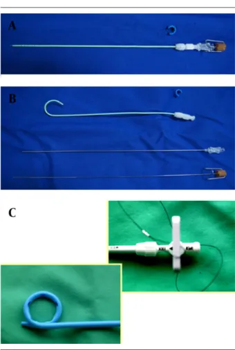
The advantage of choice-lock catheter is the 5.7 F diam-eter. The diameter of the choice-lock catheter is smaller than other draining catheters. Moreover, other draining
catheters are over 6 French sized. Choice-lock catheter con-sists of three co-axial parts. The stillet, the most inner part of the catheter can also be used for the initial puncture site. This catheter has a special lockage system that enables the catheter stability of the trocar during the puncture with one hand usage. There is a metal cannula for support-ive aim between the pigtail plastic catheter and the stillet. The supportive metallic part of the catheter is located in the catheter, it stables the other parts to be guided. This option enables the catheter to be guided safely through-out the hardened tissues. At the most through-outer part, there is a hydrophilic covered plastic part that has a pigtail lock-age system (Figure 1). The catheter can be used instead by the Seldinger technique with the help of 0.018 inch guide wire.
2. Objectives
The aim of this study was to assess the role of choice-lock catheter and trocar technique in percutaneous renal cyst treatment.
Figure 1. The structure of choice-lock catheter; A) The straight structure
of the choice-lock catheter; B) The three co-axial parts of the choice-lock catheter; C) The specific lockage system of the choice-lock catheter
3. Patients and Methods
This retrospective study consisted of 132 patients with 138 simple renal cysts who underwent percutaneous sclerotherapy and ethanol ablation treatment between February 2000 and July 2011 in our clinic. Fifty-seven pa-tients were excluded from the study due to lack of suffi-cient clinical data especially follow-up data. Most of these patients were referred to our clinic from another hospi-tal. Seventy-five patients with 88 cysts had initial criteria for the study. Forty-two patients were male and 33 were female. The mean age of the patients was 64 years (range, 44-87 years). The patients who underwent percutaneous treatment had only type I Bosniak cysts (8). Sclerother-apy indications included flank pain in 58 (77%), hydro-nephrosis in nine (10%), and hypertension in eight (9%) patients. The initial criteria for success after treatment was 60% volume reduction of the cyst. Sixty to eighty percent reduction of the cyst volume was considered as partial regression, and more than 80% was considered as complete regression. The choice-lock 5.7 F catheter with trocar technique is used for percutaneous cyst treatment and ethanol sclerotherapy. In four patients, the cysts
were septated and aspiration did not yield acceptable treatment results at the first attempt. In these patients, the choice-lock catheter was replaced by 8F pigtail cath-eter without doing a new puncture. In these cases, sub-sequently, the 6F dilatator was replaced by the puncture site with the guidance of 0.018 inch wire that was inside the choice-lock catheter. The 8 F pigtail catheter (Flexima, APDL, Boston Scientific, USA) was inserted into the cavity over a 0.035 inch guide wire (Amplatz stiff guide wire, Boston Scientific, USA). Twenty milliliter of the fluid was aspirated from the cavity with the help of the needle. The fluid was checked for further examinations. To obtain the cystogram under fluoroscopy, 50% diluted Telebrix (350 mg iodine/mL, Guerbet, France) was used. The aim of the cystogram is to assess the relationship of the cyst with the collecting system and to determine Bosniak classification. Without any extravasation or communi-cation with the collecting system, the cyst volume was aspirated. Equal to 30% of the initial cyst volume of 95% ethanol was injected into the cavity under fluoroscopic guidance. The patient was checked for any compliance of pain or other related symptoms--if the patient tolerated the session well, 95% ethanol was left in the cavity for 15 minutes. The patient was placed in at least three supine, prone and both lateral decubital positions. The reason was to allow contact of ethanol with all the cyst’s walls in order to destroy the epithelial tissue of the renal cyst. At the end of the procedure, all the injected ethanol to the cavity was aspirated and the catheter was withdrawn. The patients were called for periodic ultrasound and/or CT ex-aminations at several follow-up times. The patients were questioned about the symptoms and the volume of the treated cysts was calculated. The mean follow-up period was 23 months ranging from 3 to 58 months. Forty-two patients were followed for at least 2 years or more (29 pa-tients had 2, six papa-tients had 3, five papa-tients had 4, and two patients had 6 years follow-up).
SPSS for Windows Ver 11.5 (SPSS Inc., Chicago, Ill, USA) was used for statistical analysis. The Shapiro-Wilk test was used to determine if the distribution of the continu-ous variables were normal. The descriptive statistics for continuous variables were defined as mean±standard deviation or median (min-max). For the categorical vari-ables, the percent of patients and variables was calculat-ed. The Wilcoxon sign rank test was used for evaluation if there was a statistically significant change in the cyst volume before and after treatment. P value less than 0.05 was considered statistically significant. The Hospital Re-search Ethic Committee approved the study protocol.
4. Results
Between February 2000 and June 2011, 88 cysts among 75 patients who had percutaneous cyst aspiration and ethanol sclerotherapy were evaluated. Eighty-four cysts were treated with choice-lock with trocar technique. In four patients, the cysts were septated and aspiration did
not yield acceptable results without performing a new puncture; therefore, choice-lock catheter was changed with 8F pigtail catheter under fluoroscopic or sono-graphic guidance. All catheterization procedures were technically successful. The mean volume of the cyst be-fore treatment was 145.65 mL (39-504 mL) and it reached 15.5 mL (0-126 mL) after treatment (P<0.001). After the procedure, 57 cysts showed a higher than 80% volume reduction and 31 cysts had 60-80% volume reduction. None of the patients had any malignant cells in the cy-tological examinations.
Sixty-four cysts were located in the cortical and 24 cysts were located at the parapelvic region. Before starting the procedure, the median value of the cyst was 174.8 mL (49-504) ml in the cortical group, and 85 ml (36-175 mL) in the parapelvic group. After the procedure, the median value of the cyst volume was 17.3 mL (0.00-105 mL) in the cortical re-gion group, and 6.8 ml (0-65 mL) in the parapelvic located group. There was no statistically significant difference in the rate of regression between cortical and parapelvic lo-cated cysts (P=0.892). There was no statistically significant difference in the rate of regression between two genders and two groups of small and large volume cysts (Tables 1
and 2). A total of 75 patients who underwent percutaneous aspiration and sclerotherapy had no major complications such as renal parenchymal injury, renovascular or renal collecting system injury, pneumothorax or mortality. Pa-tients using ethanol during sclerotherapy can have minor complications such as allergic reaction, microscopic he-maturia or infection. Depending on the amount of etha-nol used during sclerotherapy patients may develop tran-sient pain and this is the restriction of ethanol usage. This is usually related with ethanol extravasation. No patient had retroperitoneal hemorrhage or hemorrhage into the cyst cavity.
Symptoms (flank pain, hydronephrosis and hyperten-sion) resolved in 66 (88%) of 75 symptomatic patients. Forty-eight (83%) of 58 patients with pain responded well to the treatment. Forty-four (76%) were free of pain, in four (6%) patients the pain decreased, whereas in four (6%) patients the pain did not change, and in six (10%) pa-tients, the pain increased. The patients’ symptoms before and after effective sclerotherapy are mentioned in Table 3. The successful treatment of a renal cyst is shown in Fig-ure 2 and FigFig-ure 3.
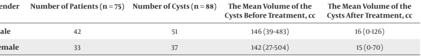
Table 1. Mean Volume of the Cysts Before and After Treatment in Two Genders
Gender Number of Patients (n = 75) Number of Cysts (n = 88) The Mean Volume of the
Cysts Before Treatment, cc Cysts After Treatment, ccThe Mean Volume of the
Male 42 51 146 (39-483) 16 (0-126)
Female 33 37 142 (27-504) 15 (0-70)
Table 2. Mean Volume of the Cysts Before and After Treatment in Two Groups of Large and Small Cysts The Mean Volume of the Cysts Before
Treatment, cc The Mean Volume of the Cysts After Treatment, cc
Cysts Larger than 300 cc (n = 20) 330 (309- 504 ) 26 (0-70)
Cysts Smaller than 300 cc (n = 68) 128.65 (39.6-294 ) 14.1 (0-126)
Table 3. Patients’ Symptoms Before and After Effective Sclerotherapy
Symptoms Number of Patients Who Described Symptoms
Before Effective Sclerotherapy Number of Patients Whose Symptoms Resolved After Effective Sclerotherapy
Flank Pain 58 44
Hydronephrosis 8 5
Hypertension 9 8
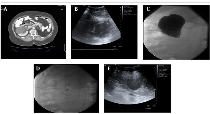
Figure 2. A 56-year-old man with Bosniak type-1 cyst in the right kidney with flank pain. A) Axial CT image shows Bosniak type-1 cyst in the right kidney
which has affected the pelvicalyceal system; B) US image shows the initial catheterization of the choice-lock catheter in the cyst during the treatment procedure; C) Cystogram shows that the cyst does not have any relationship with the pelvicalyceal system; D) Cystogram obtained after emptying of the cystic fluid; E) US image shows partial regression of the cyst one month after treatment.
Figure 3. A 72-year-old man suffering from left flank pain for 6 months, A and B) IV contrast enhanced CT images show the cyst located at the lower portion
of the left kidney which is classified at Bosniak type-1; C) Cystogram image shows that the cyst does not connect with the pelvicalyceal system; D and E) CT images obtained 3 months after the treatment show that the cyst is completely regressed and the patient had no pain.
5. Discussion
Percutaneous aspiration and sclerotherapy is the first-line treatment option for symptomatic simple renal cysts (1). Percutaneous aspiration is a simple, safe and
minimally invasive procedure. Simple drainage without sclerotherapy is associated with a high recurrence rate of 40-80%. Percutaneous sclerotherapy using a sclerosing
agent provides more satisfactory results than aspiration alone (2). In aspiration alone, the destruction of epithe-lial cell lining will not happen and the epitheepithe-lial cells will continue secreting fluid cyst. In other words the cyst fluid re-accumulates (4). Secretory epithelial lining the cyst wall must be destructed in order to prevent recurrence. For this purpose, various sclerosing agents such as bis-muth-phosphate (9), tetracyclin hydrochloride (10), ace-tic acid (11), povidone-iodine (12), n-butyl cyanoacrylate and iodized oil (13), ethanolamine oleate (14), OK-432 (15) and minocycline hydrochloride (16) have been used after the cyst fluid aspiration. Ethanol is the most commonly used sclerosing agent. Chemical properties of ethanol, leading to necrosis of epithelial cells lining the cyst wall will produce more obstacles. Secretory cells are rapidly inactivated by ethanol, but penetration of the fibrous capsule takes four to twelve hours. In this way, destruc-tion of cysts occur without affecting the renal parenchy-ma. Ethanol as a sclerosing agent has been mentioned in the literature, and there are studies that have reported success rates of over 90% (1, 2, 4). Akinci and colleagues treated 98 simple renal cysts with percutaneous ethanol sclerotherapy with a single session technique (2).
At the end of the first year follow-up, the reduction rate in cyst volume was 93.1%. In 17 patients, the cyst disappeared completely, and 83% of the patients had clinical improve-ment in the symptoms (2). Zerem et al. (17) treated 85 pa-tients and 92 cysts with percutaneous ethanol sclerother-apy. Recurrence of only six cysts occurred at the 24-month follow-up. Mohsen et al. (18) treated 64 cysts of 60 patients using sclerotherapy with 95% ethanol. In 84% the method provided complete resolution. In our study, percutaneous cyst aspiration and ethanol sclerotherapy of 88 cysts was applied. Reduction in the size of the cyst occurred in all 75 patients and 88 cysts after the procedure. In 57 cysts, 80% reduction took place in the volume of the cyst after the procedure out of which 31 had a 60-80% decrease in size. Percutaneous ethanol sclerotherapy in the treatment of symptomatic simple renal cysts were considered as suc-cessful (P < 0.001). Our success rate was similar to other studies. In the study conducted by Ozgur et al. (4), a num-ber of patients developed recurrence during follow-up while a sclerosing agent was not used for any of them. In our study, no recurrence or increase in the cyst volume was reported during follow-up. We think that this is due to the usage of ethanol as the sclerosing agent.
The single session of sclerotherapy with ethanol in the literature reported high success rates. There are also studies indicating that multiple session sclerotherapy is a more effective method of treatment. Hanna and Dahni-ya have shown increased success rates after two sessions of ethanol sclerotherapy. The recurrence rate was 80% in the group on which only aspiration was carried out and 32% in the group on which percutaneous aspiration and single-session ethanol therapy was performed. No recur-rence occurred in the group on which ethanol sclero-therapy was carried out twice. The high success rate of
sclerotherapy with ethanol depends on the injection by increasing the amount of contact time (7). Fontana and colleagues used the three-time ethanol injection method. The amount of ethanol used in the treatment was up to 30% of the volume and did not exceed 60 ml per each cyst treatment. Free drainage method of the cyst was used. As a result, recurrence of the cyst occurred in two patients and 68 cysts had complete resolution. The higher success rate in this study was based on the higher ethanol con-centration at the cyst wall. The higher ethanol concentra-tion results in the greater destrucconcentra-tion of the epithelial tis-sue. Ethanol penetrates the fibrous capsule slowly that is important in preventing systemic complication (19).
There were limitations in this study. Four of the 88 cysts had treatment with 8F caliber catheter. The number of patients was not enough for the comparison. In other words, there was not enough data to compare the choice-lock catheter and the 8F catheter. The second limitation was the pain scoring system. This is a retrospective study and we did not have a scoring system for the treatment outcome related to pain. But now in our clinic we use the pain scoring system and check before and after the pro-cedure.
In conclusion, percutaneous aspiration and ethanol sclerotherapy is an effective way of treatment of simple cysts. Our study is the first paper that describes the usage of trocar technique and choice-lock catheter.
Acknowledgements
There is no acknowledgement.
Authors’ Contribution
Dr. Burak Ozkan, Dr. Baris Emiroglu, Dr. Ilker Arer and Dr. Ali Harman developed the idea and played great roles in the result and material section. Dr. Emiroglu and Dr. Aytekin carried out statistical analysis and wrote the dis-cussion.
Financial Disclosure
There is no financial disclosure.
Funding/Support
This study was not supported by any source from any company or any grant from any hospital or such kind of institute. The funding organizations are public institu-tions and had no role in the design and conduct of the study; collection, management, and analysis of the data; or preparation, review, and approval of the manuscript.
References
1. Bozkurt FB, Boyvat F, Tekin I, Aytekin C, Coskun M, Ozkardes H. Percutaneous sclerotherapy of a giant benign renal cyst with al-cohol. Eur J Radiol. 2001;40(1):64–7.
2. Akinci D, Akhan O, Ozmen M, Gumus B, Ozkan O, Karcaaltincaba M, et al. Long-term results of single-session percutaneous
drain-age and ethanol sclerotherapy in simple renal cysts. Eur J Radiol. 2005;54(2):298–302.
3. Hulbert JC, Hunter D, Young AT, Castaneda-Zuniga W. Percutane-ous intrarenal marsupialization of a perirenal cystic collection--endocystolysis. J Urol. 1988;139(5):1039–41.
4. Ozgur S, Cetin S, Ilker Y. Percutaneous renal cyst aspiration and treatment with alcohol. Int Urol Nephrol. 1988;20(5):481–4. 5. Guazzoni G, Montorsi F, Bergamaschi F, Consonni P, Bellinzoni P,
Centemero A, et al. Laparoscopic unroofing of simple renal cysts.
Urology. 1994;43(2):154–9.
6. Chung BH, Kim JH, Hong CH, Yang SC, Lee MS. Comparison of single and multiple sessions of percutaneous sclerotherapy for simple renal cyst. BJU Int. 2000;85(6):626–7.
7. Hanna RM, Dahniya MH. Aspiration and sclerotherapy of symp-tomatic simple renal cysts: value of two injections of a sclerosing agent. AJR Am J Roentgenol. 1996;167(3):781–3.
8. Israel GM, Bosniak MA. An update of the Bosniak renal cyst clas-sification system. Urology. 2005;66(3):484–8.
9. Holmberg G, Hietala SO. Treatment of simple renal cysts by per-cutaneous puncture and instillation of bismuth-phosphate.
Scand J Urol Nephrol. 1989;23(3):207–12.
10. Kilinc M, Tufan O, Guven S, Odev K, Gurbuz R. Percutaneous in-jection sclerotherapy with tetracycline hydrochloride in simple renal cysts. Int Urol Nephrol. 2008;40(3):609–13.
11. Kwon SH, Oh JH, Seo TS, Park HC. Efficacy of single-session per-cutaneous drainage and 50% acetic Acid sclerotherapy for treatment of simple renal cysts. Cardiovasc Intervent Radiol. 2007;30(6):1227–33.
12. Phelan M, Zajko A, Hrebinko RL. Preliminary results of percuta-neous treatment of renal cysts with povidone-iodine sclerosis.
Urology. 1999;53(4):816–7.
13. Kim SH, Moon MW, Lee HJ, Sim JS, Kim SH, Ahn C. Renal cyst abla-tion with n-butyl cyanoacrylate and iodized oil in symptomatic patients with autosomal dominant polycystic kidney disease: preliminary report. Radiology. 2003;226(2):573–6.
14. Yamamoto K, Sakaguchi H, Anai H, Tanaka T, Morimoto K, Kichikawa K, et al. Sclerotherapy for simple cysts with use of eth-anolamine oleate: preliminary experience. Cardiovasc Intervent
Radiol. 2005;28(6):751–5.
15. Ham WS, Lee JH, Kim WT, Yu HS, Choi YD. Comparison of multiple session 99% ethanol and single session OK-432 sclerotherapy for the treatment of simple renal cysts. J Urol. 2008;180(6):2552–6. 16. Ohkawa M, Tokunaga S, Orito M, Shimamura M, Hirano S,
Okasho A, et al. Percutaneous injection sclerotherapy with mi-nocycline hydrochloride for simple renal cysts. Int Urol Nephrol. 1993;25(1):37–43.
17. Zerem E, Imamovic G, Omerovic S. Symptomatic simple re-nal cyst: comparison of continuous negative-pressure cath-eter drainage and single-session alcohol sclerotherapy. AJR Am J
Roentgenol. 2008;190(5):1193–7.
18. Mohsen T, Gomha MA. Treatment of symptomatic simple renal cysts by percutaneous aspiration and ethanol sclerotherapy. BJU
Int. 2005;96(9):1369–72.
19. Fontana D, Porpiglia F, Morra I, Destefanis P. Treatment of simple renal cysts by percutaneous drainage with three repeated alco-hol injection. Urology. 1999;53(5):904–7.