©Telif Hakkı 2020 Türk Yoğun Bakım Derneği Türk Yoğun Bakım Dergisi, Galenos Yayınevi tarafından yayınlanmıştır.
E-pos ta : [email protected] Tel. : +90 505 313 65 18
ORCID ID : orcid.org/0000-0002-0612-8481 Fatma İrem Yeşiler MD (✉),
Başkent University Faculty of Medicine, Department of Anesthesiology and Critical Care, Ankara, Turkey Fatma İrem Yeşiler, Helin Şahintürk, Ender Gedik, Pınar Zeyneloğlu
Başkent University Faculty of Medicine, Department of Anesthesiology and Critical Care, Ankara, Turkey Emre Günakan,
Başkent University Faculty of Medicine, Department of Obstetrics and Gynecology, Division of
Perinatology, Ankara, Turkey Geliş Tarihi/Received : 16.06.2020 Kabul Tarihi/Accepted : 13.07.2020
ABSTRACT Gebeliğin akut yağlı karaciğeri (AFLP), gebeliğin üçüncü trimesterinde veya doğum sonrası döneminde ortaya çıkan maternal karaciğer yetersizliği ile karakterize hayatı tehdit eden bir hastalıktır. Koagülopati, elektrolit anormallikleri ve çoklu organ disfonksiyon sendromu (MODS) görülebilir ve karaciğer transplantasyonu gerekebilir. Bu nedenle, MODS gelişen hamile kadınlar, uygun destekleyici tedavilerle multidisipliner olarak yoğun bakım ünitesinde (YBÜ) takip edilmelidir. Biz, YBÜ’ye sarılık ve MODS ile kabul edilen AFLP tanılı gebe bir kadının organ destek tedavilerine ve karaciğer nakline ihtiyaç duymadan başarılı şekilde yönetimini sunuyoruz. Otuz bir haftalık, ilk gebeliği olan 20 yaşında hasta bulantı, kusma ve sarılık şikayetleriyle hastaneye başvuruyor. Karaciğer nakli gerektiren olası akut karaciğer yetersizliği tanısı nedeniyle dış merkezden (608 km uzakta) YBÜ’ye sevk edildi. Yoğun bakım kabulündeki akut fizyoloji ve kronik sağlık değerlendirmesi skoru 12, sepsise bağlı organ yetersizliği skoru 8 ve Glasgow koma skalası 15 idi. AFLP tanısı düşünülen hastaya sezaryen ile acil doğum gerçekleştirildi. Doğumdan sonra karaciğer nakli ihtiyacı olmadan yoğun bakım desteği ile iyileşen hasta, 6 gün sonra YBÜ’den ve 10 gün sonra hastaneden taburcu edildi. Gebeliğin üçüncü trimesterinde veya doğum sonrası dönemde YBÜ’ye sarılık ve hiperbilirubinemi ile kabul edilen gebe bir kadının ayırıcı tanısında AFLP’den şüphelenilmelidir. Yoğun bakım uzmanı, yüksek mortalitesi ve morbid komplikasyonları olan AFLP tanısını geciktirmemelidir. Erken tanı, acil doğum ve peripartum-postpartum dönemlerde yoğun bakım desteği, maternal ve fetal sonuçları iyileştirebilir.
Anahtar Kelimeler: Sarılık, gebeliğin akut yağlı karaciğeri, yoğun bakım ünitesi
ÖZ Acute fatty liver of pregnancy (AFLP) is a life-threatening disorder characterized by maternal liver failure, and it occurs in the third trimester of pregnancy or postpartum period. The resultant effects include coagulopathy, electrolyte abnormalities, and the multiple organ dysfunction syndrome (MODS), which may require liver transplantation. Therefore, pregnant women having MODS should be managed in an intensive care unit (ICU) with multidisciplinary inputs to facilitate the appropriate supportive care. We present a successful case report of a pregnant women admitted to the ICU with jaundice and MODS without the need for liver transplantation and organ support therapies. A 20-year-old patient in her first pregnancy at 31 weeks gestation who presented with nausea, vomiting, and jaundice was admitted to our hospital. She was referred from a rural medical center (a center 608 kilometers away) to the ICU due to the possible diagnosis of acute liver failure requiring liver transplantation. Acute physiology and chronic health assessment score was 12, sepsis related organ failure score was 8, and Glasgow coma scale was 15 on ICU admission. AFLP was considered in the patient and an emergency delivery was performed by caesarean section. She recovered with intensive care support after pregnancy delivery without the need for liver transplantation. The patient was discharged from the ICU and hospital after 6 and 10 days, respectively. AFLP should be suspected in the differential diagnosis of a pregnant woman with jaundice and hyperbilirubinemia who is admitted to the ICU in the third trimester of pregnancy or postpartum period. Intensivist should not delay in the diagnosis of AFLP due to its morbid complications and high mortality. Early diagnosis, prompt pregnancy delivery, and intensive care support in the peripartum and postpartum periods may improve maternal and fetal outcomes Keywords: Jaundice, acute fatty liver of pregnancy, intensive care unit
Fatma İrem Yeşiler, Helin Şahintürk, Emre Günakan, Ender Gedik,
Pınar Zeyneloğlu
Yoğun Bakım Ünitesinde Sarılığı Olan Gebe Bir Kadın
A Pregnant Woman with Jaundice in the Intensive
Care Unit
Introduction
Jaundice is a clinical manifestation of increased serum levels of bilirubin (direct or indirect) and is associated with high maternal and perinatal mortality rates in pregnancy. One of the most common causes of jaundice in pregnancy is acute fatty liver of pregnancy (AFLP). It is an obstetric emergency characterized by maternal liver failure and occurs in the third trimester or early post-partum period (1,2). Stander and Cadden (3), first described “Acute yellow atrophy of the liver,” in 1934. The incidence ranges between 1 in 7,000 and 1 in 20,000 depending on the population studied. Clinical conditions leading to jaundice and liver dysfunction in pregnancy are intrahepatic cholestasis of pregnancy (ICP), pre-eclampsia, haemolysis elevated liver enzymes low platelet count (HELLP) syndrome and AFLP (4). The main pathogenesis is excessive accumulation of fatty acids in the maternal hepatocytes secondary to mitochondrial defects in the fetal long chain 3‐hydroxyacyl‐ coenzyme A dehydrogenase (4,5). AFLP may have serious complications- acute liver failure, encephalopathy, mechanical ventilation requirement, renal failure, and coagulopathy- and mortality risk both mother and fetus. So, women suspected of having AFLP should be hospitalized in an intensive care unit (ICU) during peripartum and postpartum periods (4-6). Early diagnosis and prompt delivery are critical to maternal and fetal outcomes (7). We presented a successful case report without the need for liver transplantation and organ support therapies of a pregnant women with AFLP admitted to ICU.
Case Report
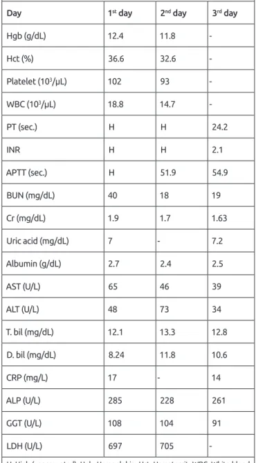
A 20-year-old primipar pregnant woman with gestational age of 31 weeks was admitted to theICU as a referral from a rural medical center - a center 608 kilometers away- due to possible diagnosis of acute liver failure requiring liver transplantation. She had nausea and vomiting for 6 days and yellowness of the eyes for one day with no pruritus. Laboratory values of the rural medical center were included in Table 1. There were no history of diarrhea or flu like symptoms, no history of travel to a malaria endemic area, no chronic illnesses and no history of paracetamol, acetylsalicylic acid, sodium valproate or herbal medicine ingestion. On physical examination, she was awake at ICU admission. Her vitals were as follows: heart rate 90 beats/minute, respiratory rate 20/minute, and blood pressure 122/70 mmHg and body temperature of (tympanic
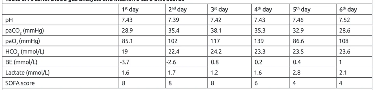
measurement) 35.9 ˚C. Acute physiology and chronic health assessment score was 12, sepsis related organ failure score was 8 and Glasgow coma scale was 15. Arterial blood gas analysis pH: 7.43, partial oxygen pressure: 85.1 mmHg, partial carbondioxide pressure: 28.9 mmHg, bicarbonate: 19 mmol/L, lactate 1.6 mmol/L, oxygen saturation: 97.4 % on room air at ICU admission. Respiratory and cardiovascular examinations were
Table 1. Laboratory parameters of rural medical center
Day 1st day 2nd day 3rd day
Hgb (g/dL) 12.4 11.8 -Hct (%) 36.6 32.6 -Platelet (103/µL) 102 93 -WBC (103/µL) 18.8 14.7 -PT (sec.) H H 24.2 INR H H 2.1 APTT (sec.) H 51.9 54.9 BUN (mg/dL) 40 18 19 Cr (mg/dL) 1.9 1.7 1.63 Uric acid (mg/dL) 7 - 7.2 Albumin (g/dL) 2.7 2.4 2.5 AST (U/L) 65 46 39 ALT (U/L) 48 73 34 T. bil (mg/dL) 12.1 13.3 12.8 D. bil (mg/dL) 8.24 11.8 10.6 CRP (mg/L) 17 - 14 ALP (U/L) 285 228 261 GGT (U/L) 108 104 91 LDH (U/L) 697 705
-H: High (unaccounted), Hgb: Hemoglobin, Hct: Hematocrit, WBC: White blood cell, PT: Prothrombin time, INR: International normalized ratio, APTT: Activated partial thromboplastin time, BUN: Blood urea nitrogen, Cr: Creatinine, AST: Aspartate aminotransferase, ALT: Alanin aminotransferase, T. bil: Total bilirubin, D. bil: Direct bilirubin, CRP: C-reactive protein, ALP: Alkaline phosphatase, GGT: Gamma glutamyl transpeptidase, LDH: Lactate dehydrogenase, sec.: Second
normal. The abdomen was non-tender without hepatomegaly and splenomegaly. The height of fundus was 31 weeks and the foetal heart was present and normal. An emergency delivery was decided by the perinatology team to the patient who was diagnosed possible acute liver failure due to pregnancy. She was transfused 1unit packed red blood cell (RBC) due to low hemoglobin level, 1 unit of platelet concentrate (PC) due to thrombocytopenia, 6 bags of cryoprecipitate, and 1 g of fibrinogen concentrate (Haemocomplettan®) due to
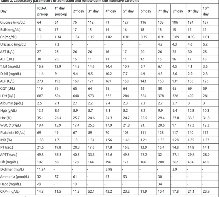
hypofibrinogenemia to prevent bleeding during the delivery at preoperative period in ICU and 2 units of fresh frozen plasma (FFP) due to coagulopathy during operation. A baby girl with an Apgar score of 4/8 and birth weight of 1,400 gram was delivered by caesarean section and meconium stained amniotic fluid was seen. The baby was admitted to the neonatal intensive care unit (NICU).On ICU admission and follow-up laboratory parameters were presented in Table 2 and 3: increased white blood cell (WBC) count, decreased
Table 2. Laboratory parameters of admission and follow-up in the intensive care unit
Day ICU-A
pre-op
1st day
post-op 2
nd day 3rd day 4th day 5th day 6th day 7th day 8th day 9th day 10 th day Glucose (mg/dL) 64 51 76 112 71 127 116 103 106 124 137 BUN (mg/dL) 18 17 17 15 14 16 18 18 15 12 12 Cr (mg/dL) 1.5 1.34 1.34 1.19 1.02 0.81 0.79 0.91 0.89 0.93 1.01 Uric acid (mg/dL) - 7.3 - - - 4.2 4.3 4.6 5.2 AST (U/L) 27 25 26 26 16 17 20 26 25 30 25 ALT (U/L) 30 23 16 11 11 11 12 15 16 17 18 T. bil (mg/dL) 16.9 12.9 14.5 14.6 14.4 10.7 6.7 6.1 4.5 4.1 3.6 D. bil (mg/dL) 11.6 9 9.4 9.5 10.2 7.7 4.9 4.5 3.6 2.9 2.8 ALP (U/L) 273 192 169 171 161 158 143 158 131 136 126 GGT (U/L) 119 79 65 64 63 64 66 80 65 69 59 LDH (U/L) 687 594 640 573 335 284 324 378 326 409 281 Albumin (g/dL) 2.5 2.1 2.1 2.2 2.4 2.3 2.3 2.7 2.7 3 3 Hgb (g/dL) 12.1 8.6 8.9 8.7 8.1 8.2 8.2 9.9 9.4 10.8 10.3 Htc (%) 35.1 26.4 25.7 24.6 24.3 24.7 25.5 29.4 27.8 33.5 31.8 WBC (103/µL) 19.4 15.9 17.4 25.5 17.9 21.8 21. 20.6 17 17.2 12.3 Platelet (103/µL) 69 49 67 89 70 103 111 128 117 140 172 INR (%) 1.88 1.7 1.8 1.54 1.56 1.46 1.21 1.35 1.28 1.25 1.23 PT (sec.) 21.5 19.8 20.3 17.6 17.8 16.8 13.9 15.4 14.8 14.8 14.1 APTT (sec.) 49.3 38.3 40.5 33.3 32.6 49.3 27.2 32 27.1 29.8 28.9 Fib (mg/dL) 102 58 128 144 196 171 166 208 262 434 418 D-dimer (mg/L) 11.24 - - - 3.98 - - - 3.9 - -Ammonia (µmol/L) 32 57 61 - 43 53 - 30 - - -Hapt (mg/dL) <8 - 10 - - - - 34 - - -CRP (mg/dL) 14.8 11.5 11.5 52.1 42.2 23.2 11.9 10.4 17.8 21.1 23.9
ICU-A: Admission of Intensive Care Unit, Pre-op: Pre-operative, Post-op: Post-operative, BUN: Blood urea nitrogen, Cr: Creatinine, AST: Aspartate aminotransferase, ALT: Alanin aminotransferase, T. bil: Total bilirubin, D. bil: Direct bilirubin, ALP: Alkaline phosphatase, GGT: Gamma glutamyl transpeptidase, LDH: Lactate dehidrogenase, Hgb: Hemoglobin, Hct: Hematocrit, WBC: White blood cell, INR: International normalized ratio, PT: Prothrombin time, APTT: Activated partial thromboplastin time, Fib: Fibrinogen, Hapt: Haptoglobin, CRP: C-reactive protein, sec.: Second
haemoglobin and platelets, prolonged coagulation tests [prothrombin time, activated partial thromboplastin time, international normalized ratio increased serum creatinine, elevated total and direct bilirubin, alkaline phosphatase, gamma glutamyl transpeptidase, lactate dehydrogenase, uric acid with normal aspartate aminotransferase (AST) and alanine aminotransferase (ALT)]. But, there were minimally elevated AST and ALT 2 days ago. A virology screen performed for hepatitis A, B, C, E viruses; cytomegalovirus, herpes simplex virus, human immunodeficiency virus, brucella agglutination and Epstein-Barr virus were all negative. Immunological analysis, extractible nuclear antigens, anti-smooth muscle antibody, antimitochondrial antibodies, antineutrophil cytoplasmic antibodies, and liver kidney microsome type-1, were negative but anti-nuclear antibody was positive at 1/100 titer. Polymerase chain reaction of influenza type A-B, respiratory syncytial virus
and blood-urine cultures were all negative. Serum copper and ceruloplasmin levels were within the reference values. There was no protein and bilirubin value was high in the urine analysis. She had several episodes of hypoglycaemia (3-3.5 mmol/L) which were corrected with 10% dextrose.
Intravenous ceftriaxone 1x2 g and metronidazole 3x500 mg were administered empirically due to chorioamnionitis. Then, oral cefixime 1x400 mg and metronidazole 3x500 mg for 1 week were changed empirically due to possible diagnosis of listeria monocytogenesis chronic chorioamnionitis as the pathology result of placenta. The abdominal ultrasonography (USG) include increased minimally diffuse liver parenchymal echogenicity (bright liver), appearance of minimal gall mud and moderate dilatation in the right urinary collecting system. There was no pathology in vascular structures, biliary tract, spleen and no ascites. Her hepatobiliary and pancreatic systems were normal in the magnetic resonance
cholangiopancreatography on the 2nd day postdelivery.
She was transfused totally 1unit packed RBC, 6 bags of cryoprecipitate, 1 unit of PC, and 7 units of FFP for six days in the ICU.
Her peripheral smear never showed any evidence of hemolysis as microangiopathic hemolytic anemia (MAHA) and ADAMTS-13 activity (>94%) was normal (range of 40 to 130%).
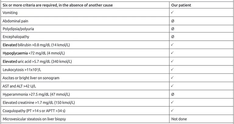
The Swansea criteria, which defines AFLP at 6 points or more (1,4,7), scored at least 9 points at admission (vomiting, bright liver on ultrasound scan, renal impairment, minimally elevated transaminase, increased uric acid, hyperbilirubinemia, coagulopathy, leukocytosis and hypoglycemia were positive; liver biopsy were not examined; abdominal pain, polydipsia or polyuria, hyperammonemia, encephalopathy and ascites were negative). Liver biopsy was not performed in consideration of coagulopathy. According to these criteria, AFLP was considered in the patient and emergency delivery was done by cesarean section. After delivery, she was followed and had supportive care in the ICU.
She did not need intracranial pressure monitoring, invasive hemodynamic monitoring, invasive or non-invasive mechanical ventilation, renal replacement therapy, liver support therapies and liver transplantation. Coagulation tests and complete blood counts were followed twice a day and blood-blood product replacement were performed for coagulopathy. She was administered optimal fluid resuscitation for acute renal injury. Normoglycaemia was maintained by close blood sugar monitoring and intravenous glucose for hypoglycemia. Liver function tests and bilirubin values were followed twice a day for acute liver failure and they regressed without the need liver support therapies and liver transplantation.
Table 3. Arterial blood gas analysis and intensive care unit scores
1st day 2nd day 3st day 4th day 5th day 6th day
pH 7.43 7.39 7.42 7.43 7.46 7.52 paCO2 (mmHg) 28.9 35.4 38.1 35.3 32.9 28.6 paO2 (mmHg) 85.1 102 117 139 86.6 108 HCO3 (mmol/L) 19 22.4 24.2 23.3 23.5 23.6 BE (mmol/L) -3.7 -2.6 0.8 0.2 0.4 1 Lactate (mmol/L) 1.6 1.7 1.2 1.6 2.8 2.1 SOFA score 8 8 8 6 4 4
ICU: Intensive Care Unit, PaCO2: Partial carbondioxide pressure, PaO2: Partial oxygen pressure, HCO3: Bicarbonate BE: Base excess, SOFA: Sequential Organ Failure Assessment
Our patient was referred from a rural medical center
to our ICU due to possible diagnosis of acute liver failure requiring liver transplantation. The ICU team consulted her to the transplantation team for liver transplantation. She recovered with intensive care support after pregnancy delivery without the need for liver transplantation. The patient was discharged from ICU and hospital after 6 and 10 days, respectively. One week after discharge, the listeria monocytogenesis blood culture was negative and other laboratory values were normal. The baby died on the 14th day postdelivery in the NICU because of acute congestive cardiac failure and cardiogenic shock.
Discussion
AFLP is one of the lethal conditions that need intensive
care during the third trimester or postpartum period of
pregnancy. Women with AFLP mostly present with non-specific gastrointestinal symptoms, such as jaundice, vomiting, nausea and abdominal pain (8). Other differential diagnoses of these symptoms occurring during pregnancy include viral hepatitis, gastroenteritis, autoimmune hepatitis, preeclampsia, hyperemesis gravidarum (HG), cholelithiasis, ICP and HELLP syndrome (7). The clinical presentation
and laboratory findings of AFLP are vague and non-specific and pose a diagnostic challenge (9). AFLP has a similar presentation particular the HELLP syndrome. Whilst HELLP syndrome and AFLP usually complicate the third trimester of pregnancy, HELLP syndrome (1 in 5,000) is seen more frequently than AFLP (1 in 13,000) (2,7). The Swansea criteria are commonly used to diagnose AFLP (Table 4) (2,10). A patient positive for at least 6 of the Swansea criteria in the absence of another explanation of liver dysfunction, mainly HELLP syndrome, should be considered for a diagnosis of AFLP. In our case, the patient was both young and nulliparous unlike HELLP in terms of risk factors. The blood pressure was however normal and urinalysis was negative for proteinuria. Key features of jaundice, vomiting and the episodes of hypoglycaemia, mild elevated ALT, AST, high WBC and uric acid, coagulopathy made the diagnosis of AFLP more likely.
Viral hepatitis is the most common cause of jaundice in pregnancy. It also presents with jaundice together fever, nausea, vomiting, arthralgia and myalgia but is characterised by a patient with markedly elevated aminotransferases. Patients with acute viral hepatitis typically have aminotransferase levels greater than 500 IU/L with ALT greater than or equal to AST. Gastroenteritis only slightly
Table 4. Swansea criteria for diagnosis of acute fatty liver of pregnancy
Six or more criteria are required, in the absence of another cause Our patient
Vomiting
Abdominal pain Ø
Polydipsia/polyuria Ø
Encephalopathy Ø
Elevated bilirubin >0.8 mg/dL (14 kmol/L)
Hypoglycaemia <72 mg/dL (4 mmol/L)
Elevated uric acid >5.7 mg/dL (340 kmol/L)
Leukocytosis >11x103/L
Ascites or bright liver on sonogram
AST and ALT >42 U/L
Hyperammonia >27.5 mg/dL (47 mmol/L) Ø
Elevated creatinine >1.7 mg/dL (150 kmol/L)
Coagulopathy (PT >14 s or APTT >34 s)
Microvesicular steatosis on liver biopsy Not done
elevates liver enzymes, without affection on coagulation function (6,11). In our patient, all viral hepatitis markers were negative, she had not fever and arthralgia or myalgia and she had also mild increased liver enzymes and severe coagulopathy.
Autoimmune hepatitis can be newly diagnosed or flares of its can be seen during pregnancy. Flares can be observed in 20% of women in pregnancy and can occur postpartum in 30% to 50% (11,12). All autoimmune markers were negative in our patient and there was no feature in her history.
Preeclampsia is defined as high blood pressure >140/90 mmHg on at least two occasions, 4 hours apart, with proteinuria, which occurs after 20 weeks of pregnancy (11,13). Although our patient was >20 weeks of pregnancy, her blood pressure was normal and there was no proteinuria.
HG is a condition in which nausea and vomiting occurs to such an extent causing dehydration, ketosis, and electrolyte imbalance leading to liver damage. It usually occurs in first trimester around 10 -12th week of gestation and resolves
by 20th week. ALT is a sensitive marker, may increase up
to 1,000 IU/L and mild hyperbilirubinaemia may also occur (7,11,13). Our patient had 31 weeks of gestation and severe hyperbilirubinemia.
Patients with cholelithiasis, in addition to the jaundice, also have pain the right upper quadrant as well as fever and an USG scan aids in the diagnosis. Cholelithiasis may occur at any time during pregnancy unlike AFLP, is usually diagnosed in the third trimester as noted earlier (4,11). ICP is a cause of jaundice in pregnancy. Pruritus (without rash) is the chief complaint, serum bilirubin levels do not usually exceed 6mg/ dl and coagulopathy rarely happens in ICP (2,11). Jaundice may occur and typically develops one to four weeks after the onset of symptoms. In our patient, the absence of pruritus and 12 mg/dL of bilirubin excluded the diagnosis of ICP.
Ingestion of drugs and herbal remedies that could lead to hypoglycaemia were ruled out from the history. Sepsis was unlikely as the patient had no tachycardia, hyperlactatemia or hypotension and remained normothermic. Other differential diagnoses were excluded in our case based on the symptoms, the timing of the presentation and investigations that were available (12,14).
In the differential diagnosis of a pregnant woman with thrombocytopenia, thrombotic thrombocytopenic purpura (TTP), HELLP syndrome and hemolytic uremic syndrome (HUS) should be considered. TTP/HUS is characterized by thrombosis, which is localized in HUS or systemic in
TTP. Coagulation studies are normal. TTP is characterized by the classic pentad of neurologic symptoms, MAHA, thrombocytopenia, renal impairment and fever, but all 5 features are infrequently present. HUS presents with a triad of symptoms including severe renal impairment, variable thrombocytopenia, and thrombotic MAHA. The definitive marker for TTP is undetectable ADAMTS-13 activity (15,16). In our patient, the peripheral smear never showed any evidence of hemolysis and there weren’t neurologic symptoms and fever. In addition to, she had coagulopathy and normal ADAMTS-13 activity (>94%).
Her hemoglobin level decreased from 12.1 to 8.6 g/dL in the post-operative period. The amount of pelvic drainage was 800, 950, 640, 450, 200 mL per day respectively. While the drainage was hemorrhagic at first, then it turned into sero-hemorrhagic. It was explained by post-operative blood loss and coagulopathy.
In patients with severe AFLP, multiorgan dysfunctions may occur (3,14,17,18). They are; cerebral edema and intracranial hypertension may require intracranial pressure monitoring. Acute respiratory distress syndrome and pulmonary edema may require intubation and mechanical ventilation. Acute cardiac and circulatory dysfunction may require invasive hemodynamic monitoring. Acute renal failure may require renal replacement therapy. Acute liver failure may require extracorporeal liver support systems (i.e. molecular adsorbent recirculating system and high-volume plasmapheresis). Severe coagulopathy and hemorrhage may require aggressive blood product replacement. Severe sepsis, metabolic derangements and electrolyte abnormalities may require close follow-up. Severe hyperdynamic systemic inflammatory response may require plasma exchange that may improve clinical symptoms and laboratory results (19). And then, an emergency liver transplantation may be required in a pregnant woman with fulminant liver failure (4,18). So, AFLP may require intensive care management during the peripartum and postpartum periods. A multidisciplinary approach is required to safely and effectively manage these complex patients. Early detection coupled with advancements in critical care management has changed AFLP from being a highly fatal complication of pregnancy to a treatable entity (3,14,17,20,21). Maternal mortality has decreased from 85% to 12.5% (8). The definitive management of AFLP involves rapid delivery of the foetus and supportive intensive care. Organ support therapies and liver transplantation were not needed in our patient with
prompt delivery and intensive care management. Our patient was delivered 6 days after the onset of symptoms and her symptoms and laboratory findings improved by intensive care management after delivery.
AFLP should be suspected in differential diagnosis of a pregnant woman with jaundice and hyperbilirubinemia who was admitted to ICU in the third trimester of pregnancy or post-partum period. It is recommended that patients who are critically ill at the time of clinical presentation, who develop complications, or who continue to deteriorate despite emergency delivery, should be managed in the ICU. Intensivists should not delay in the diagnosis of AFLP with morbid complications with high mortality. Early recognition, prompt delivery, and intensive care support are essential to optimize maternal and fetal prognosis. Patients diagnosed with possible AFLP in rural medical centers can either be transferred to any advanced center with ICU and transplantation ability or followed-up at any local tertiary ICU
after early delivery. This decision should be made individually for each patient and fetus.
Ethics
Informed Consent: The patient informed consent is received verbally. A written informed consent could not obtained due to the pandemic period and discharge of the patient. Also, no information was shared to identify the patient.
Peer-review: Externally peer-reviewed.
Authorship Contributions
Surgical and Medical Practices: F.İ.Y., H.Ş., E.G., P.Z., En.G., Concept: En.G., P.Z., Design: En.G., P.Z., Data Collection and Process: F.İ.Y., H.Ş., Analysis or Interpretation: En.G., P.Z., Literature Search: F.İ.Y., H.Ş., Writing: F.İ.Y., En.G., P.Z.
Conflict of Interest: No conflict of interest was declared by the authors.
Financial Disclosure: No financial disclosure was declared by the authors.
References
1. Sheehan H. The pathology of acute yellow atrophy and delayed chloroform poisoning. J Obstetrical Gyecol 1940;47:49-62.
2. Liu J, Ghaziani TT, Wolf JL. Acute fatty liver disease of pregnancy: U p d at e s i n p at h o ge n e s i s, diagnosis, and management. Am J Gastroenterol 2017;112:838-46. 3. Stander H, Cadden B. Acute yellow
atrophy of the liver in pregnancy. Am J Obstet Gynecol 1934;28:61-9.
4. Hammad AI and Porter TF. Acute Fatty Liver of Pregnancy-Chapter 30. In: Phelan JP, Pacheco LD, Foley MR, Saade GR, Dildy GA, Belfort MA, editors. Critical Care Obstetrics. 6th ed. USA, CN: John Wiley and Sons; 2018. p. 471-7. 5. Kushner T, Tholey D, Kaçma J, Saberi
B, Schiano T, Terrault N. Outcomes of liver transplantation for acute fatty liver disease of pregnancy. Am J Transplant. 2019; 19: 2101-7.
6. Kalpana SV, Veena RS and Geeta PP. Acute fatty liver of pregnancy: A case report of an uncommon disease. Indian J Crit Care Med 2009;13:34-6.
7. Tran TT, Ahn J, Reau NS. ACG clinical guideline: Liver disease and pregnancy. Am J Gastroenterol 2016;111:176-94. 8. Zhong Y, Zhu F and Ding Y. Early
diagnostic test for acute fatty liver of pregnancy: a retrospective case control
study. BMC Pregnancy Childbirth 2020; 20:162-7
9. English N, Rao J. Acute fatty liver of pregnancy with hypoglycaemia, diabetes insipidus and pancreatitis, preceded by intrahepatic cholestasis of pregnancy. BMJ Case Rep 2015;2015:bcr2015209649. doi: 10.1136/bcr-2015-209649.
10. Gorginzadeh M, Safari S, Alavian SM. Acute fatty liver of pregnancy: a life-threatening condition requiring a life-saving approach. Hepat Mon 2016; 16:e35256. doi: 10.5812/ hepatmon.35256
11. Westbrook RH, Dusheiko G, Williamson C. Pregnancy and liver disease. J Hepatol 2016;64:933-45.
12. Brady CW. Liver disease in pregnancy: W h a t ’ s n e w. H e p a t o l Commun 2020;4:145-56.
13. Lee NM, Brady CW. Liver disease in pregnancy. World J Gastroenterol 2009;15:897-906.
14. Watkins L, Soens M. Chapter 37 -Acute fatty liver of pregnancy. In: Van de Velde M, Scholefield H, Plante LA, editors. Maternal Critical Care: A Multidisciplinary Approach. 1st ed. Cambridge University Press; 2013. p. 418-27.
15. Batinelli EM. TTP and pregnancy. Blood 2014;123:1624-5.
16. Sibai BM. Diagnosis, controversies, and management of the syndrome of hemolysis, elevated liver enzymes,
and low platelet count. Obstet Gynecol 2004;103:981-91.
17. Naoum EE, Leffert LR, Chitilian HV, Gray KJ, Bateman BT. Acute fatty liver of pregnancy: Pathophysiology, anesthetic implications, and o b s t e t r i c a l m a n a g e m e n t . Anesthesiology 2019;130:446-61.
18. Nanchal R, Subramanian R, Karvellas CJ, Hollenberg SM, Peppard WJ, Singbartl K et al. Guidelines for the management of adult acute and acute-on-chronic liver failure in the ICU: Cardiovascular, endocrine, hematologic, pulmonary and renal considerations: Executive summary. Crit Care Med 2020;48:415-9.
19. Yu CB, Chen JJ, Du WB, Chen P, Huang JR, Chen YM, et al. Effects of plasma exchange combined with continuous renal replacement therapy on acute fatty liver of pregnancy. Hepatobiliary Pancreat Dis Int 2014;13:179-83.
20. Chen G, Huang K, Ji B, Chen C, Liu C, Wang X, et al. Acute fatty liver of pregnancy in a Chinese Tertiary Care Center: a retrospective study. Arch Gynecol Obstet 2019;300:897-901. 21. Gao Q, Qu X, Chen X, Zhang J, Liu
F, Tian S,et al. Outcomes and risk factors of patients with acute fatty liver of pregnancy: a multicentre retrospective study. Singapore Med J 2018;59:425-430.